Methods and devices for providing anti-infective activity to a medical device

Inactive Publication Date: 2005-01-04
OXIBIO
View PDF58 Cites 20 Cited by
  • Summary
  • Abstract
  • Description
  • Claims
  • Application Information

AI Technical Summary

Benefits of technology

The method of the invention provides improved anti-infective activity to medical devices, due to the anti-infective oxidant which is formed in solution rapidly and in sufficient quantity to deposit controllable, desired concentrations of the oxidant onto the lumenal walls of the medical device, or loading rod, whence transfer across the polymer wall ensues. The ability to exploit chemical formation of free iodine de novo, and positioning and transfer of elemental iodine using this chemistry within the lumenal cavity at a specific site for the treatment and prevention of infections, is of great advantage because it allows this technology to be used on existing

Problems solved by technology

Introduction of medical devices implanted into the body can lead to serious nosocomial infections.
Implanted medical devices (e.g., venous and arterial catheters, neurological prostheses, wound drains, urinary “Foley” catheters, peritoneal catheters, and other lumenal indwelling devices), while sterilized and carefully packaged to guard against introduction of pathogens during implantation, pose a risk during insertion, and subsequently.
In the case of urinary and venous catheters, especially those used long term, there is a significant threat of microbial growth along the exterior surface of the catheter.
This can lead to chronic urinary tract infections (CUTI), or septicemia in the case of venous and arterial catheters, thrombolytic emboli caused with infections introduced by the catheter, and other life-threatening complications, especially among the elderly.
Despite the foregoing attempts at preventing infections associated with the implantation of catheters and lumenal indwelling devices in various body cavities, these methods of preventing and treating infections have not proven satisfactory.
For example, the long term use, and misuse, of antibiotics often results in the selection of antibiotic resistant strains.
Hence, in general, systemic antibiotic therapy is ill advised and ineffective in warding off CUTI, for example.
The secondary side effects of systemic antibiotic treatments can also pose a serious risk to many patients.
Furthermore, in many implant sites, the formation of fibrous tissue around the implant site reduces the supply of blood to the implant cavity thereby precluding systemic antibiotic treatment of the critical space between the implant and capsular endothelial wall.
The problem is particularly acute since it is known that when catheters, and other indwelling lumenal devices, are inserted into body cavities such as the urinary tract, venous or arterial vessels, a biofilm forms rapidly on the walls of the implant device.
On the other hand, because of its high degree of diffusion through water, air and lipids, and its reactivity as an oxidizing agent, elemental iodine is difficult to handle in a clinical setting.
This formulation has been tried without satisfactory success in conferring to catheters anti-infective properties.
Povidone-iodine washes free of devices as a coating, and is consequently present an insufficient duration to significantly reduce the incidence of infections brought on following implantation, particularly in complex biological media Jansen et al.
While they were able to demonstrate weak anti-infective activity in aqueous buffered solutions, this strategy proved unsatisfactory in complex media Jansen reported that the activity conferred by this technique lasted for less than 3 hours in serum.
PVP, the binding agent used in trapping iodine in aqueous solutions in a bound form, is also problematic in retarding wound-healing (LeVeen et al.
While they were able to

Method used


Image

  • Methods and devices for providing anti-infective activity to a medical device
  • Methods and devices for providing anti-infective activity to a medical device
  • Methods and devices for providing anti-infective activity to a medical device

Examples


specific examples

Example 1

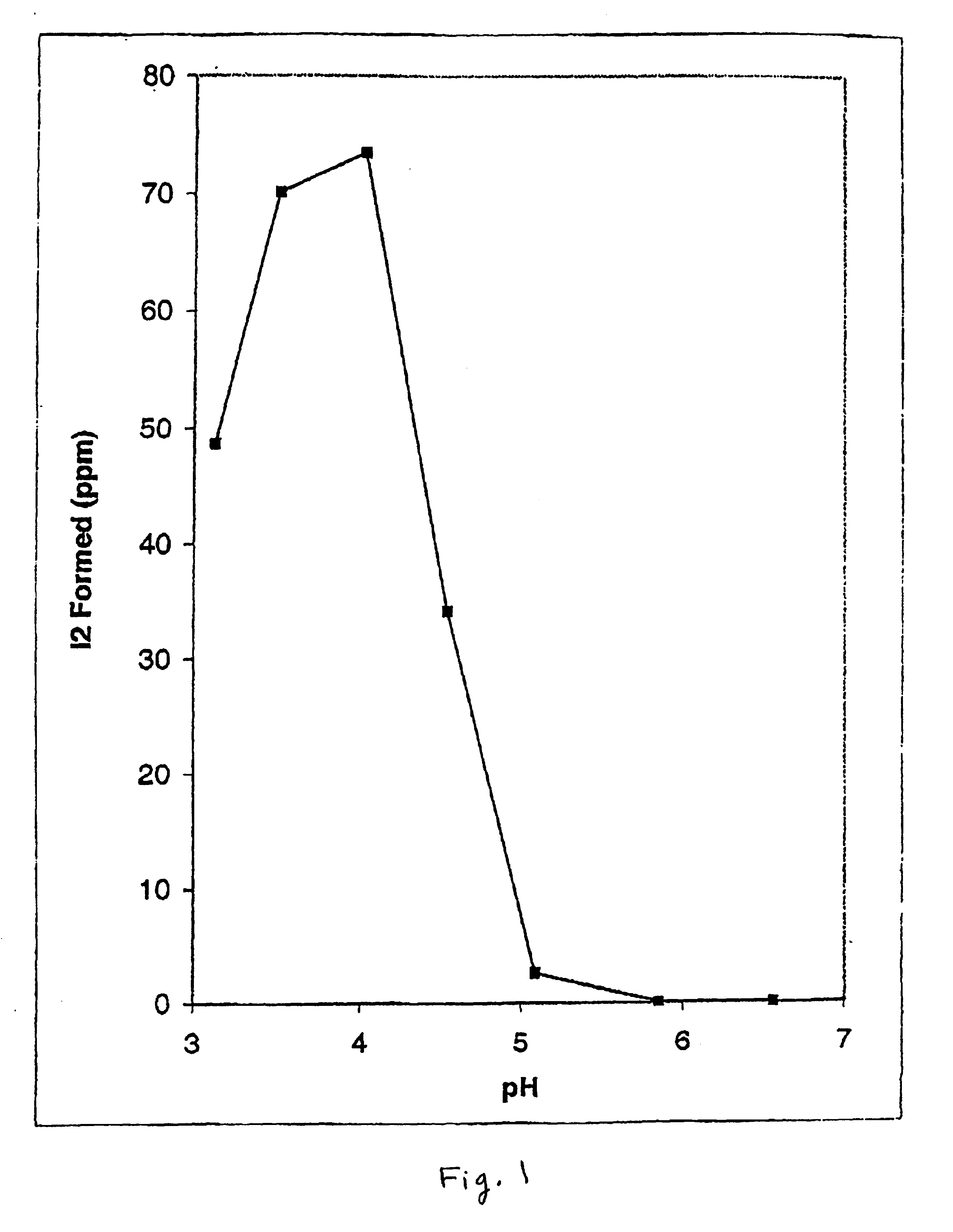
Effect of pH on De Novo Formation of Elemental Iodine Produced from Inorganic Iodide and Iodate. FIG. 1 shows the effect of varying the pH of buffered citrate solutions (50 mM) on the rate of free elemental iodine formation using potassium iodide and sodium iodate as precursors. Iodine was quantitated as the triiodide complex formed by transfer of aliquots of test reaction mixtures to stock 10 mM potassium iodide made up in distilled water measured at 350 nm on a double-beam UV-265 double beam Shimadzu spectrometer. A calibration curve was prepared using crystalline iodine dissolved in the same stock potassium iodide solution. Citrate buffered solutions were made up to known pH values as indicated, determined by titration and measurement on a standard pH meter. Aliquots of the citrate buffered solutions were mixed with silicone discs impregnated with a mixture of 2% potassium iodide and 10% sodium iodate (by weight relative to the disc). Following varying incubation periods...

example 2

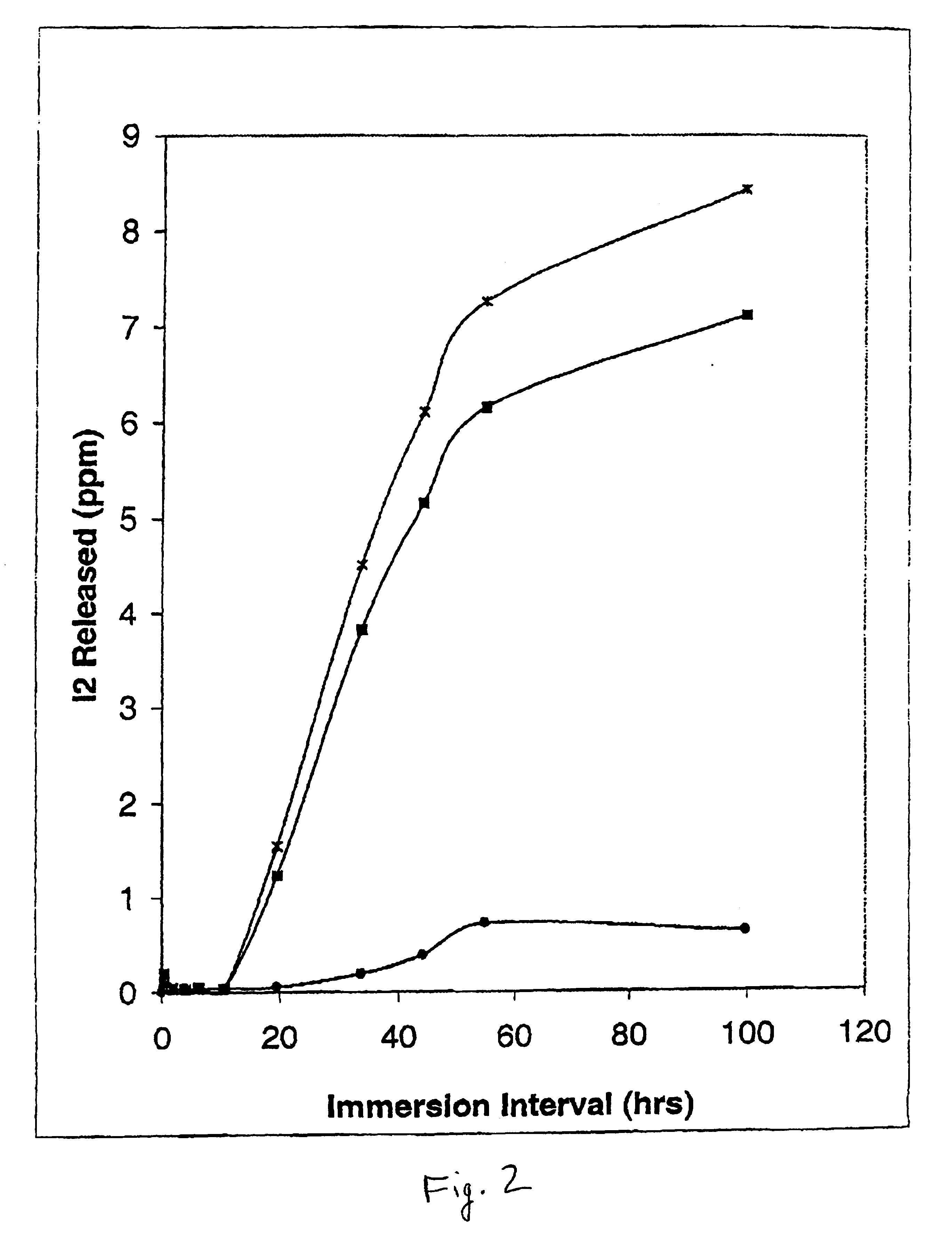
Egress of Iodine from Polyvinylchloride (PVC)-Loaded Catheter Tubing by ICAT. FIG. 2 shows the recovery of iodine released from PVC tubing preloaded with varying concentrations of iodine using in-situ chemical anti-infective loading technology (ICAT) for iodine generating formulations versus immersion times of the catheter in fluid. Five cm lengths of PVC tubing (outer diameter, 5.5 mm; inner diameter, 3.0 mm) were preloaded with iodine generating formulations made up of parts A and B in which the final concentration of inorganic sodium iodide ranged from 2.5 (solid circles), to 12.5 mM (solid squares) to 50 mM (“x” symbols), respectively. The final concentration of Carbopol C971 PNF was made up as 0.25%, and sodium iodate was held constant at 12.5 mM. The pH of the iodine generating formulation was 4.6. Immediately after preloading the inner lumen of the PVC tubing, the ends were capped with glass rods (total ICAT loading volume, 0.353 ml), and the catheter tubing was rinsed with d...

example 3

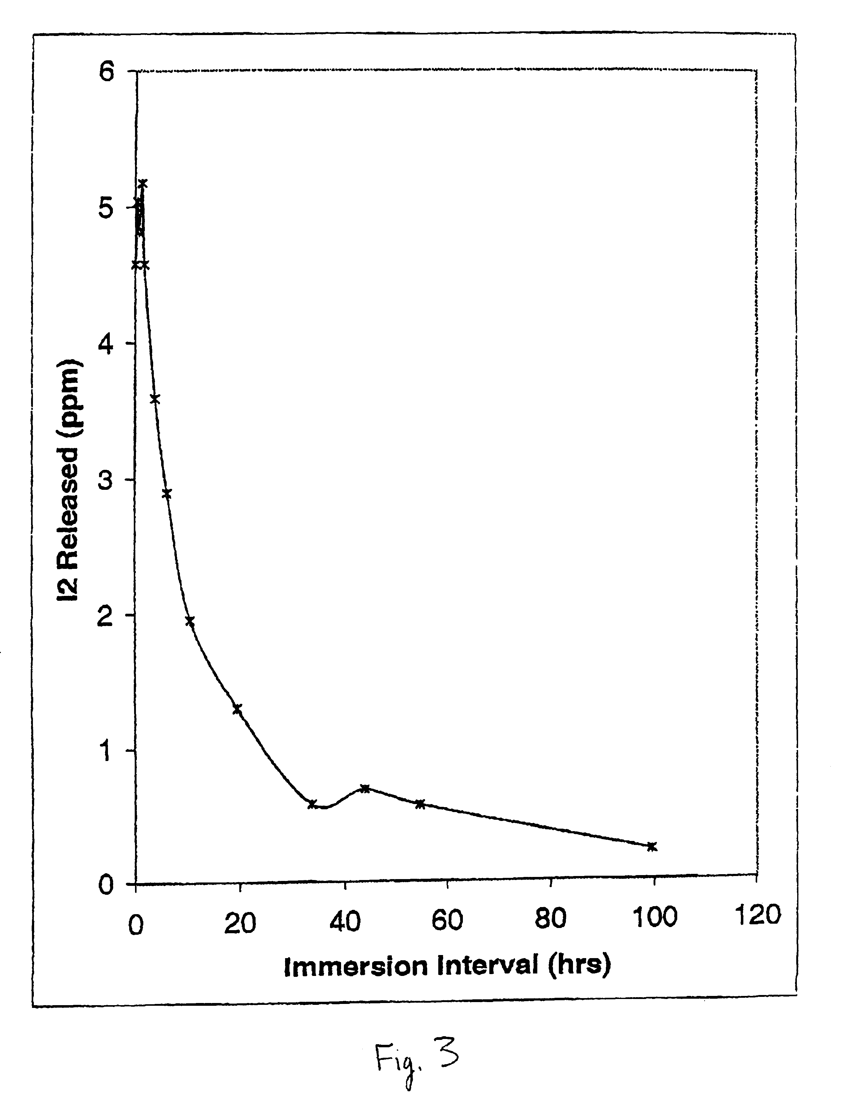
Uptake and Transport of Free Iodine into the Catheter Wall of ICAT-Loaded PVC Tubing. FIG. 3 shows the corresponding uptake of free iodine formed with loading of ICAT into the same PVC tubing using an identical experimental setup as in Example 2. In these experiments, ICAT loading was made using a final concentration of sodium iodide of 25 mM with sodium iodate held constant at 12.5 mM, Carbopol C971 PNF held at 0.25%, and the final pH set at 4.6. Following end capping of the PVC catheter tubing, and rinsing of the capped tubing in distilled water, the tubing was again placed in 10 ml of 10 mM KI. However, rather than monitoring the egress of iodine, at the points indicated in FIG. 3, 10 μl aliquots of the ICAT fluid were withdrawn from the internal lumen and the level of free iodine in the latter solution determined following 100-fold dilution in 10 mM potassium iodide solution. The concentrations shown in FIG. 3 thus represent {fraction (1 / 100)}th of the actual concentration of fe...


PUM


Abstract

Methods of providing anti-infective activity to a medical device including the steps of exposing the medical device to an anti-infective oxidant, and transferring the anti-infective oxidant into a wall of the medical device. One embodiment includes the step of exposing a medical device that is at least in part within a patient to the anti-infective oxidant. Another embodiment includes the steps of exposing a medical device to an aqueous solution which produces the anti-infective oxidant and transferring a sufficient amount of the anti-infective oxidant into the medical device wall to provide the medical device with anti-infective activity. Also provided is an oxidant releasing member that has anti-infective oxidant releasably contained therein and that is configured to be disposed adjacent to the medical device. Exemplary devices include catheters such as venous, arterial, and urinary catheters, generally containing an elongated catheter shaft having a lumen extending therein, and optionally having a balloon on the shaft in fluid communication with the catheter lumen.

Description

FIELD OF THE INVENTIONThe invention is directed to methods and devices for providing anti-infective activity to a medical device, and more particularly to methods and devices for transferring anti-infective activity through in-situ presentation of formulations which generate anti-infective oxidant such as elemental iodine and allow for its transfer to the polymer base of urinary, venous, and other lumen indwelling medical devices, conferring to such devices prophylactic and therapeutic treatment of implant-linked infections.REFERENCESU.S. Patents:U.S. Pat. No. 4,278,548 Bettinger et al. (1981) Control of biological growth in reverse osmosis permeators.U.S. Pat. No. 4,312,833 Clough et al. (1982) Sterilizing hydrophilic contact lenses.U.S. Pat. No. 4,476,108 Kessler et al. (1984) Bactericidal method.U.S. Pat. No. 4,576,817 Montgomery and Pellico (1986) Enzymatic bandages and pads.U.S. Pat. No. 4,937,072 Kessler et al. (1990) In situ sporicidal disinfectantU.S. Pat. No. 5,227,161 Kess...

Claims


Application Information

Patent Timeline
IPC IPC(8): A61L27/00A61L27/54A61L29/00A61L2/18A61L29/16A61L31/14A61L31/16
CPCA61L2/18A61L29/16A61L27/54A61L2300/404
Inventor GREEN, TERRENCE R.FELLMAN, JACK
Owner OXIBIO
Why Eureka
Try Eureka