Methods for tissue welding using laser-activated protein solders

a technology of laser activation and soldering, which is applied in the field of medical glues and adhesives, can solve the problems of inflammatory reactions, drawbacks of alternative tissue adhesives, and difficulty in healing and sealing tissue wounds

Inactive Publication Date: 2006-07-18
THE GENERAL HOSPITAL CORP
View PDF31 Cites 36 Cited by
  • Summary
  • Abstract
  • Description
  • Claims
  • Application Information

AI Technical Summary

Benefits of technology

[0114]A particular advantage of the present invention is the precision with which it can be used. Other glues that have been used stick wherever they are applied and much care must be taken to avoid the glue spilling over into areas where it is not desired. In the present invention, the glue is activated only where the practitioner applies the laser light and this can be a very small area, i.e., on the order of about 2 mm diameter. Thus any excess glue can be ignored or washed off. In this way the area of actual tissue adhesion is precisely controlled and is determined by the steadiness of the application of the laser energy. It is contemplated that even more precision is possible through the application of the laser energy by electronically controlled means. The glue composition may also be applied in layers to gradually fill in a defect or gradually strengthen the wound. The electromagnetic radiation may be delivered within the glue matrix through special delivery devices or simply applied on top of the glue layer.
[0115]The electromagnetic radiation necessary to achieve photoactivation will generally have a wavelength from about 10 nm to about 810 nm and will be within the visual, infra red or ultra violet spectra. The radiation will be supplied in the form of a monochromatic laser beam or other form of electromagnetic radiation source. The choice of energy source will generally be made in conjunction with the choice of photosensitizer employed in the composition. For example, an excimer laser is suitable for refractive surgeries. Suitable combinations of lasers and photosensitizers will be known to those of skill in the art, and are exemplified, but not limited to, those shown in Table 2.
[0116]TABLE 2PhotosensitizerLaser Source WavelengthAny tetrapyrrole including but not limitedBlue diode (460 nm)to: hematoporphyrin, chlorin,bacteriochlorin, phthalocyanine ornaphthalocyanineRose bengal or riboflavinBlue-Green Argon488–514 nmPorphyrins or cyaninesGreen diode (532 nm)Hematoporphyrin, protoporphyrin, otherRed diode (630 nm)porphyrins, toluidine blue, malachite green,azure A, azure B, brilliant green, patentblue VFChlorins, chlorine6, tetrahydroxyphenylRed diode 660 nmchlorin, methylene blue, Janus green,Benzoporphyrin derivativeRed diode (690 nm)All of the dye listed under red diodesTunable dye laser(600–700 nm)
[0117]Tunable dye lasers are also used with the present invention. These lasers can be tuned to emit generally any wavelength within a broad spectrum and allow for exact matching of dye and laser wavelength. In other embodiments, the source of irradiation may be non-laser light (i.e., polychromatic light) from an incandescent, fluorescent or other source which could be used to activate the glue / matrix composition. In particular embodiments the non-laser light source may be from a xenon bulb devices using filters and liquid light guides to deliver defined wavelength ranges depending on the filter. Application of these technologies will be known to those of skill in the art.
[0118]The present invention is envisioned to be suitable for use in a variety of surgical embodiments where one desires to seal, close, appose or otherwise join two or more portions of soft tissue, or to fill in cavities or damage to soft or hard tissues. The invention is considered to be particularly suitable for use in microsurgery, such as, for example, in surgical operations or maneuvers concerning the eye, small vascular tissue, gastro-intestinal tract, nerve sheaths, small ducts (e.g., urethra, ureter, bile ducts, thoracic duct) or even the inner ear, teeth or gums. It can be used in areas where use of a suture in a non-sterile part of the body would help foster an infection such as in the oral cavity. The invention may also be used in conjunction with ordinary sutures to provide a better cosmetic closure, such as to provide a smooth surface on top of the suture surface. It is particularly suitable for procedures involving laparoscopic operations or interventions such as laparoscopic (LP) cholecystectomy, LP nephrectomy, LP thoracic procedures, LP appendectomy, LP hernia repairs, LP tubal ligations and LP orbital surgeries. The present invention will also be useful in retractive surgeries. However, these examples are clearly not limiting and the use of the methods and compositions described herein in connection with any type of wound closure or surgical procedure is encompassed by the invention.
[0119]In yet further embodiments, the present invention concerns methods for cross-linking proteinaceous compounds, active agents and / or lipids. Such methods comprise forming an effective amount of a proteinaceous material or lipid cross-linker combination at the proteins, polypeptides, peptides, active agents and / or lipids to be cross-linked and irradiating the combination with electromagnetic radiation in a manner effective to promote the formation of cross-links between the proteins, polypeptides, peptides, active agents and / or lipids. The at least one proteinaceous compound, active agent and / or lipid cross-linker combination may be formed by preparing a composition in accordance herewith and applying the composition to the materials to be cross-linked, or by applying only the photosensitizer component itself to the proteins, polypeptides, peptides, active agents and / or lipids. In certain embodiments, dimers, such as an albumin dimer may form during crosslinking. In further embodiments, albumin-collagen cross-linking may occur. It is also contemplated that in certain embodiments, type I and / or type II photoprocesses may contribute to the process of crosslinking. The choice of electromagnetic radiation and photosensitizer will be coordinated as discussed above and as exemplified by the combinations listed in Table 2.

Problems solved by technology

Healing and sealing tissue wounds remains a problem in medical practice.
Alternative tissue adhesives have drawbacks.
Cyanoacrylate glues, which have been most frequently used in ophthalmology (Leahey et al., 1993) can be toxic, causing inflammatory reactions and are nonbiodegradable (Siegal and Zaidman, 1989).
Fibrin sealants (Spontnitz, 1995) are not particularly effective, form bonds of insufficient strength (Basu et al., 1995; Siedentop et al., 1988), present the possibility of viral infection if prepared from pooled human plasma, and may inhibit would healing (van der Ham et al., 1993).
Resorcinol gelatin sealants (Albes et al., 1993) can damage tissue because they contain formaldehyde (Ennker et al., 1994).
However, laser-activated tissue solders allows safe preparation and sterilization of the material, because it is activated only under laser illumination and is thought unlikely to lead to tissue toxicity (Bass and Treat, 1995).

Method used


Image

  • Methods for tissue welding using laser-activated protein solders
  • Methods for tissue welding using laser-activated protein solders
  • Methods for tissue welding using laser-activated protein solders

Examples


example 1

Photodynamic Tissue Adhesion with Chlorine6 Protein Conjugates

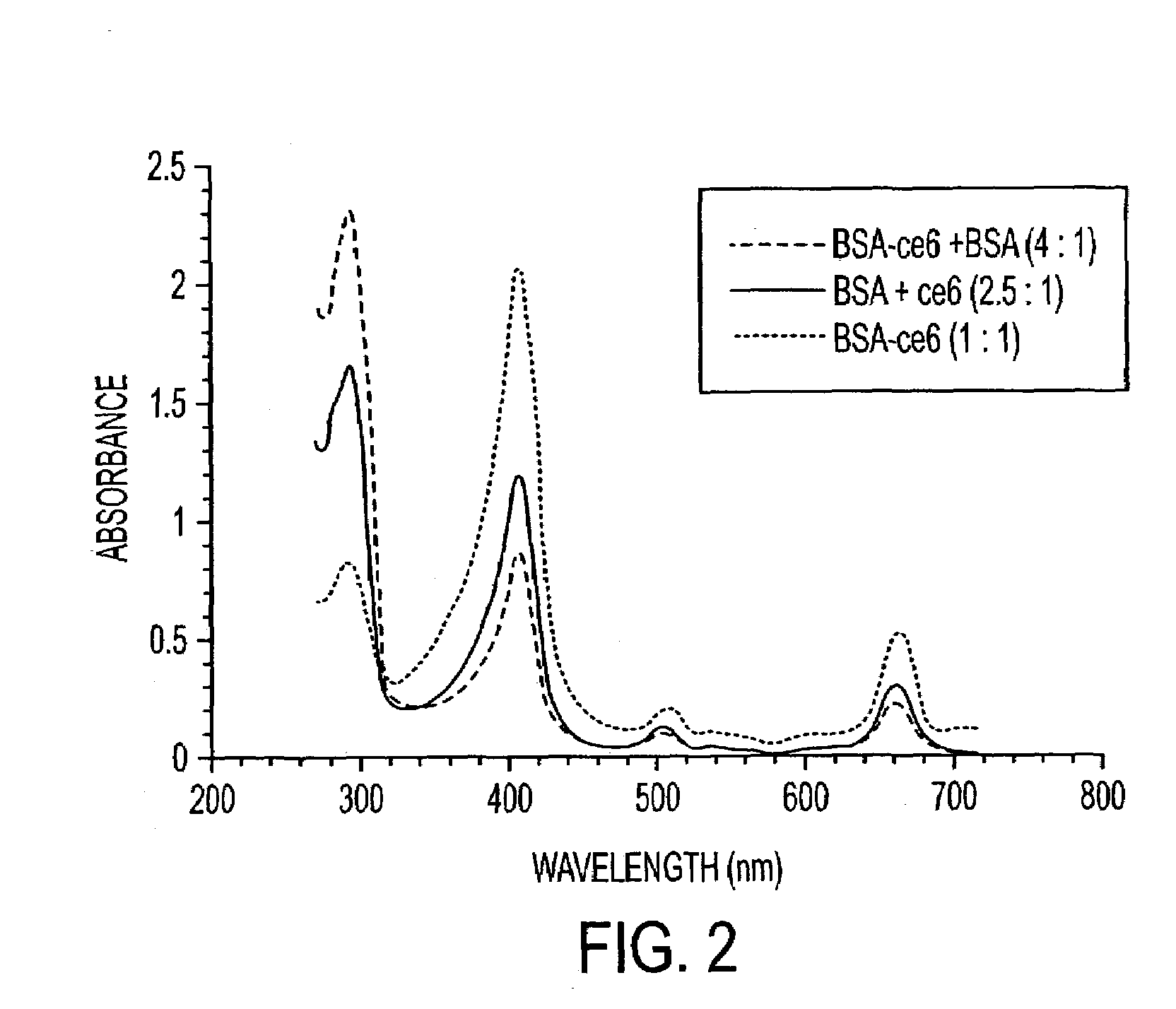
[0130]This example demonstrates that the use of a chromophore, such as for example, chlorine6 (Ce6) may be effective in cross-linking proteins by a photodynamic mechanism. Covalent conjugates between Ce6 and proteins used as laser-activated solders form stronger tissue bonds than noncovalent mixtures. The finding that addition of sodium azide to the glue preparation reduces the leaking strength by more than 50% is attributed to quenching of singlet oxygen.

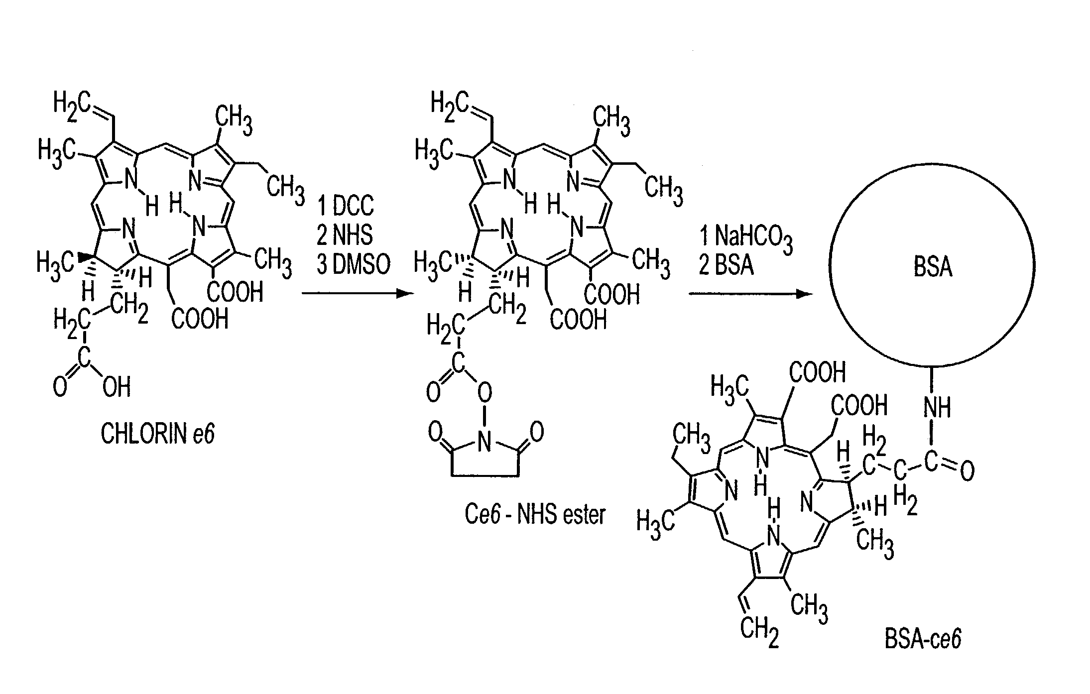
[0131]Preparation of Conjugates: Ce6 was obtained from Porphyrin Products (Logan, Utah), N-hydroxy succinimide (NHS), dicyclohexylcarbodiimide, bovine fibrinogen, bovine serum albumin (BSA), and gelatin were from Sigma (St. Louis, Mo.). Frozen, nonpreserved, human cadaveric eyes were obtained from the Illinois Eye Bank (Chicago). The reaction sequence used to attach the Ce6 molecules to the proteins covalently is shown in FIG. 1. All reactions were performed in the dark ...

example 2

Photodynamic Tissue Adhesion with Chlorine6 Protein Conjugates

[0142]The adhesive strength of covalent conjugates to non-covalent mixtures between chlorine6 and proteins in a laser activated soldering process are compared in this example.

[0143]The covalent and non-covalent mixtures of Ce6 photosensitizer contained different molar ratios of albumin, gelatin and fibrinogen. These photodynamic glues were applied to 5 mm incisions along the equatorial sclera and activated with an Argon blue-green laser over a two minutes period. Intra-ocular pressure was increased in 5 mmHg increments until fluid or air escaped from the wound.

[0144]The albumin-chlorine6 conjugate welds were significantly stronger than non-covalent mixtures. The albumin-Ce6 with added albumin provided the highest bursting pressures (mean=207.1 mmHg) in comparison to the other covalent conjugates (mean=88.8 mmHg) and non-covalent mixtures (mean=76.7 mmHg). The order of strength for the most effective photodynamic glues was...

example 3

Tissue Glue as a Slow Release Biomatrix

[0146]In this example, a dye containing protein glue mixture containing a phamacologically active amount of the appropriate growth factor will be formulated. Experiments will be conducted in vitro to test the extent of inactivation of the growth factor during the laser activation of the glue. It is expected that although some of the growth factor will be inactivated, enough will remain of the amount introduced to stimulate the tissue repair or wound healing.

[0147]Preliminary experiments in the rat cornea show that the laser activated glue remains in the incision for 48 hours, although much less than at time zero. This apparently fulfills the requirement of being a slow release vehicle with a defined rate of biodegradation.


PUM

PropertyMeasurementUnit
viscosityaaaaaaaaaa
temperatureaaaaaaaaaa

Abstract

Various tissue glues have drawback such as toxicity, causing inflammatory reactions or insufficient bonding strength. The present invention is directed to methods of form tissue adhesion by administering to tissues compositions comprising proteins conjugated to one or more novel photosensitizers and irradiating the composition. The composition may further comprise one or more proteins not conjugated to the photosensitizer. Additionally, the present invention relates to compositions and methods wherein increased ratios of protein to photosensitizer enhance weld strength.

Description

[0001]This Application is a continuation of prior U.S. application Ser. No. 09 / 527,369, filed Mar. 16, 2000 now, U.S. Pat No 6,607,522 B1.BACKGROUND OF THE INVENTION[0002]1. Field of the Invention[0003]The present invention relates generally to the fields of medical glues and adhesives. More particularly, it concerns methods and compositions for sealing of wounds and incisions. In certain aspects, the invention concerns adhesion of two or more tissue samples using proteinaceous and / or lipoproteinaceous compositions conjugated and / or mixed with a photosensitizer or dye upon irradiation.[0004]2. Description of Related Art[0005]Healing and sealing tissue wounds remains a problem in medical practice. To enhance tissue healing, much effort has gone towards producing a biocompatible slow release formulation which can be introduced into a tissue defect and that will release biologically active growth factor at a steady rate for the required time. These formulations were designed to promote...

Claims


Application Information

Patent Timeline
Patent Type & Authority Patents(United States)
IPC IPC(8): A61B18/18A61B17/00A61L24/00A61L24/04A61L24/10A61L31/04A61L31/14
CPCA61L31/14A61L31/043A61B2017/00517
Inventor HAMBLIN, MICHAEL R.KHADEM, JOHN
Owner THE GENERAL HOSPITAL CORP
Why Eureka
Try Eureka