Nanoparticles for protein drug delivery

a technology of nanoparticles and proteins, applied in the direction of depsipeptides, peptide/protein ingredients, powder delivery, etc., can solve the problems of limited hydrophilic drug transport through paracellular pathway, inability to easily diffuse hydrophilic drugs across cells, and inability to achieve the effect of enhancing intestinal paracellular transport, maintaining protein bioactivity, and adequate solubility

Inactive Publication Date: 2007-10-16
NANOMEGA MEDICAL CORP
View PDF14 Cites 48 Cited by
  • Summary
  • Abstract
  • Description
  • Claims
  • Application Information

AI Technical Summary

Benefits of technology

[0010]It is one object of the present invention to provide a novel nanoparticle system and methods of preparation for paracellular transport drug delivery using a simple and mild ionic-gelation method upon addition of a poly-γ-glutamic acid (γ-PGA) solution into a low molecular weight chitosan (low-MW CS) solution. In one embodiment, the molecular weight of a low-MW CS of the present invention is about 80 kDa or less, preferably at about 40 kDa, adapted for adequate solubility at a pH that maintains the bioactivity of protein and peptide drugs. It is stipulated that a chitosan particle with about 30-50 kDa molecular weight is kidney inert. The particle size and the zeta potential value of the prepared nanoparticles are controlled by their constituted compositions. The results obtained by the TEM (transmission electron microscopy) and AFM (atomic force microscopy) examinations showed that the morphology of the prepared nanoparticles was generally spherical in shape. Evaluation of the prepared nanoparticles in enhancing intestinal paracellular transport was investigated in vitro in Caco-2 cell monolayers. In some aspects of the present invention, it provides the nanoparticles with CS dominated on the surfaces to effectively reduce the transepithelial electrical resistance (TEER) of Caco-2 cell monolayers. The confocal laser scanning microscopy (CLSM) observations confirm that the nanoparticles with CS dominated on the surface are able to open the tight junctions between Caco-2 cells and allows transport of the nanoparticles via the paracellular pathways.

Problems solved by technology

This is because hydrophilic drugs cannot easily diffuse across the cells through the lipid-bilayer cell membranes.
The transport of hydrophilic molecules via the paracellular pathway is, however, severely restricted by the presence of tight junctions that are located at the luminal aspect of adjacent epithelial cells (Annu. Rev. Nutr. 1995;15:35-55).
However, these nanoparticles are not ideal carriers for hydrophilic drugs because of their hydrophobic property.
The absorption of protein drugs following oral administration is challenging due to their high molecular weight, hydrophilicity, and susceptibility to enzymatic inactivation.
However, the tight junction forms a barrier that limits the paracellular diffusion of hydrophilic molecules.
Most commercially available CSs have a quite large molecular weight (MW) and need to be dissolved in an acetic acid solution at a pH value of approximately 4.0 or lower that is sometimes impractical.

Method used


Image

  • Nanoparticles for protein drug delivery
  • Nanoparticles for protein drug delivery
  • Nanoparticles for protein drug delivery

Examples


example no.1

EXAMPLE NO. 1

[0043]Materials and Methods of Nanoparticles Preparation

[0044]CS (MW ˜2.8×10⁵) with a degree of deacetylation of approximately 85% was acquired from Challenge Bioproducts Co. (Taichung, Taiwan). Acetic acid, cellulase (1.92 units / mg), fluorescein isothiocyanate (FITC), phosphate buffered saline (PBS), periodic acid, sodium acetate, formaldehyde, bismuth subnitrate, and Hanks balanced salt solution (HBSS) were purchased from Sigma Chemical Co. (St. Louis, Mo.). Ethanol absolute anhydrous and potassium sodium tartrate were obtained from Merck (Darmstadt, Germany). Non-essential amino acid (NEAA) solution, fetal bovine serum (FBS), gentamicin and trypsin-EDTA were acquired from Gibco (Grand Island, N.Y.). Eagle's minimal essential medium (MEM) was purchased from Bio West (Nuaille, France). All other chemicals and reagents used were of analytical grade.

example no.2

EXAMPLE NO. 2

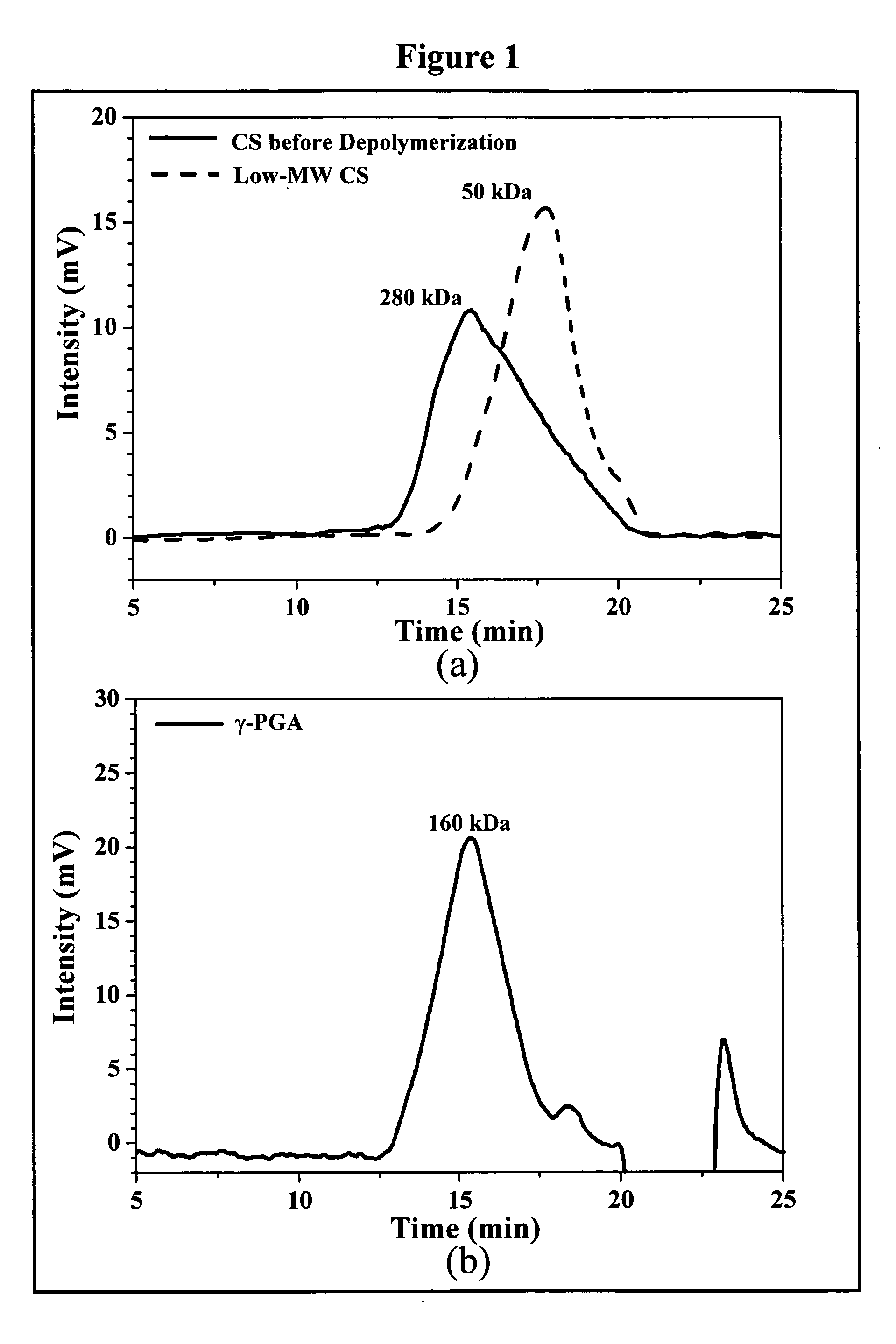
[0045]Depolymerization of CS by Enzymatic Hydrolysis

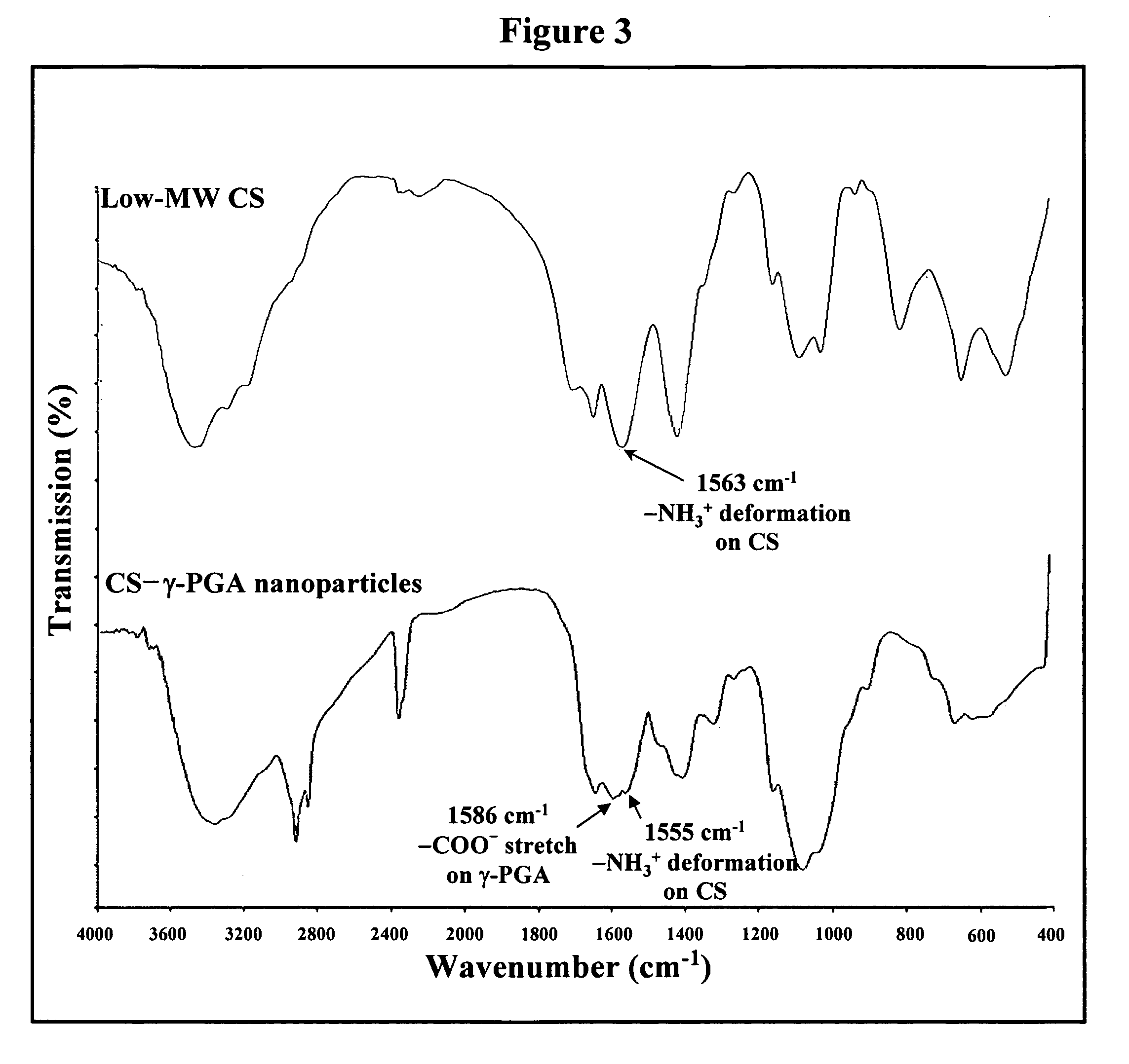
[0046]Regular CS was treated with enzyme (cellulase) to produce low-MW CS according to a method described by Qin et al. with some modifications (Food Chem. 2004;84:107-115). A solution of CS (20 g / l) was prepared by dissolving CS in 2% acetic acid. Care was taken to ensure total solubility of CS. Then, the CS solution was introduced into a vessel and adjusted to the desired pH 5.0 with 2N aqueous NaOH. Subsequently, cellulase (0.1 g) was added into the CS solution (100 ml) and continuously stirred at 37° C. for 12 hours. Afterward, the depolymerized CS was precipitated with aqueous NaOH at pH 7.0-7.2 and the precipitated CS was washed three times with deionized water. The resulting low-MW CS was lyophilized in a freeze dryer (Eyela Co. Ltd, Tokyo, Japan).

[0047]The average molecular weight of the depolymerized CS was determined by a gel permeation chromatography (GPC) system equipped with a series of PL aquagel-OH colum...

example no.3

EXAMPLE NO. 3

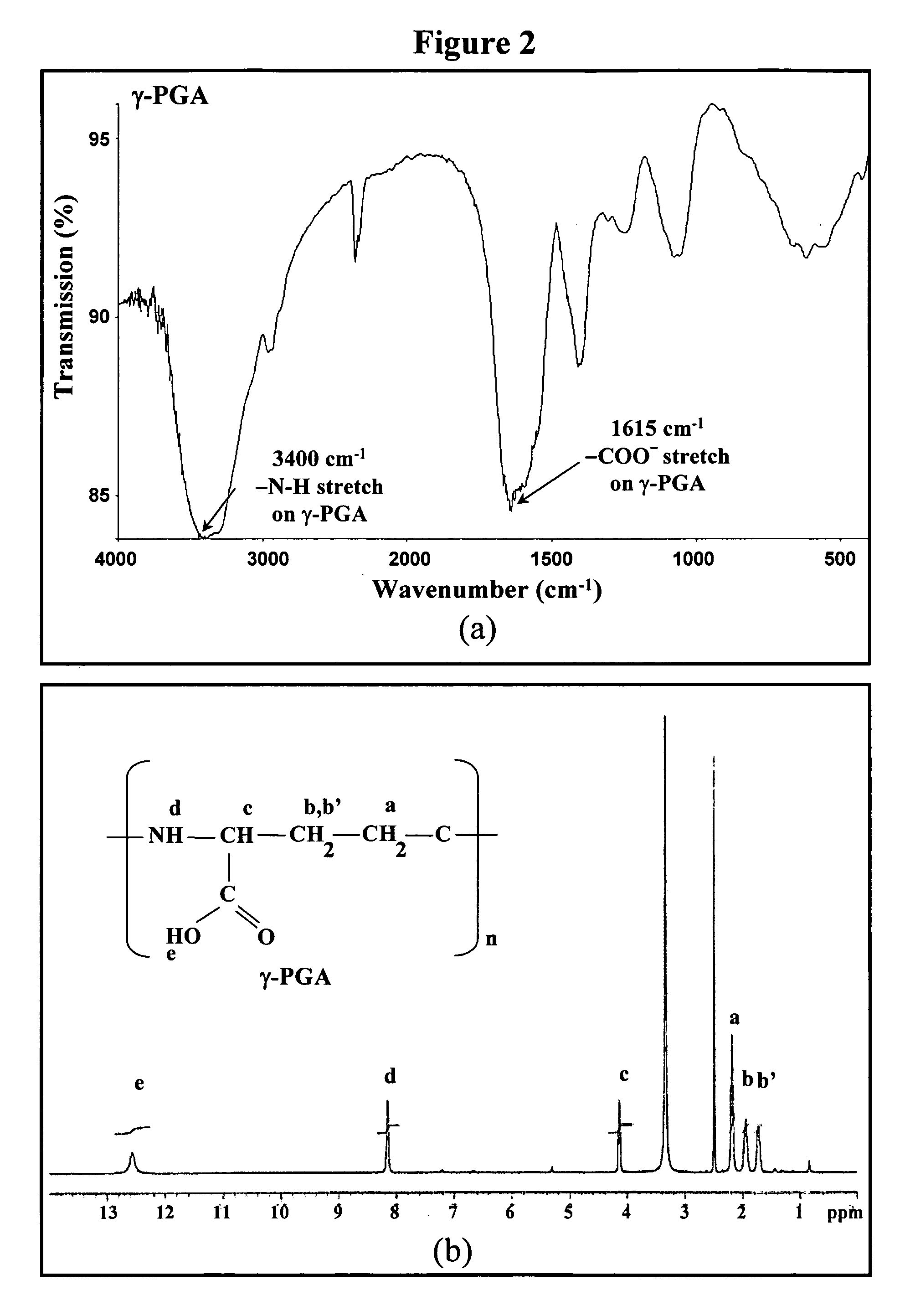
[0050]Production and Purification of γ-PGA

[0051]γ-PGA was produced by Bacillus licheniformis (ATCC 9945, Bioresources Collection and Research Center, Hsinchu, Taiwan) as per a method reported by Yoon et al. with slight modifications (Biotechnol. Lett. 2000;22:585-588). Highly mucoid colonies (ATCC 9945a) were selected from Bacillus licheniformis (ATCC 9945) cultured on the E medium (ingredients comprising L-glutamic acid, 20.0 g / i; citric acid, 12.0 g / l; glycerol, 80.0 g / l; NH4Cl, 7.0 g / l; K2HPO4, 0.5 g / l; MgSO4·7H2O, 0.5 g / l; FeCl3·6H2O, 0.04 g / l; CaCl2·2H2O, 0.15 g / l; MnSO4·H2O, 0.104 g / l, pH 6.5) agar plates at 37° C. for several times. Subsequently, young mucoid colonies were transferred into 10 ml E medium and grown at 37° C. in a shaking incubator at 250 rpm for 24 hours. Afterward, 500 μl of culture broth was mixed with 50 ml E medium and was transferred into a 2.5-1 jar-fermentor (KMJ-2B, Mituwa Co., Osaka, Japan) containing 950 ml of E medium. Cells were cultur...


PUM

PropertyMeasurementUnit
concentrationaaaaaaaaaa

Abstract

The invention discloses the nanoparticles composed of chitosan, poly-γ-glutamic acid, and at least one bioactive agent characterized with a positive surface charge and their enhanced permeability for paracellular drug delivery.

Description

CROSS-REFERENCE TO RELATED APPLICATIONS[0001]This application is a continuation-in-part application of U.S. patent application Ser. No. 11 / 029,082, filed Jan. 4, 2005, entitled “Nan particles for Paracellular Drug Delivery”, now U.S. Pat. No. 7,265,090, which is a continuation in part application of U.S. patent application Ser. No. 10 / 958,864, filed Oct. 5, 2004, pending, the entire contents of which are incorporated herein by reference.FIELD OF THE INVENTION[0002]The present invention is related to medical uses of nanoparticles composed of chitosan / poly-γ-glutamic acid with protein drugs and their permeability for enhanced paracellular drug delivery.BACKGROUND OF THE INVENTION[0003]Production of pharmaceutically active peptides and proteins in large quantities has become feasible (Biomacromolecules 2004;5:1917-1925). The oral route is considered the most convenient way of drug administrations for patients. Nevertheless, the intestinal epithelium is a major barrier to the absorption...

Claims


Application Information

Patent Timeline
Patent Type & Authority Patents(United States)
IPC IPC(8): A61K51/00A61K9/50
CPCA61K9/0019A61K9/5161A61K9/5192A61K31/13A61K31/473A61K31/55A61K31/7008A61K31/704A61K31/727A61K33/24A61K38/28A61K45/06A61K47/4823A61K9/5146Y10S977/773A61K2300/00A61K47/61
Inventor SUNG, HSING-WENLIN, YU-HSINLIANG, HSIANG-FATU, HOSHENG
Owner NANOMEGA MEDICAL CORP
Why Eureka
Try Eureka