Self-contained microelectrochemical bioassay platforms and methods

a microelectrochemical and bioassay technology, applied in the direction of fluid speed measurement, positive displacement liquid engine, chemical vapor deposition coating, etc., can solve the problems of not including a separation step, unfavorable immunoactive physisorption, and unfavorable immunoactive physisorption, so as to improve detection limits, sensitivity and speed, and improve detection accuracy. , the effect of improving the detection limi

Inactive Publication Date: 2010-01-12
THE BOARD OF TRUSTEES OF THE UNIV OF ARKANSAS
View PDF3 Cites 3 Cited by
  • Summary
  • Abstract
  • Description
  • Claims
  • Application Information

AI Technical Summary

Benefits of technology

[0021]The methods used to form these microstructures allow them to be extremely small. Micropores and microcavities may be formed that are less than a hundred micrometers wide. In fact they may be less than 10 μm wide. The depth of the microstructures ranges anywhere from less than 10 μm to over 100 μm. Microstructures are combined with known chemical detection assays. Surface immobilization assays are especially well suited for these microstructures, although any assays susceptible to electrochemical detection may be combined with these microstructures. Surface immobilization assays are well known to those skilled in the art and include, but are not limited to, immunoassays, northern and southern blots, western blots and incorporation of proteins into lipid layers. Surface immobilization assays are especially advantageous for use in microstructures because the small size of the structures allows for a very short distance between the analyte being captured or recognized and the electrodes being used for electrochemical detection. In addition, the short distance between the electrodes used for detecting the analyte also accelerates both detection and amplification by means of redox cycling. Another benefit of combining surface immobilization assays with electrochemical microstructures is that physisorption of materials used for surface immobilization may be regulated within the microstructure. By applying electrical currents to various electrodes, the location of analyte binding materials within the microstructure may be controlled. This allows a certain material, such as protein binding styrene or primary antibodies, to bind to insulating layers or specific electrodes while preventing physisorption of these molecules to working electrodes.
[0031]This invention makes smaller volumes no more difficult to analyze than macrovolumes because all of the electrodes are prefabricated within the same small volume, thereby allowing self-contained electrochemistry to occur. Not only can heterogeneous immunoassays in such electrochemical systems be applied to smaller sample volumes than previously possible, but they also offer better detection limits, sensitivity, and speed because of the close proximity of electrodes and modified surfaces. In addition, a new polyimide passivation protocol protects the detecting electrodes until they are needed and prevents immuno active physisorption to undesirable locations. These procedures allow fabrication of accurate and reliable immunosensor arrays. This procedure is also promising for modification in enclosed microfluidic devices where photo patterning may not be convenient.

Problems solved by technology

This does not necessarily demand better detection limits.
The latter device, however, does not include a separation step, and therefore may be susceptible to interferences from other sample species.
Low detection limits (pg / mL or fM) were not possible.
Consequently, this setup is not well-suited for integration with small volume handling or automation and has added complexity due to the SECM instrumentation and operation.
Even while in a laboratory, many chemical detection assays take a relatively long period of time.
The disadvantages of immunofluorescence assays (IFA) include their low recovery efficiency, long processing times, the need for highly trained analysts and high cost.
In addition, IFA detection often involves the time consuming and skill intensive step of filtering and analyzing water sludge for analytes that have been labeled with a fluorescent antibody.
It is also often difficult to distinguish microorganisms from debris bound non-specifically by the antibodies.
The procedure is expensive and often takes days to complete.
Flow cytometry techniques can quantify microorganisms but involves much preparation, and time and require extremely expensive equipment.
Numerous problems are associated with prior art methods of detecting microorganisms and biological molecules in water and environmental samples.
In addition to those mentioned and the general lack of precise, reliable assays, prior art techniques generally require that samples be transferred to a laboratory or to another remote location for the conduct of the assay.
Prior art techniques lack the requisite reliability, speed and sensitivity to accurately detect microorganisms and biological compounds in contaminated water samples.
The known methods of enzyme immunoassays and immunofluorescence do not fulfill these requirements.
The detection, viability or quantity of microorganisms found in water or other environmental samples cannot be reliably determined using prior art methods.

Method used


Image

  • Self-contained microelectrochemical bioassay platforms and methods
  • Self-contained microelectrochemical bioassay platforms and methods
  • Self-contained microelectrochemical bioassay platforms and methods

Examples


example 1

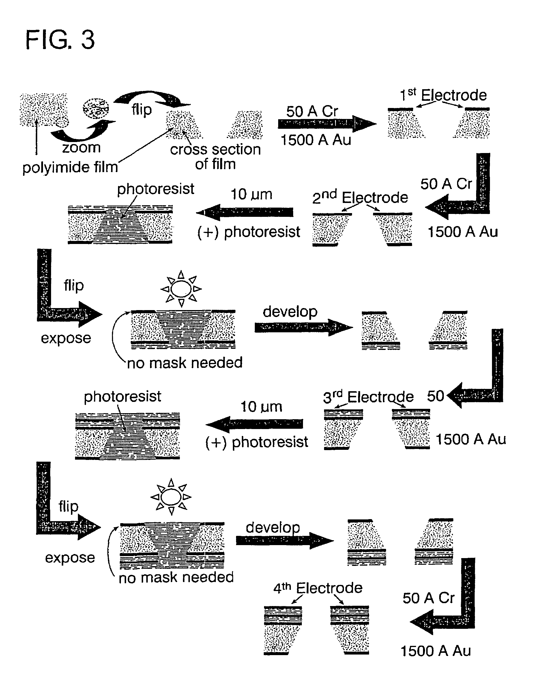
[0101]Fabrication of macrochips. Au macrochips (approximately 1.4 cm×2 cm, where the electroactive area is about 0.6 to 1 cm2) were made from a 125 mm diameter silicon wafer substrate that had 1.4 to 1.8 m SiO2 deposited on both sides at 250° C. by plasma enhanced chemical vapor deposition (PEVCD, Plasma Therm System VII). Deposition of a 15 μM adhesion layer of Cr and 1000 μm of Au was carried out using an Edwards Auto 306 TURBO thermal evaporator (Edwards High Vacuum Instrument International, West Sussex, UK). The Au macrochips were diced to size by hand using a diamond scribe. Polyimide (PI) macrochips were made using the Au macrochips as the starting substrate and spin coating a 4 μm thick layer of polyimide followed by cross-linking with UV light at 350 nm for 12 s and curing at 150° C. for 30 min and at 250° C. for another 30 min. The Au coated silicon wafer was spin-rinsed-dried (SRD) using ST 270D (Semitool, Calif.) for a total of 400 s before spin coating the PI. This proce...

example 2

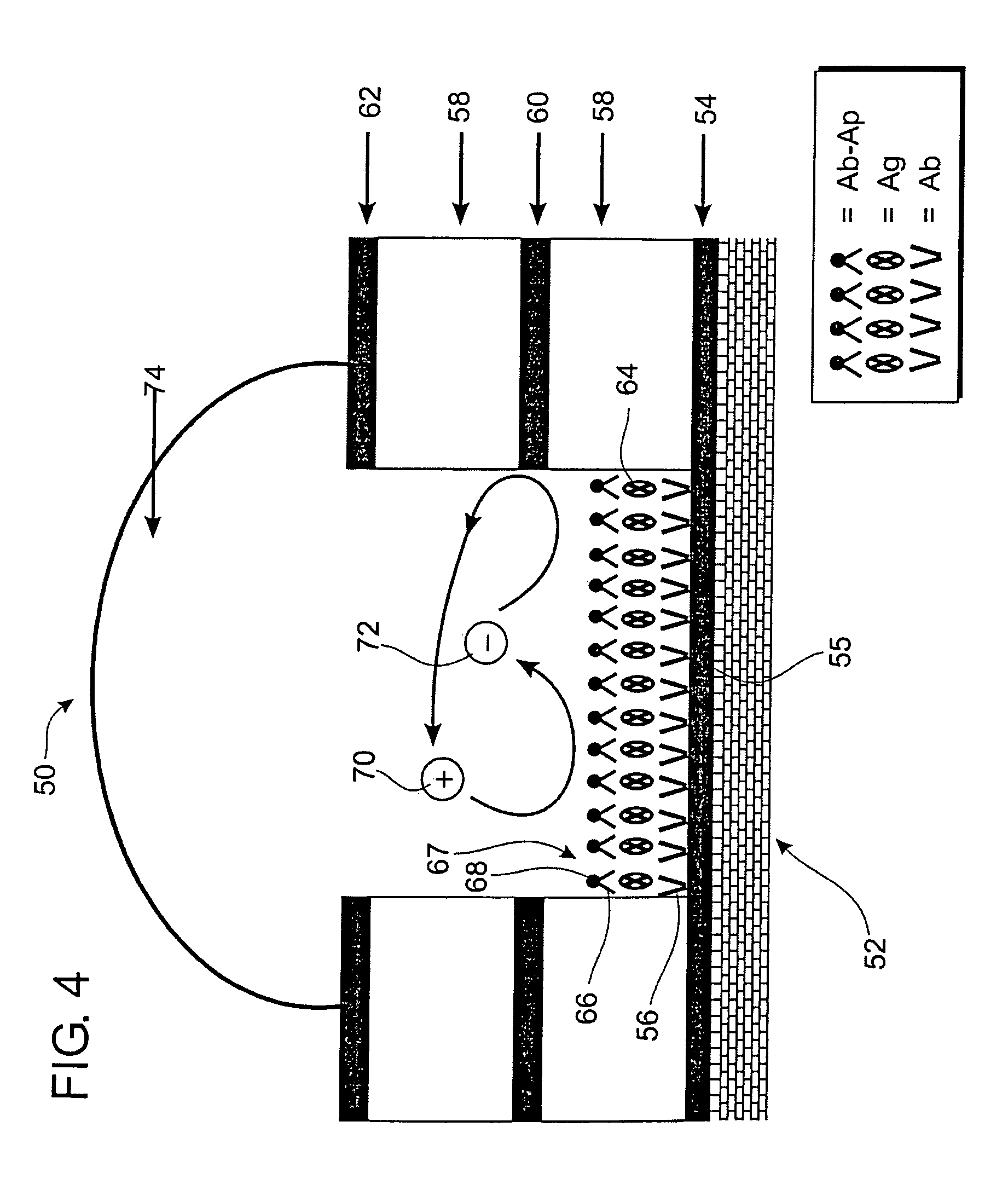
[0143]The following novel technique was designed because there is no existing electrochemical immunoassay for Cryptosporidium parvum-ZPA. Although the method described below is macroscale, those skilled in the art will recognize that it may easily be scaled down to operate within a microstructure as shown in FIG. 10 and may be adapted for detection of a variety of microorganisms simply by changing the antibodies used.

[0144]1) Deposit 50 Å(angstrom) chromium (Cr) followed by 1000 Å gold (Au) on 5 on oxidized silicon wafers using thermal vapor deposition.

[0145]2) Using a diamond scribe cut the wafer into 1.2×1.2 cm2 Au chips.

[0146]3) Clean with piranha (3 parts 30% H2O2 hydrogen peroxide: 7 parts concentrated H2SO4 sulfuric acid) and rinse thoroughly with running dI water.

[0147]4) Deposit self assembled monolayers (SAMs) using 25 mL of 4 mM MUA (mercaptoundecanoic acid) or MUOL (mercaptoundecanol) in ethanol overnight inside an Ar (argon) purged glove bag.

[0148]5) Rinse thoroughly wit...

example 3

[0156]The novel DNA-hybridization Assay for Cryptosporidium parvum-Macro system described below was developed to demonstrate the feasibility of the viability assay for microorganisms. Those skilled in the art will appreciate that this method is easily scaled down to work inside a microstructure, as shown in FIG. 11B.

[0157]Make sure all solutions and solvents are filtered with 0.2 μm filter. Vortex all solutions before to use. Autoclave deionized (dI) water for all solutions. Use only RNAse and DNAase free reagents without autoclaving.

[0158]Macro Chips

[0159]1) Deposit ˜100 Å Cr / 1000 Å Au on 5 in silicon wafer (Si / SiO2) using BOC Edwards Thermal Evaporator.

[0160]2) Dice with a diamond tip scrib to 1.2×1.2 cm2 pieces or macro chips.

[0161]3) Soak macro chips in piranha solution for 30 min then wash thoroughly with running dl water.

[0162]MUOL SAMs

[0163]1) Inside an AR filled glovebag, soak the chips in 4 mM MUOL in ethanol overnight.

[0164]2) Rinse three times with ethanol followed by dI ...


PUM

PropertyMeasurementUnit
volumeaaaaaaaaaa
pHaaaaaaaaaa
volumesaaaaaaaaaa

Abstract

Methods and devices for improved chemical and biological detection assays combined well defined microstructures having independently addressable electrodes with various surface immobilization electrochemical assays. Combining known chemical detection immobilization assays, electrochemically active moieties with microstructures having independently addressable electrodes provides for vastly improved methods of detecting microorganisms, chemical compounds, and measuring membrane transport.

Description

CROSS-REFERENCE TO RELATED APPLICATIONS[0001]This application is a divisional of U.S. patent application Ser. No. 10 / 253,187, filed Sep. 24, 2002, and claims priority to U.S. patent application Ser. No. 09 / 978,734, now U.S. Pat. No. 6,887,714, filed Oct. 15, 2001, and claims priority to U.S. Provisional Application Ser. No. 60 / 240,691, filed Oct. 16, 2000.BACKGROUND OF THE INVENTION[0002]1. Field of the Invention[0003]The present invention relates to a microvolume electrochemical detection assay. More specifically, the present invention relates to microscale structures having an analyte immobilizing surface and at least one electrode separate from the immobilizing surface. The microscale devices are used in conjunction with an immobilization assay that utilizes an electroactive complex to generate a current that may be detected by the electrode. The small volumes and sensitivity of the assays used in conjunction with microstructures provides an extremely rapid detection method havin...

Claims


Application Information

Patent Timeline
Patent Type & Authority Patents(United States)
IPC IPC(8): G01N33/543B01F13/00B01J19/00B01L3/00B81B1/00B81C1/00F04B17/00G01N27/403
CPCB01F13/0077G01N27/3278B01L3/5027B01L3/5085F04B17/00G01N33/5438B01J19/0046B01J2219/00317B01J2219/00511B01J2219/00585B01J2219/00596B01J2219/00653B01J2219/00659B01J2219/00704B01L3/502707B01L2200/0605B01L2400/0415B01L2400/043C12Q2565/607B01F33/3032
Inventor FRITSCH, INGRIDBEITLE, JR., ROBERTAGUILAR, ZORAIDA
Owner THE BOARD OF TRUSTEES OF THE UNIV OF ARKANSAS
Why Eureka
Try Eureka