
Muscle-derived cells (MDCs) for treating muscle- or bone-related injury or dysfunction

a technology of muscle- or bone-related injury or dysfunction, which is applied in the field of myogenic or muscle-derived cells, can solve the problems of affecting the delivery of such quantities of protein, affecting the effect of urethra wall bulking, and general undetectable agents, etc., and achieves the effect of large bulking

Inactive Publication Date: 2011-02-15
UNIVERSITY OF PITTSBURGH
View PDF53 Cites 16 Cited by
  • Summary
  • Abstract
  • Description
  • Claims
  • Application Information

AI Technical Summary

Benefits of technology

[0039]It is an object of the present invention to provide new and effective methods and compositions for the treatment of various types of disease conditions and defects of the musculoskeletal system and the bone, using genetically engineered muscle-derived cells, e.g., myoblasts and muscle-derived stem cells, (also referred to as MDCs herein) in the cell-mediated delivery of exogenous genes for the expression and production of encoded gene products. The present invention affords a stable gene delivery vehicle to afflicted areas, e.g., the joint (ligament, meniscus, and cartilage), smooth muscle, skeletal muscle and bone, which sustains the production of proteins that ameliorate pathological muscle-related conditions, e.g., musculoskeletal and bone conditions. Examples of pathological conditions of the musculoskeletal system include arthritis and damage to ligaments, cartilage and meniscus, resulting in general muscle weakness and / or dysfunction, such as in the face and hands, as a nonlimiting example. Disease conditions of the bone include segmental bone fractures, defects, weakness, non-unions and any type of bone augmentation. The present invention overcomes the problem of transient gene expression, which reduces the efficacy of ex vivo gene transfer to the joint, for example, using synovial cells.

Problems solved by technology

Although synovial cells have been used to deliver potentially therapeutic agents into the joint, the expression of such agents has declined over time, thereby causing these agents generally to become undetectable after about four to six weeks.
Segmental bone defects and non-unions are relatively common problems facing all orthopedic surgeons.
Moreover, current modes of delivering such quantities of protein, i.e., a biological allograft or a synthetic carrier, are hampered by limited availability, possible disease transmission and the need for further research and investigation.
However, this reparative process is usually incomplete and accompanied by a fibrous reaction producing scar tissue.
Unfortunately, current techniques of autograft, allograft, and electrical stimulation are often suboptimal.
However, neither the mechanism of formation nor suppression of ectopic bone is clearly understood.
Unfortunately, the ACL has a low healing capacity, possibly secondary to its encompassing synovial sheath or the surrounding synovial fluid.
Because complete tears of the ACL are incapable of spontaneous healing, current treatment options are limited to surgical reconstruction using autograft or allograft.
Transient, low levels of these growth factors resulting from their direct injection into the injured ligament are unlikely to produce a significant response.
Urinary incontinence is a devastating medical and social condition.
Presently, the estimated annual cost for treating people with urinary incontinence is over $16 billion in the United States.
Overflow incontinence may be due to impaired bladder contractility or to bladder outlet obstruction leading to overdistension and overflow.
Another common and serious cause of urinary incontinence (urge and overflow type) is impaired bladder contractility.
With inadequate contractility, the bladder cannot empty its content of urine; this causes not only incontinence, but also urinary tract infection and renal insufficiency.
Presently, clinicians are very limited in their ability to treat impaired detrusor contractility.
There are no effective medications to improve detrusor contractility.
However, there are no drugs approved or effective for stress urinary incontinence.
In addition, most patients stop the exercise and drop out from the protocol because of the very long time and daily discipline required.
The plug is associated with over 20% urinary tract infection and, unfortunately, does not cure incontinence.
However, these methods are time-consuming and expensive and the results are only moderately better than Kegel exercise.
More specifically, there are significant reservations among those in the medical community concerning the use of Teflon.
Collagen injections generally employ bovine collagen, which is expensive and is often reabsorbed, resulting in the need for repeated injections.
A further disadvantage of collagen is that about 5% of patients are allergic to bovine source collagen and develop antibodies.
Autologous fat grafting as an injectable bulking agent has a significant drawback in that most of the injected fat is resorbed.
In addition, the extent and duration of the survival of an autologous fat graft remains controversial.

Method used


Image

  • Muscle-derived cells (MDCs) for treating muscle- or bone-related injury or dysfunction
  • Muscle-derived cells (MDCs) for treating muscle- or bone-related injury or dysfunction
  • Muscle-derived cells (MDCs) for treating muscle- or bone-related injury or dysfunction

Examples


example 1

Materials and Methods:

[0109]The materials and methods described in Example 1 pertain to the description of the invention and the other examples as set forth hereinbelow.

[0110]Animals and Myoblast Injection: Adult Sprague Dawley (S.D.) female rats (Hilltop Laboratories, Pittsburgh, Pa.), weighing approximately 250 g were used in the experiments described. Only certified viral free animals were used. The animals were housed in an approved viral gene therapy P-2 facility at the University of Pittsburgh Medical Center. Strict adherence to P-2 protocol was followed. The animals were anesthetized with pentobarbital (50 mg / kg) for myoblast harvesting, myoblast injection, and during measurement of bladder pressure.

[0111]After surgical preparation, a low midline incision was made to expose the bladder and proximal urethra. During injection using a Hamilton syringe, 10-20 microliter (μl) of myoblast suspension (approximately 1-2×10⁶ cells per 10 μl) were injected into the bladder or dorsal pr...

example 2

[0158]Experiments were performed using myoblast injection into the urethral wall as a treatment for stress urinary incontinence.

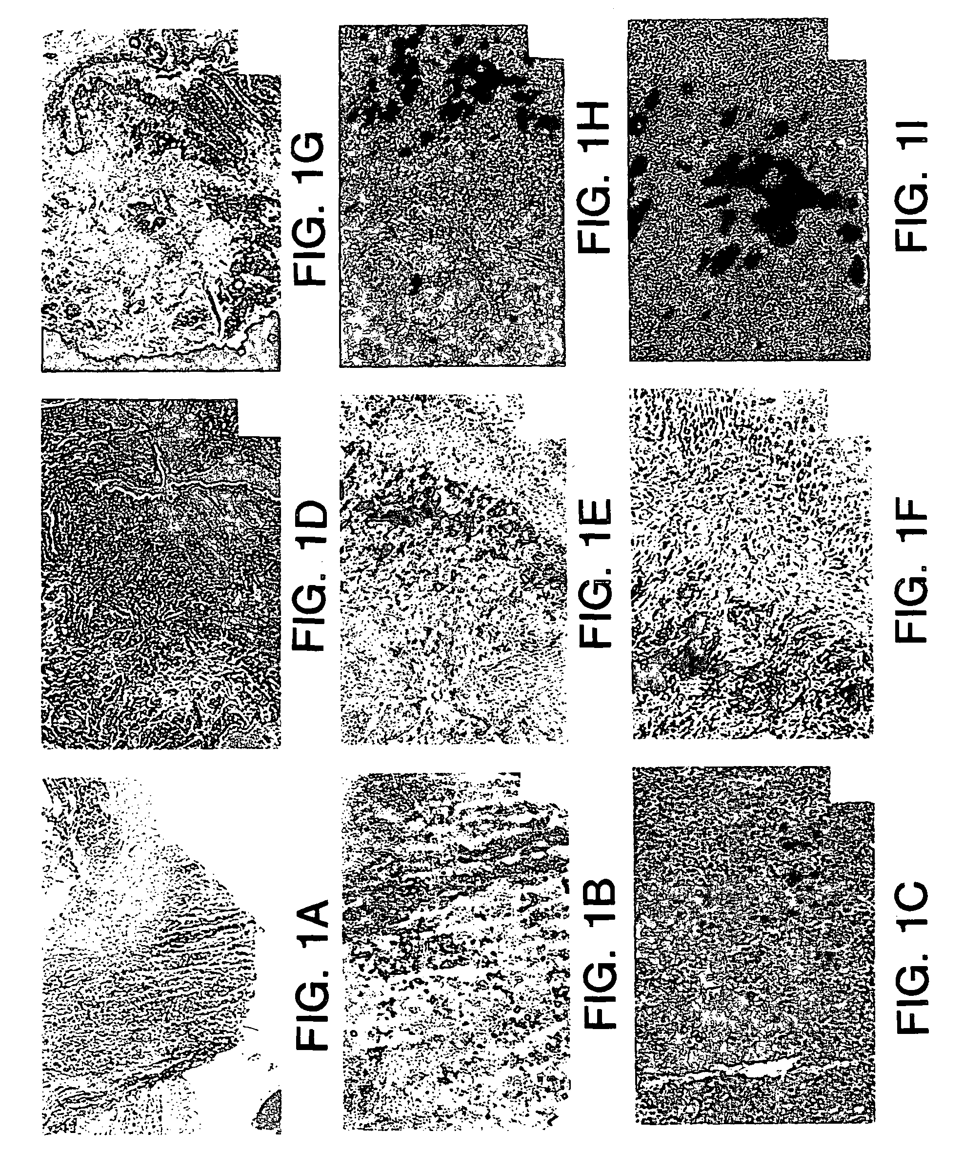
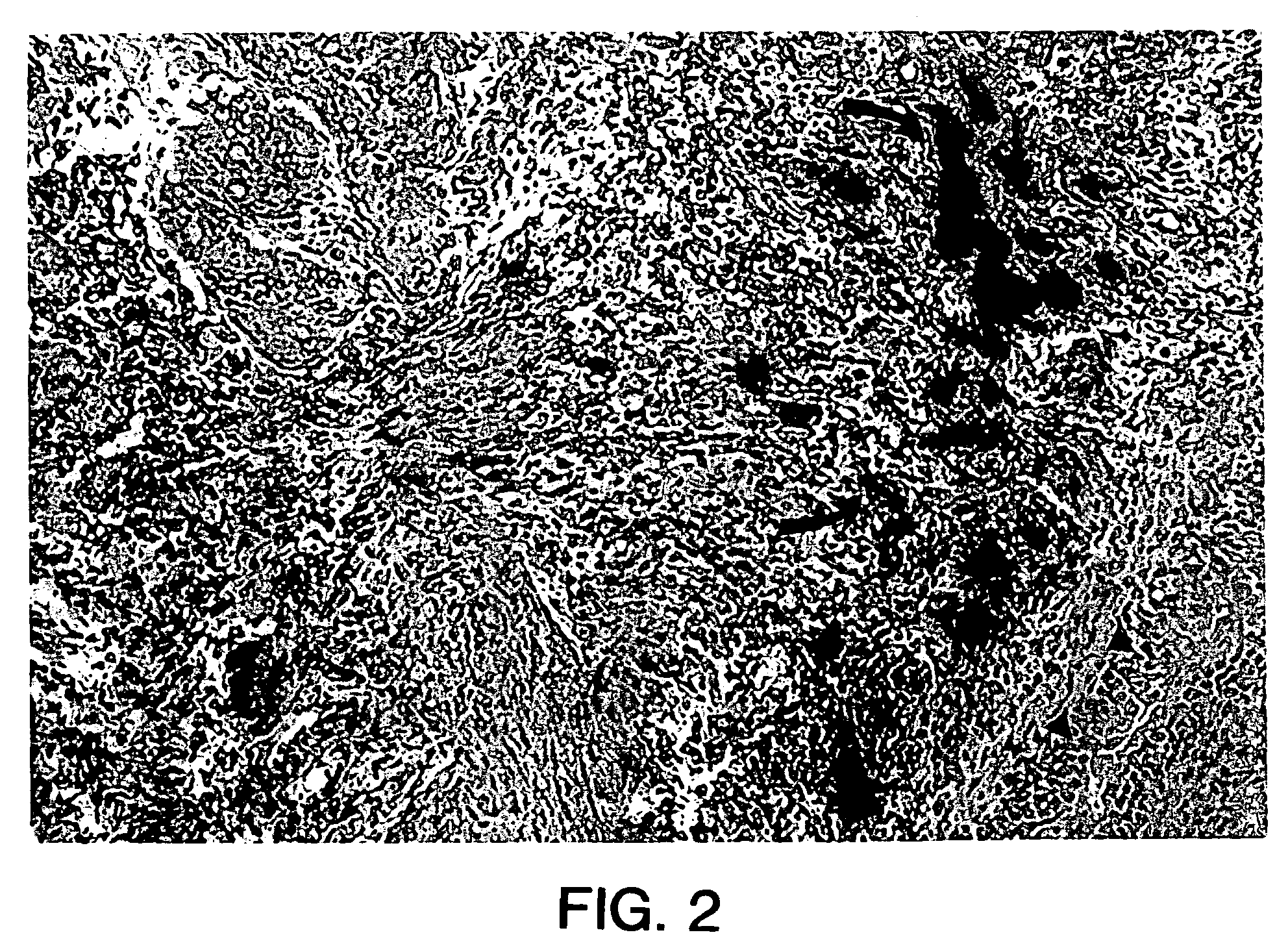
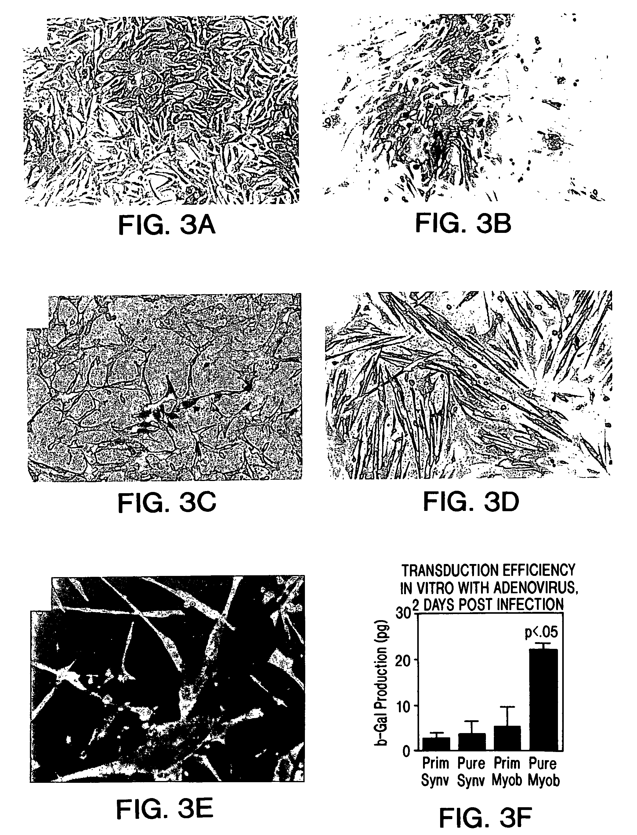
[0159]In these experiments, myoblasts from the GH8 myoblast cell line (available from the American Type Cell Culture depository) transduced with an adenoviral vector harboring a reporter gene encoding β-galactosidase (E1-E3 deleted first generation adenovirus), were cultured and injected into the proximal urethral wall of the female S.D. rat.

[0160]In addition, the cells were incubated with fluorescent latex microspheres (FLM) to follow the fate of the injected cells (see Example 1). The transduced, FLM-labeled cells were injected into adult female S.D. rats (n=20). A midline incision was made and myoblasts were injected into the proximal urethral wall at two sites with a 10 μl Hamilton micro syringe. The myoblast concentration ranged from 1.33×10⁵ to 1×10⁶. The tissue was harvested 2-4 days after injection and flash frozen in liquid nitrogen. The tissue was...

example 3

[0169]Experiments were performed to demonstrate the feasibility of myoblast injection into the bladder wall to improve detrusor contractility. A myoblast cell line transduced with an adenovirus vector carrying a β-galactosidase reporter gene as described hereinabove was used for these experiments. The cells were incubated with fluorescent latex microspheres (FLM) to follow the fate of the injected cells. Cells were injected into adult male and female S.D. rats (n=18). The myoblasts were injected into the dome of the bladder and into the right and left lateral walls near the dome with a 10 μl Hamilton microsyringe. The myoblast concentration ranged from 1.33×10⁵ to 1×10⁶ cells. The tissue was harvested after 2-5 days, sectioned and assayed for β-galactosidase expression. For gene therapy experiments, myoblasts were transduced with the adenovirus containing the human inducible nitric oxide synthase (iNOS) gene at a multiplicity of infection of 50 (MOI=50) and were injected into the bl...


PUM

PropertyMeasurementUnit
diameteraaaaaaaaaa
volumeaaaaaaaaaa
tensionaaaaaaaaaa

Abstract

The present invention provides muscle-derived cells, preferably myoblasts and muscle-derived stem cells, genetically engineered to contain and express one or more heterologous genes or functional segments of such genes, for delivery of the encoded gene products at or near sites of musculoskeletal, bone, ligament, meniscus, cartilage or genitourinary disease, injury, defect, or dysfunction. Ex vivo myoblast mediated gene delivery of human inducible nitric oxide synthase, and the resulting production of nitric oxide at and around the site of injury, are particularly provided by the invention as a treatment for lower genitourinary tract dysfunctions. Ex vivo gene transfer for the musculoskeletal system includes genes encoding acidic fibroblast growth factor, basic fibroblast growth factor, epidermal growth factor, insulin-like growth factor, platelet derived growth factor, transforming growth factor-β, transforming growth factor-α, nerve growth factor and interleukin-1 receptor antagonist protein (IRAP), bone morphogenetic protein (BMPs), cartilage derived morphogenetic protein (CDMPs), vascular endothelial growth factor (VEGF), and sonic hedgehog proteins.

Description

CROSS REFERENCE TO RELATED APPLICATIONS[0001]This application is a divisional of U.S. patent application Ser. No. 09 / 302,896 filed on Apr. 30, 1999, which claims the benefit of Provisional Patent Application U.S. Ser. No. 60 / 083,917, filed May 1, 1998, the entire disclosures of which are hereby incorporated by reference.GOVERNMENT INTERESTS[0002]This invention was made with Government support under Grant No. DK055387 awarded by the National Institutes of Health. The Government has certain rights to this invention.FIELD OF THE INVENTION[0003]The present invention relates generally to compositions and methods comprising myogenic or muscle-derived cells, including myoblasts and muscle-derived stem cells (also termed MDC herein) for tissue engineering and cell-mediated gene therapy. The invention further relates to the introduction of exogenous nucleic acids into muscle-derived cells, including myoblasts and muscle-derived stem cells, resulting in the expression of one or more gene prod...

Claims


Application Information

Patent Timeline
Patent Type & Authority Patents(United States)
IPC IPC(8): A61K48/00A01N63/00C12P21/06C12N5/00C12N15/00A01N43/04A61K45/00C12N5/077C12N15/09C12N15/63
CPCC12N5/0658C12N5/0659C12N2501/105C12N2510/04C12N2501/115C12N2501/13C12N2510/00C12N2501/113A61P13/10A61P19/02A61P19/04A61P19/08A61P21/00A61P29/00A61K35/34
Inventor CHANCELLOR, MICHAEL B.HUARD, JOHNNY
Owner UNIVERSITY OF PITTSBURGH
Learn More