Method for treating systemic bacterial, fungal and protozoan infection

a systemic bacterial, fungal and protozoan infection technology, applied in the field of medical treatment of diseases, can solve the problems of not being able to justify widespread use, not having enough clinical efficacy, and no therapeutic method developed to efficiently target extracellular blood dna in systemic bacterial, fungi or protozoa infection

Active Publication Date: 2013-04-30
CLS THERAPEUTICS
View PDF65 Cites 42 Cited by
  • Summary
  • Abstract
  • Description
  • Claims
  • Application Information

AI Technical Summary

Benefits of technology

[0013]The object of this invention is to develop high-performance and low-toxic method for treatment of systemic infections that are accompanied by quantitative and / or qualitative change of composition of blood plasma extracellular DNA and caused by bacteria, fungi and protozoa.
[0015]The present invention suggests that systemic infections caused by bacteria, fungi and protozoa can be treated by reducing circulating extracellular blood DNA levels.
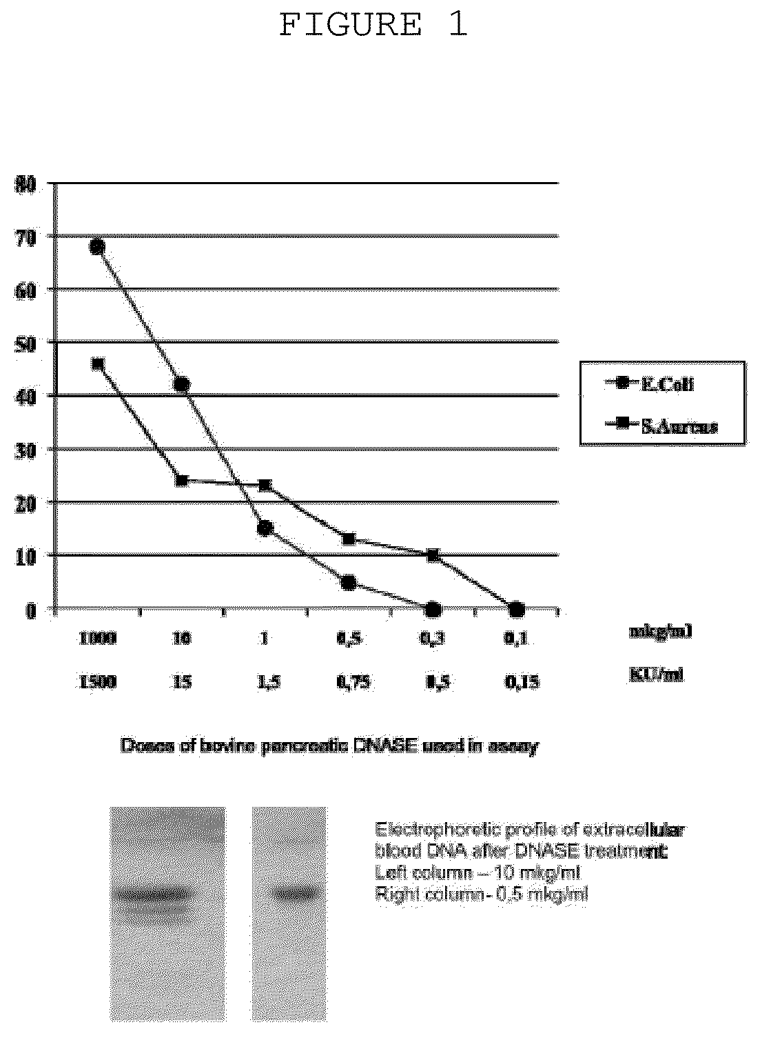
[0019]It was established that enzymatic destruction of extracellular blood DNA by DNase enzyme when applied in certain surprisingly high specific doses has significant therapeutic effect on the course of systemic infections.

Problems solved by technology

Reports indicate that this product had some clinical efficacy, but not enough to justify its widespread use.
Bovine pancreatic DNase has been sold under the tradename Dornavac (Merck), but this product was withdrawn from the market mainly due to efficacy concern.
Accordingly, no therapeutic method was developed which efficiently targets extracellular blood DNA in systemic bacterial, fungi or protozoan infection.
Thus it makes impossible to take any technical solution as prototype.

Method used


Image

  • Method for treating systemic bacterial, fungal and protozoan infection
  • Method for treating systemic bacterial, fungal and protozoan infection
  • Method for treating systemic bacterial, fungal and protozoan infection

Examples


example 1

SEQUENCING of Extracellular Blood DNA from the Patient Suffering From Bacillary Dysentery, Shiqelosis Caused by Shiqella Flexneri

[0031]The probes of patient's extracellular blood DNA were taken before initiation of antibacterial chemotherapy and week after completion of the therapy. The extracellular DNA was cloned by the method which allows to get non amplified plasmid libraries of blood extracellular DNA with representativeness up to one million of clones with the average size of 300-500 base pairs. The DNA which has been isolated using the protocol specified above was treated with Proteinase K (Sigma) at 65° C. and subjected to additional phenol-chloroform extraction step with further overnight precipitation by ethanol. The DNA fraction was than treated by Eco RI restrictase or by Pfu polymerase (Stratagene) in presence of 300 mkM of all desoxynucleotidetriphosphates for sticky-ends elimination. The completed DNA was phosphorylated by polynucleotidkinase T4 and ligated to pBluesc...

example 2

Extracellular Blood DNA Promotes Pathogenic Behavior of Microorganism

[0034]The extracellular blood plasma DNA were obtained as follows: S. aureus VT38 and Escherichia coli K1 was injected intravenously at LD50 dose to 10 SHR mice (5 mice for each pathogen) to induce sepsis. On day 3 the diseased mice were sacrificed and extrcellular blood DNA were collected as specified above.

[0035]Biofilm formation assay was performed as follows: Escherichia coli K1 and Staphylococcus aureus VT38 pathogenic strains were grown at 37° C., and liquid cultures were incubated without shaking. Before use in biofilm formation assay, cells were harvested and washed twice with 0.15M isotonic phosphate-buffered saline (pH7.2), and the cell suspensions were standardized to an optical density (OD) of 0.8 at 520 nm. The inoculums containing 7.53+0.22 log10CFU / mL, was added to the wells of 96-well plates (200 mkL / well) and plates were incubated for 24 hours at 37° C. Different amounts of extracellular blood plas...

example 3

Extracellular Blood DNA Promotes Spread of Antibiotic Resistance and Clonogenic Potential of Microorganisms

[0039]The extracellular blood plasma DNA were obtained as follows: Escherichia coil K1 was injected intravenously at LD50 dose to 10 SHR mice to induce sepsis. On day 3 animals were treated with subtherapeutic doses of ampicillin and kanamycin for 3 consecutive days. On day 6 the diseased mice were sacrificed and extrcellular blood DNA were collected as specified above. Biofilms of E. coli ATCC 25922 were grown as described in example 2 with or without extracellular blood DNA from E. coli K1 infected animals treated with antibiotics (50 mkg / well) on selection media containing ampicillin or kanamycin or both.

[0040]Colony-forming assay was performed as follows: The term “the number of CFU” means the number of viable bacteria that grow on nutrient media in the current conditions. Biofilms were grown in 96-well plates for 24 or 48 h. at 37° C. Liquid medium with bacteria was aspira...


PUM

PropertyMeasurementUnit
pHaaaaaaaaaa
pHaaaaaaaaaa
volumeaaaaaaaaaa

Abstract

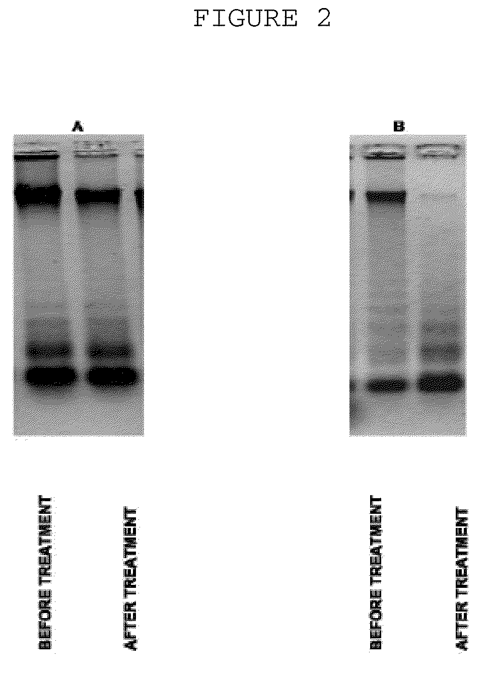
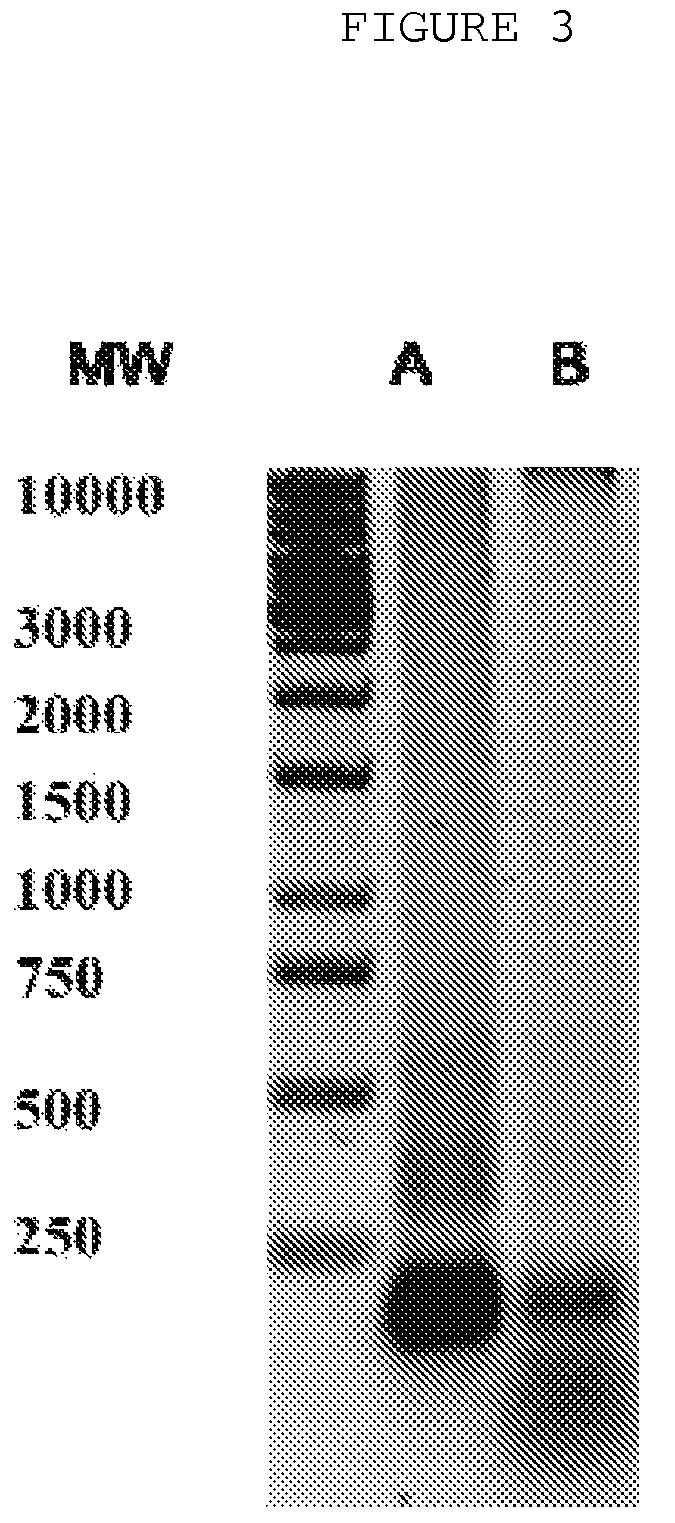
The invention is directed to a treatment of diseases that are accompanied by quantitative and / or qualitative changes of blood extracellular DNA and, more particularly, to a treatment of systemic bacterial, fungal and protozoan infections. The inventive method comprises introducing a treatment agent into a circulating blood system of a patient diagnosed with systemic infection caused by bacteria, fungi or protozoa, wherein said treatment agent destroys extracellular DNA in said blood of said patient and wherein said treatment agent used to destroy said extracellular DNA is a DNase enzyme: said agent being administered in doses and regimens which are sufficient to decrease the average molecular weight of circulating extracellular blood DNA in the blood of said patient; such decrease in the average molecular weight can be measured by gel electrophoresis of extracellular blood DNA fraction from the blood of said patient. A DNase enzyme may be applied in a dose and regimen that provide a DNase DNA hydrolytic activity measured in blood plasma that exceeds 1.5 Kunitz units per 1 ml of blood plasma for more than 12 hours within a period of 24 hours.

Description

CROSS-REFERENCE TO RELATED APPLICATIONS[0001]This application is a Continuation-in-Part of U.S. application Ser. No. 10 / 564,609, filed on Jan. 12, 2006, now abandoned, which is a U.S. national phase application under 35 U.S.C. §371 of International Patent Application No. PCT / RU2004 / 000260, filed Jul. 1, 2004 (published in Russian on Jan. 20, 2005 as WO 2005 / 004789), which claims priority of Russian Federation Patent Application No. RU2004108057, filed Mar. 12, 2004, and International Patent Application No. PCT / RU2003 / 000304, filed Jul. 14, 2003, all of which are incorporated by reference as if fully rewritten herein.BACKGROUND OF THE INVENTION[0002]1. Field of the Invention[0003]The present invention relates generally to medical treatment of the diseases that are accompanied by quantitative and qualitative changes of blood extracellular DNA and, more particularly, to a treatment of systemic bacterial, fungal and protozoan infections.[0004]2. Description of the Related Art[0005]Oppos...

Claims


Application Information

Patent Timeline
Patent Type & Authority Patents(United States)
IPC IPC(8): A61K38/43C12Q1/34C12Q1/68C12N9/16C07K14/00
CPCA61K38/46A61K38/465Y02A50/30
Inventor GENKIN, DMITRY DMITRIEVICHTETS
Owner CLS THERAPEUTICS
Why Eureka
Try Eureka