

Nano bionic wound-surface cover and preparation method thereof
A nano-bionic and covering technology, applied in the field of biomedicine, can solve the problems of general effect, limited repair effect, poor mechanical properties of artificial materials, etc., and achieve the effect of avoiding inconvenience
- Summary
- Abstract
- Description
- Claims
- Application Information
AI Technical Summary
Problems solved by technology
Method used
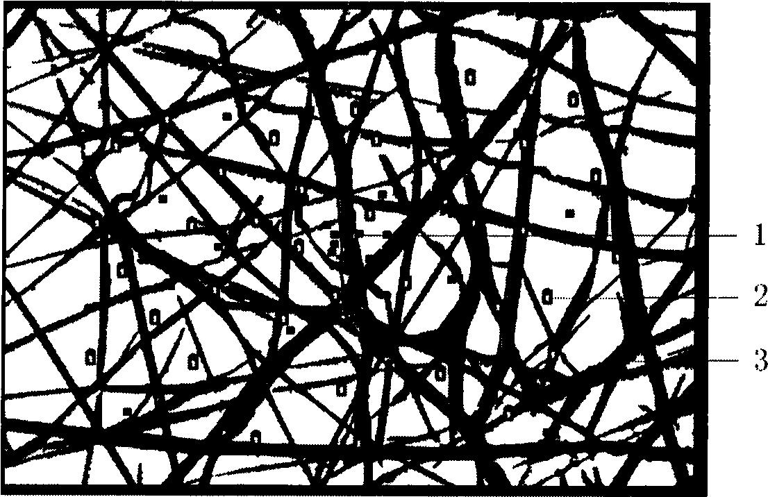
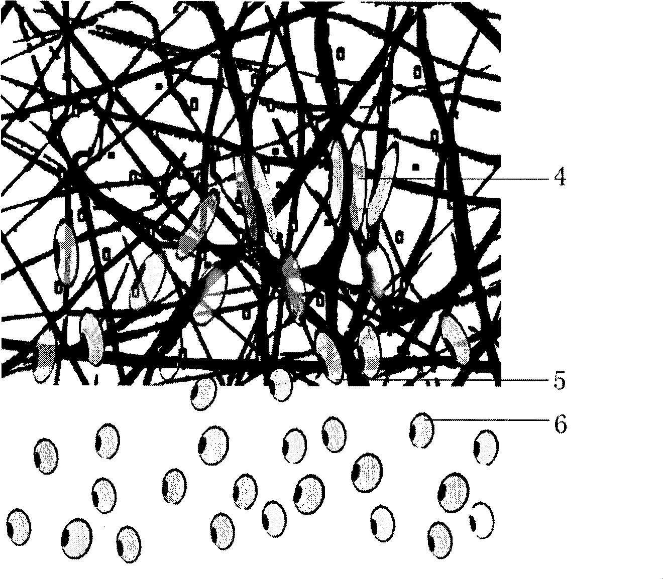
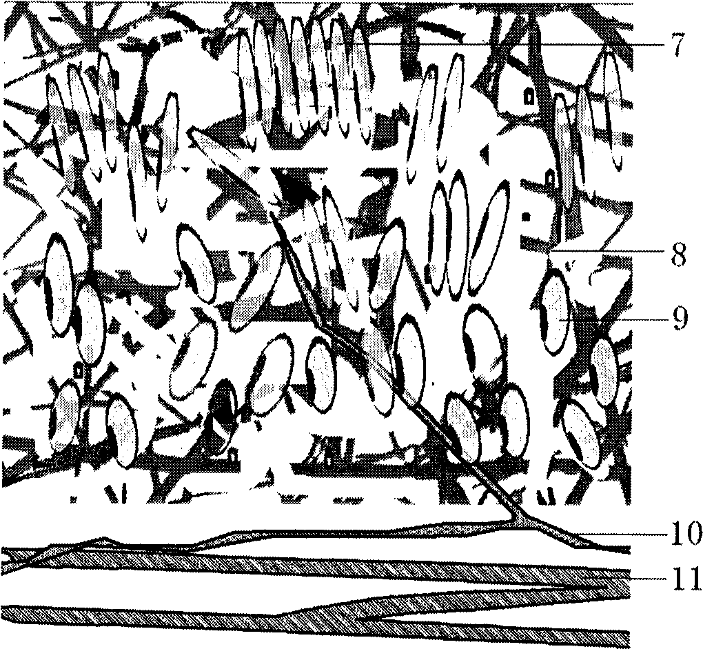
Image

Examples
Embodiment 1
[0073] (1) Prepare electrospinning solution, hydrosol solution containing cytokines and cross-linking agent solution;
[0074] The electrospinning solution adopts poly-DL-lactic acid with a viscosity average molecular weight of 10000 and a dichloromethane solution of polyethylene glycol with a molecular weight of 400; the weight percentages of the poly-DL-lactic acid and polyethylene glycol are respectively 80 % and 20%, the solvent accounts for 85% by mass.
[0075] The cross-linking agent solution is 0.1M calcium chloride solution.
[0076] The hydrosol solution containing the cytokine is a cytokine alginate solution, and the concentration of the cytokine SDF-1 in the cytokine alginate solution is 10 ppm by mass.
[0077] Put the prepared 0.1M calcium chloride solution into a cell culture dish with a diameter of 150 mm, and place it on the flat receiver shared by the electrospinning device and the printer. The Hewlett-Packard 550C inkjet printer was refitted according to e...
Embodiment 2
[0085] Implementation steps are the same as in Example 1.
[0086] The electrospinning solution adopts poly-DL-lactic acid with a viscosity average molecular weight of 10000 and a dichloromethane solution of polyethylene glycol with a molecular weight of 400; the weight percentages of the poly-DL-lactic acid and polyethylene glycol are respectively 80 % and 20%, the solvent accounts for 85% by mass.
[0087] The cross-linking agent solution is 100IU / ml thrombin solution;
[0088] The hydrosol solution containing cytokines adopts cytokine fibrinogen solution, the fibrinogen concentration is 40mg / ml, and the mass percent concentration of epidermal growth factor and SDF-1 factor in the fibrinogen solution is 500ppm.
[0089] The specific operation is to put the configured thrombin solution into a cell culture dish with a diameter of 90 mm, and place it on the flat receiver shared by the electrospinning device and the printer. The Hewlett-Packard 550C inkjet printer was refitted...
Embodiment 3
[0093] Implementation steps are the same as in Example 1.
[0094] The electrospinning solution adopts polycaprolactone with a viscosity average molecular weight of 50000 and a hexafluoroisopropanol solution of polyethylene glycol with a molecular weight of 400; the weight percentages of the polycaprolactone and polyethylene glycol are respectively 90 % and 10%. The weight percent concentration of the hexafluoroisopropanol solution is 85%.
[0095] The crosslinking agent solution is selected from 100 mg / ml water-soluble carbodiimide solution;
[0096] The hydrosol solution containing cytokines adopts collagen and polyanion solution of cytokines, wherein the collagen concentration is 1%, polyanion adopts polyglutamic acid with a concentration of 50 mg / ml, and the cytokines include cell directional migration factor SDF-1, The concentration of epidermal growth factor, fibroblast growth factor, interleukin IL-3 and basic fibroblast growth factor is 1%.
[0097] The specific ope...
PUM
| Property | Measurement | Unit |
|---|---|---|
| thickness | aaaaa | aaaaa |
| thickness | aaaaa | aaaaa |
| thickness | aaaaa | aaaaa |
Abstract
Description
Claims
Application Information