Method and device for preparing three-dimensional porous support for tissue engineering

A three-dimensional porous and tissue engineering technology, applied in the fields of biomedical engineering and advanced manufacturing, can solve the problems of wide particle size distribution of microcapsules, the inability to realize industrialization, and difficulty in satisfying the biological activity of biological scaffolds.

Inactive Publication Date: 2010-11-17
HEBEI UNIV OF TECH
View PDF3 Cites 25 Cited by
  • Summary
  • Abstract
  • Description
  • Claims
  • Application Information

AI Technical Summary

Problems solved by technology

The gas foaming method can avoid the use of organic solvents in the preparation of scaffolds. Combining this method with particle leaching technology, porous scaffolds with connected open-pore structures can be prepared, but the disadvantage of this method is that the pores in the scaffolds cannot control
The microcapsules prepared by the above method usually need to be subjected to high temperature conditions or use violently reactive destructive organic solvents. The prepared microcapsules have a wide particle size distribution, and it is difficult to meet the requirements for maintaining the activity of biological substances in biological scaffolds. Screening is required. filter
The electrostatic method is to prepare microcapsules through the reaction between ionic substances in the electric field. Although the manufacturing process is relatively mild, the production scale is small, and the particle size of the produced microcapsules is often above 200 μm, which cannot meet the actual use requirements.
There are few reports on microcapsule manufacturing technology at home and abroad. Bnenedetti et al. used supercritical object technology to produce microcapsules with a particle size of less than 20 μm. However, supercritical object technology has strict requirements on experimental equipment, experimental environment and fluid properties. unable to achieve industrialization
The cell-encapsulated sodium alginate microcapsules produced by the Biomedical Materials Engineering Group of Dalian Institute of Chemical Physics, Chinese Academy of Sciences have an average diameter of about 240 μm, and the particle size is also large, which cannot meet the actual use requirements.

Method used


Image

  • Method and device for preparing three-dimensional porous support for tissue engineering
  • Method and device for preparing three-dimensional porous support for tissue engineering
  • Method and device for preparing three-dimensional porous support for tissue engineering

Examples


preparation example Construction

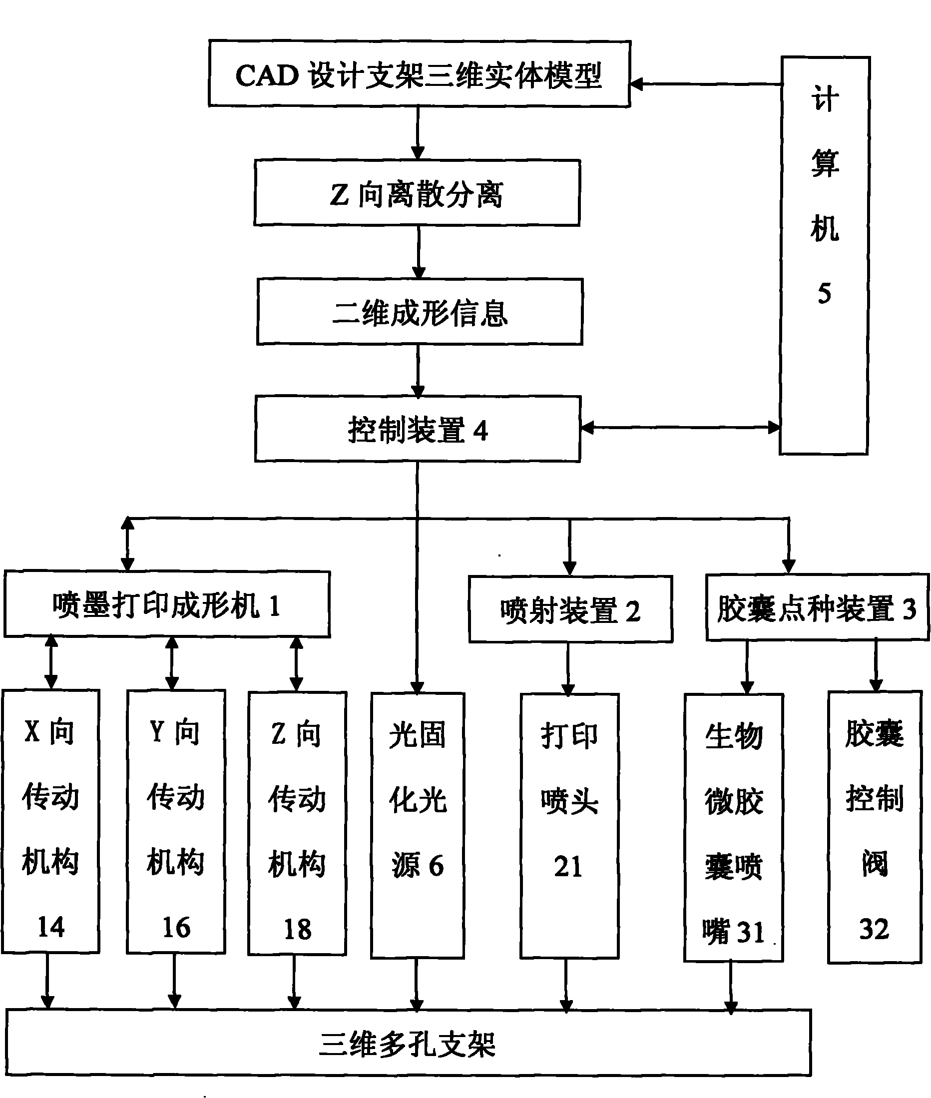
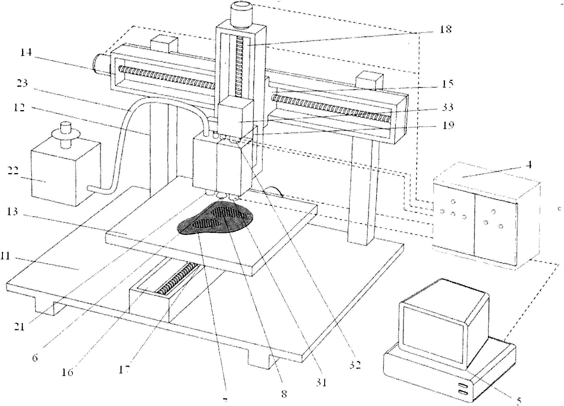
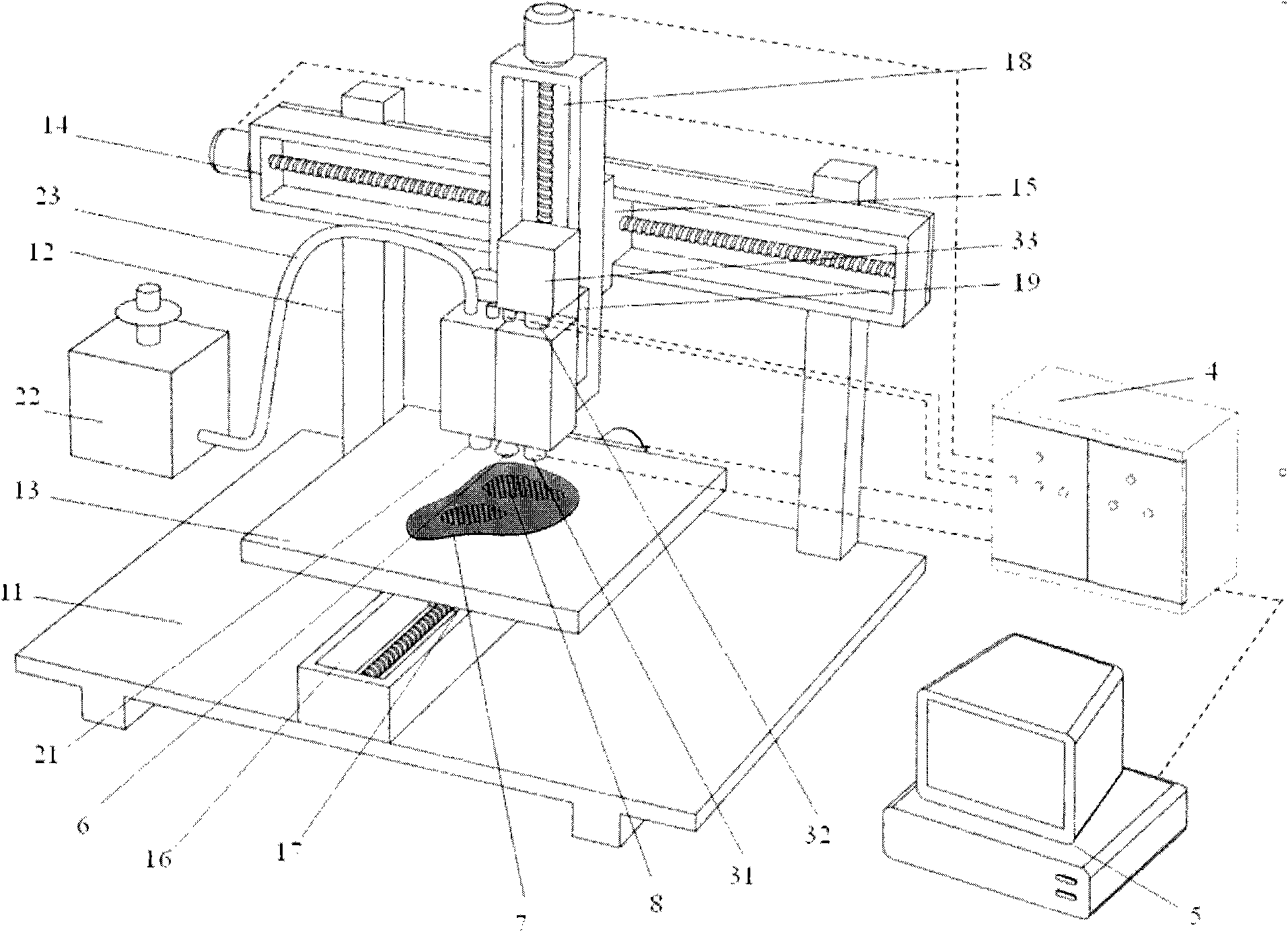
[0021] The preparation method (referred to as the preparation method) of the three-dimensional porous scaffold (abbreviated as the scaffold) for tissue engineering designed by the present invention, see figure 1 ) is based on the organic combination of three-dimensional inkjet printing method and biological microcapsule spotting method. Take the following steps:

[0022] (1) First, the shape of the bracket is designed by 3D CAD software, and then according to the process requirements of the product, the bracket is sliced ​​and layered according to a certain thickness, and the two-dimensional cross-sectional information of each layer of the bracket is obtained;

[0023] (2) Under the control of CAD software, the three-dimensional inkjet printing nozzle runs along the X-axis and Y-axis direction, completes the printing movement set by the cross-sectional information of the first layer of the bracket, and sprays the liquid polymer according to the cross-sectional information to m...

Embodiment 1

[0039] Three-dimensional porous scaffolds for bone tissue engineering with a length of 50 mm were prepared from unsaturated polyphosphate (UPPE) materials. The particle size of the biocapsule particles is 20 μm, and the number of scaffold slices is 2500 pieces.

[0040] First use the 3D CAD software to design the 3D solid model of the bracket (see figure 1 ), and then according to the product design process requirements, it is sliced ​​and layered according to a certain thickness to form a series of two-dimensional slices, and the slice thickness of each layer is 20 μm. The computer 5 controls each control mechanism to perform coordinated movement according to the two-dimensional forming information of each layer. Specifically, when the preparation starts, the computer 5 transmits the two-dimensional forming information of the first layer to the control device 4, and the capsule control valve 32 in the biological microcapsule spotting device 3 opens the valve under the contro...

Embodiment 2

[0042] It is basically the same as in Example 1, except that the particle size of the biocapsule particles is 18 μm, and active pharmaceutical molecules are added into the capsules, and the prepared scaffold has a pore size of 200-400 μm and a porosity of 90%.


PUM

PropertyMeasurementUnit
particle diameteraaaaaaaaaa
sizeaaaaaaaaaa
sizeaaaaaaaaaa

Abstract

The invention discloses a method and device for preparing a three-dimensional porous support for tissue engineering. The preparation method comprises the following steps of: 1, slicing and layering a support according to a certain thickness firstly to obtain two-dimensional cross section information of each layer of the support; 2, preparing a first layer slice of the support by using a three-dimensional inkjet printing spray head under the control of CAD software, dibbling microcapsule particles on the first layer slice of the support by using a bio-microcapsule preparation nozzle, and stacking the microcapsule particles to form gaps required by a support design; 3, moving the three-dimensional inkjet printing spray head and the bio-microcapsule preparation nozzle for the height of one layer slice of the support to finish the forming preparation of a second layer of the support; and 4, repeating the method layer by layer until the three-dimensional porous support is prepared. The device for preparing the three-dimensional porous support is suitable for the preparation method, and comprises a three-dimensional inkjet printing forming machine, a jetting device, a bio-microcapsule dibbling device and a control system.

Description

technical field [0001] The invention belongs to the technical field of biomedical engineering and advanced manufacturing, and specifically relates to a preparation method and equipment for a three-dimensional porous scaffold for tissue engineering. Background technique [0002] In recent years, with the rapid development of cell biology, molecular biology and biomaterials research, tissue engineering, as an emerging interdisciplinary subject, has also made great progress in its research and application. The basic principle and method of tissue engineering is to plant normal tissue cells cultured and expanded in vitro on tissue engineering porous scaffolds that have good biocompatibility and can be gradually degraded and absorbed in vivo to form cell-scaffold complexes. Proliferate and differentiate on the scaffold, and then implant the complex into the lesion of the body tissue, continue to proliferate and secrete extracellular matrix in the body, and along with the gradual ...

Claims


Application Information

Patent Timeline
Patent Type & Authority Applications(China)
IPC IPC(8): A61F2/82A61L27/22A61L27/20A61L27/18A61L27/56
Inventor 徐安平汪建明朱东彬曲云霞
Owner HEBEI UNIV OF TECH
Why Eureka
Try Eureka