Method for detecting DNA (deoxyribonucleic acid) glycosylase activity on basis of single quantum dot level

A glycosylase, level detection technology, applied in the field of biological analysis, can solve the problems of complex operation, low detection limit, long time consumption, etc., to achieve the effect of good specificity, high specificity and reducing non-specificity

Active Publication Date: 2016-07-13
SHANDONG NORMAL UNIV
View PDF7 Cites 25 Cited by
  • Summary
  • Abstract
  • Description
  • Claims
  • Application Information

AI Technical Summary

Problems solved by technology

These detection methods are very effective, but very time-consuming, cumbersome to operate, and have potential safety hazards
[0004] In order to overcome the above shortcomings, in recent years, colorimetric detection methods based on nanotechnology and fluorescent probe detection methods based on various fluorescent dyes have also been developed. Highly sensitive detection of uracil DNA glycosylase, using DNA glycosylase UDG as a model, developed the recognition mechanism of hairpin remodeling, and transformed the action of DNA modifying enzymes on substrates into the triggering process of hybridization chain reaction , thereby causing the self-assembly process of DNA, and realizing the construction of a universal DNA nano-device, which can be used for highly sensitive detection of DNA modifying enzymes (especially UDG), wherein the detection limit of UDG reaches 0.000043U/mL; CN104630363A A meth

Method used


Image

  • Method for detecting DNA (deoxyribonucleic acid) glycosylase activity on basis of single quantum dot level
  • Method for detecting DNA (deoxyribonucleic acid) glycosylase activity on basis of single quantum dot level
  • Method for detecting DNA (deoxyribonucleic acid) glycosylase activity on basis of single quantum dot level

Examples


Embodiment 1

[0035] Preparation of the incubation buffer: 100 millimoles per liter of tris(hydroxymethyl)aminomethane-hydrochloric acid (Tris-HCl), 10 millimoles per liter of ammonium acid, 3 millimoles per liter of magnesium chloride, 0.83 nanosulfide per liter Liter of quantum dots (605QDs), pH 8.0.

[0036] Preparation of anti-quench buffer: 67 millimoles per liter of glycine-potassium hydroxide (pH 9.4), 2.5 millimoles per liter of magnesium chloride, 50 micrograms per milliliter of bovine serum albumin, 1 mg per milliliter of glucose oxidation Enzyme, 0.04% mg / ml catalase, 0.4% (mass / volume) D-glucose.

[0037] Cell extract preparation: Human lung adenocarcinoma cell (A549) medium is Dulbecco's modified Eagle's medium (DMEM) containing 10% fetal bovine serum (FBS) and 1% penicillin-streptomycin. Cultivate in an incubator with 5% carbon dioxide and 37 degrees. When the cells grow to the logarithmic growth phase, they are digested with trypsin, washed twice with phosphate buffered saline (...

Embodiment 2

[0043] 2.1 Experimental verification of principle

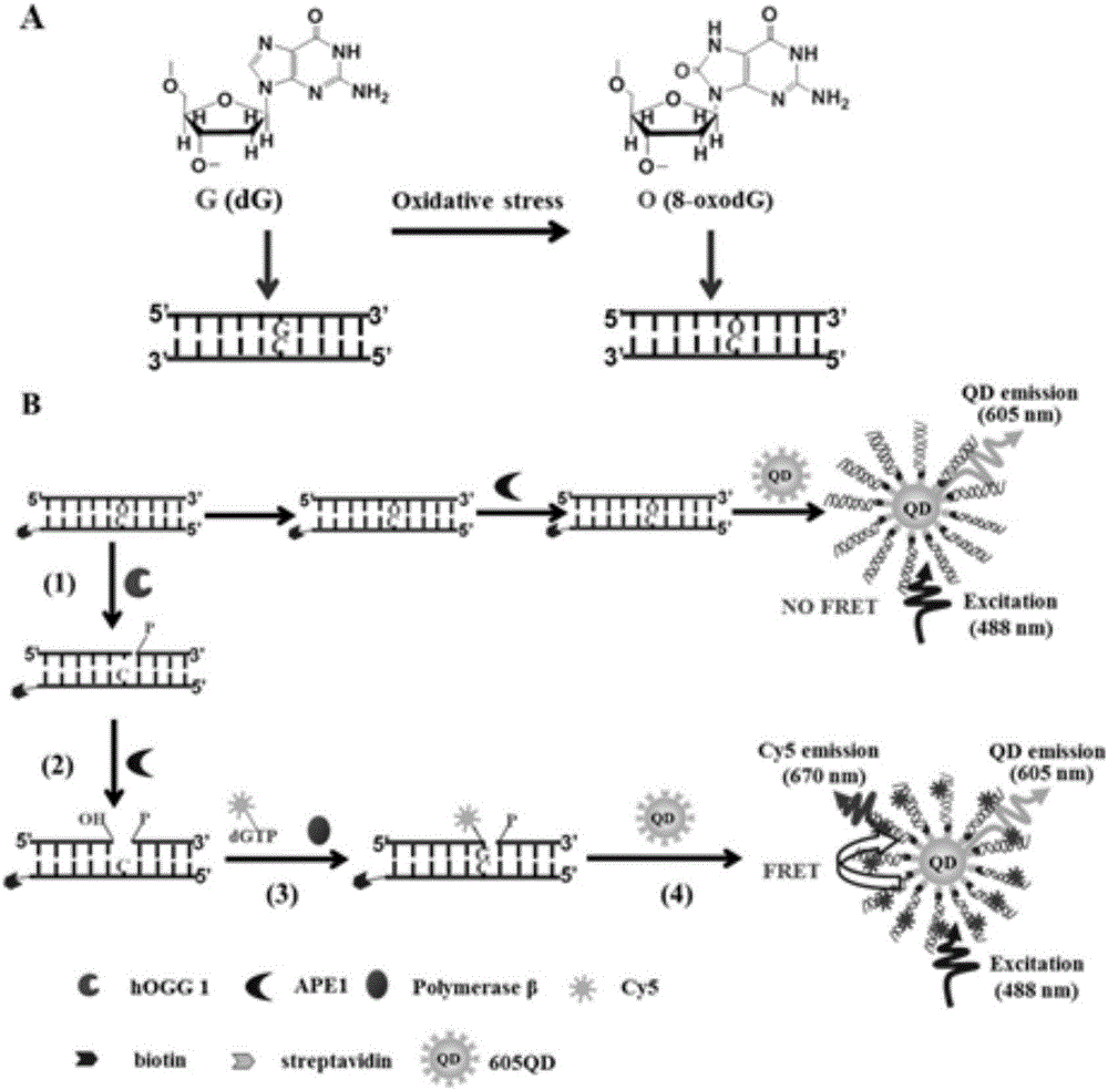
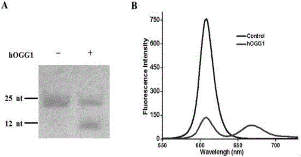
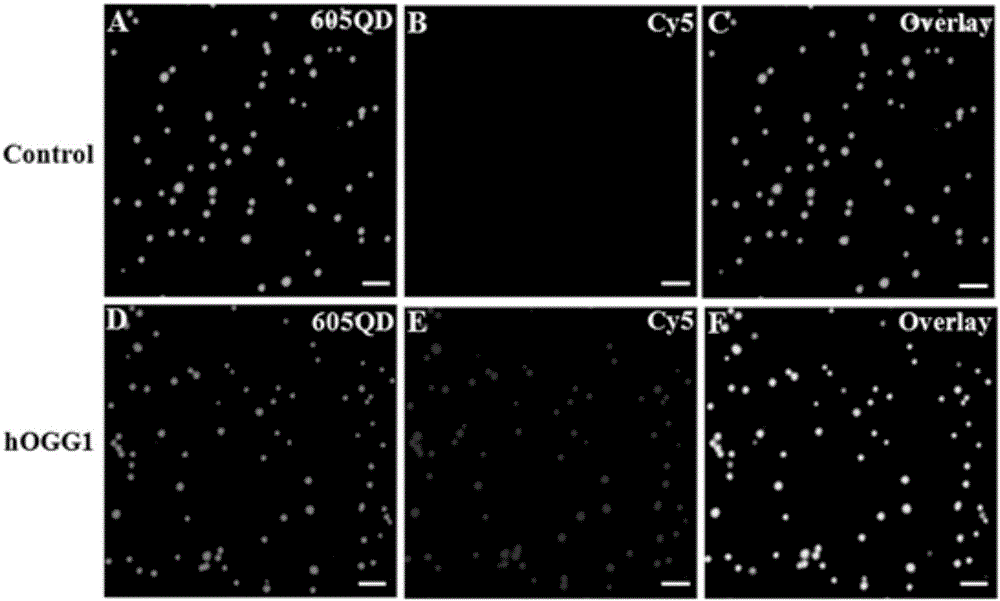
[0044] In order to verify the feasibility of DNA glycosylase hOGG1 in extracellular base excision repair, the inventors tested and analyzed the repair reaction products, and the results are as follows figure 2 Shown. First, the present invention uses non-denaturing polyacrylamide gel (PAGE) electrophoresis for verification analysis. From figure 2 It can be seen from A that when there is no DNA glycosylase hOGG1, there is only a 25bp band, indicating that the excision repair reaction did not occur. When there is DNA glycosylase hOGG1, two bands can be seen, 25bp and 12nt in length, indicating that one of the DNA strands is cut and a small 12nt fragment is produced. The above results show that DNA glycosylase hOGG1 can specifically recognize and excise 8-oxoguanine (8-oxoG), and under the cleavage of apurinic endonuclease-1 (APE1), the abasic site (APsite ) Leaves a gap in nucleotides. Cy5-dGTP is added and DNA polymerase β p...


PUM


Abstract

The invention discloses a method for detecting DNA (deoxyribonucleic acid) glycosylase activity on basis of single quantum dot level.In detection, DNA glycosylase hOGG1 specifically recognizes and excises damaged guanine to leave an abasic site, apurinic endonuclease-1 further excises the abasic site to leave a nucleotide gap, and DNA polymerase beta polymerizes Cy5-dGTP at the gap to generate a double-tagging double-strand nucleotide substrate; by specific reaction between biotin and streptavidin, the DNA substrate is combined to the surface, covered with the streptavidin, of quantum dots to form a QD-DNA-Cy5 compound; due to reduction of spatial distance, fluorescence resonance energy transfer occurs between the quantum dots and Cy5, and Cy5 fluorescence signals can be observed in the unimolecular level.The method has the advantages of simplicity, quickness and sensitivity, and the lower limit of detection can reach 1.8*10<-6>U/microliter.

Description

technical field [0001] The invention belongs to the technical field of biological analysis, and in particular relates to a method for detecting DNA glycosylase activity based on a single quantum dot level. Background technique [0002] The maintenance of genomic DNA integrity is of great significance to the stability of species, but in real life, genomic DNA is inevitably affected by various factors from inside and outside the body, such as exogenous sources such as radiation (UV), chemical mutagens, etc., and endogenous sources such as reactive oxidizing substances (ROS), etc. These factors will cause DNA single- and double-strand breaks, mismatches, and base deletions, thereby destroying the integrity of the genome and affecting the survival of species. In order to reduce DNA damage, organisms have formed a variety of damage repair mechanisms during evolution. For single-base mutations, there are mismatch repair (MMR), base excision repair (BER), nucleotide excision repair ...

Claims


Application Information

Patent Timeline
IPC IPC(8): C12Q1/34
CPCC12Q1/34
Inventor 张春阳唐波王黎娟马飞
Owner SHANDONG NORMAL UNIV
Why Eureka
Try Eureka