
A method for inducing the transdifferentiation of fibroblasts into Leydig cells by a combination of transcription factors

A technology of Leydig cells and fibroblasts, applied in chemical instruments and methods, bone/connective tissue cells, biochemical equipment and methods, etc., can solve the problems of loss of division characteristics, immune rejection, limited quantity, etc.

Active Publication Date: 2019-07-19
BIOPHARM RES & DEV CENT JINAN
View PDF0 Cites 1 Cited by
  • Summary
  • Abstract
  • Description
  • Claims
  • Application Information

AI Technical Summary

Problems solved by technology

However, Leydig cells only exist in the testis, and the number is limited (accounting for only 2-5% of the total testicular cells). Although there are relatively mature separation methods for Leydig cells, cell loss during the separation process is inevitable , and adult Leydig cells can no longer undergo mitosis, and immature Leydig cells will lose their division characteristics in subsequent cultures, these factors exacerbate the scarcity of Leydig cells
In addition, this kind of transplantation generally relies on the supply of allografts, and patients need to take immunosuppressants for a long time after surgery, which has a lot of side effects and also bears a series of ethical controversies, which greatly limits the wide application of this technology in clinical practice
[0004] Although mesenchymal stem cells or embryonic stem cells can also be used to induce differentiation into testis-like Leydig cells that can produce steroids, there are still the following defects in the use of these cells in clinical research: (1) induced stem cells or iPSCs ( The differentiation efficiency and purity of induced pluripotent stem cells (i.e., induced pluripotent stem cells) into Leydig cells are very low, and the ability of the resulting cells to produce steroids is very limited; (2) Embryonic stem cells face immune rejection, ethical and moral problems and the risk of teratoma formation at the implantation site; (3) the phenotype of mesenchymal stem cells is heterogeneous, there is no clear standard for its identification, and technical difficulties are faced in isolation, purification and large-scale expansion; (4) although iPSCs avoid the However, the reliability of its source and technical safety are still the biggest obstacles in clinical application
However, there is no report on which transcription factor or combination of transcription factors can be used to induce Leydig-like cells

Method used


Image

  • A method for inducing the transdifferentiation of fibroblasts into Leydig cells by a combination of transcription factors
  • A method for inducing the transdifferentiation of fibroblasts into Leydig cells by a combination of transcription factors
  • A method for inducing the transdifferentiation of fibroblasts into Leydig cells by a combination of transcription factors

Examples


Embodiment 1、11

[0097] Example 1. Construction of 11 kinds of transcription factor lentiviral expression vectors and viral packaging

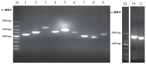
[0098] First, total RNA was obtained from mouse testes using an RNA extraction kit (Cat.#: 74134, Qiagen, purchased from Guangzhou Jianlun Biotechnology Co., Ltd., China). Then use PrimeScript TM II 1st Strand cDNASynthesis Kit (Cat.#: 6210A, Dalian Bao Biotechnology Co., Ltd., China) was used to synthesize cDNA and stored at -80°C. The upstream and downstream primers used to amplify transcription factors Dmrt1, Nr0b1, Sp1, Wt1, Nr4a1, Nr5a2, AP1, Creb1, Smad3, Nr5a1 and Gata4 genes were designed and synthesized by BGI Corporation respectively, and the forward and reverse primers Restriction sites were introduced at both ends (underlined part, Table 2).

[0099]

[0100] According to the conventional PCR method, using the mouse testis cDNA as a template, adding upstream and downstream primers (Table 2), and then setting up the PCR reaction conditions, 94°...

Embodiment 2

[0102] Example 2. Separation of embryonic fibroblasts and adult fibroblasts for transdifferentiation

[0103] In order to obtain embryonic fibroblasts (mouse embryonic fibroblasts, MEFs), pregnant mice at 12.5-14.5 days of pregnancy were first sacrificed, and then the abdomen and uterus were cut open with scissors to take out the embryos. Then use sterile forceps to remove the envelope around the embryo, the limbs, head and viscera of the embryo. After washing with phosphate buffer solution, transfer the embryonic tissue pieces to a penicillin bottle and mince them with scissors, add 2 ml of trypsin, and incubate at 37°C for 10 minutes. Centrifuge at 250xg for 5 minutes, discard the supernatant, resuspend the cells with DMEM medium (Cat.#: 12100-046, invitrogen Inc., USA) containing 10% bovine serum and antibiotics, and inoculate them in a petri dish; cultivate for 24 hours After that, replace with fresh DMEM medium; when the cells are full, they can be harvested and frozen i...

Embodiment 3

[0105] Embodiment 3, construct the mouse fibroblast cell line (cyp11a1-mcherry-Fibroblasts) expressing reporter gene cyp11a1-mcherry

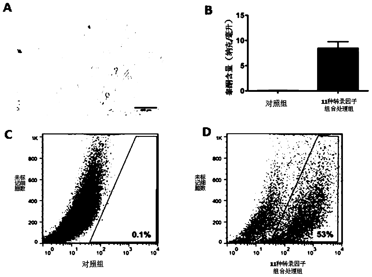
[0106] A sufficient amount of lentiviral plasmid pEZX-cyp11a1-mcherry was obtained from Example 1, and then 1×10 8 U titer of lentivirus was added to the containing 1×10 6 MEFs or TTFs (isolated in Example 2) culture dish and cultivated for 24 hours. The virus-containing DMEM medium (Cat.#: 12100-046, invitrogen Inc., USA) was discarded, replaced with fresh DMEM medium, and after continuing to cultivate for 72 hours, a final concentration of 2 micrograms / milliliter (μg / ml) was added. Puromycin was used for selection, continuous selection for 1 week ( figure 2 A). Positive clones were randomly selected for amplification, and verified by molecular biology methods such as PCR and protein hybridization.


PUM


Abstract

The invention relates to a method for inducing transdifferentiation of fibroblast into similar testicular interstitial cells. The method is characterized in that required transcription factors are respectively cloned to lentiviral vectors, and packaged to be virus particles for transfection to fibroblast; by utilizing an overexpression transcription factor method, inducing transdifferentiation of the fibroblast into inducible testicular interstitial cells, wherein the transcription factors are sourced from human beings, mice or rats, and the transcription factors are a part selected from a group of Dmrt1, Nr0b1, Sp1, Wt1, Nr4a1, Nr5a2, AP1, Creb1, Smad3, Nr5a1 and Gata4,or a combination of all transcription factors. The cells after transdifferentiation have the androgen synthesis related gene expression profile similar to that of mature testicular interstitial cells, and can synthesize and secrete testosterone. The similar testicular interstitial cells prepared with the method can be used for treating male hypogonadism syndrome.

Description

technical field [0001] The invention belongs to the field of biomedicine, and relates to the process of cell transdifferentiation and its application, in particular to a method for using lentiviral vectors to express multiple transcription factors to induce fibroblasts to transdifferentiate into testis-like Leydig cells with androgen synthesis and secretion functions. At the same time, the testis-like Leydig cells are transplanted into the male testis to provide a new treatment method for the treatment of male hypogonadism syndrome. Background technique [0002] Leydig cells are the main source of testosterone in male individuals and play a vital role in male spermatogenesis, sex differentiation, and the development and maintenance of secondary sexual characteristics. The testosterone synthesis ability of Leydig cells gradually declines with age after puberty. At the same time, factors such as environmental pollution, accelerated pace of life, and population aging make the ...

Claims


Application Information

Patent Timeline
Patent Type & Authority Patents(China)
IPC IPC(8): C12N15/867C12N5/10A61K35/52A61P5/26
CPCA61K35/52C07K14/47C12N5/0683C12N15/86C12N2506/02C12N2506/1307C12N2510/00C12N2740/15043
Inventor 黄亚东苏志坚杨艳葛仁山项琪张齐好
Owner BIOPHARM RES & DEV CENT JINAN
Learn More