Detection method of content of vitamin D in dried blood spot

A detection method and a technology for dried blood spots, applied in the field of chemical analysis, can solve the problems of inability to distinguish vitamin D2 and vitamin D3, inconvenient detection, long analysis time, etc., and achieve shortened extraction time, shortened detection time, and less blood consumption. Effect

Inactive Publication Date: 2017-06-30
北京爱普益医学检验中心有限公司
View PDF4 Cites 13 Cited by
  • Summary
  • Abstract
  • Description
  • Claims
  • Application Information

AI Technical Summary

Problems solved by technology

[0003] Since the discovery of vitamin D two centuries ago, traditional vitamin D detection methods mainly include enzyme-linked immunoassay, radioimmunoassay, immunochemiluminescence, competitive protein binding method, and high-performance liquid chromatography (HPLC). They mainly have the following problems: (1) The immunoassay and competing protein binding method will be interfered by the matrix of the high-affinity binding protein in the biological sample, whi

Method used


Image

  • Detection method of content of vitamin D in dried blood spot
  • Detection method of content of vitamin D in dried blood spot
  • Detection method of content of vitamin D in dried blood spot

Examples


Embodiment 1

[0051] I. Preparation of dried blood spot samples:

[0052] (1) Preparation of dried blood spot calibration curve samples: add a series of 25-hydroxyvitamin D2 and 25-hydroxyvitamin D3 standard solutions to whole blood, dilute 10 times, so that the final concentrations of the two 25OHDs are 5, 10, 25, 50, 100ng / mL, take 50μL of blood of each concentration and the corresponding unspiked whole blood, drip onto filter paper, dry it, and store it at -20°C.

[0053] (2) Preparation of dried blood spot samples to be tested: puncture the skin at the fingertip or heel with a blood collection needle, apply the blood on the blood collection filter paper, dry at room temperature overnight, and put it in a sealed bag containing a desiccant at -20°C save.

[0054] (3) Preparation of quality control dried blood spots: Add 18 μL and 35 μL of 1 μg / mL 25OHD2 and 25OHD3 mixed standards to 1 mL of whole blood respectively, so that the final concentration of both 25OHDs is 17.7 ng / mL (Q1) , 33....

Embodiment 2

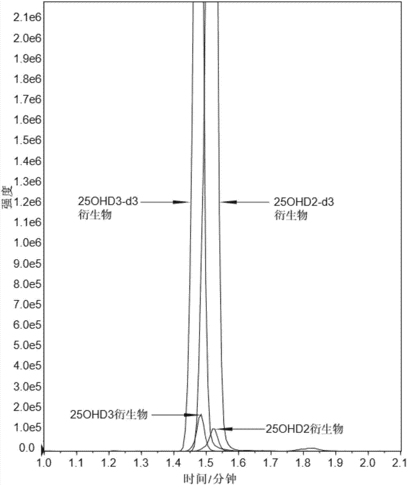
[0069] The difference between this example and Example 1 is that in the pretreatment of dried blood spot samples, the supernatant transferred to the 96-well plate in step (1) is 250 μL, and 50 μL containing 0.1 mg / Ethyl acetate of mL PTAD, room temperature 60min; When using high performance liquid chromatography tandem mass spectrometry, take step (2) and use 50 μ L of 40% acetonitrile aqueous solution to redissolve, 25 μ L of sample detection, the formic acid buffer added in acetonitrile and water Proportion is 0.2%, except that, all the other conditions are identical with embodiment 1.

Embodiment 3

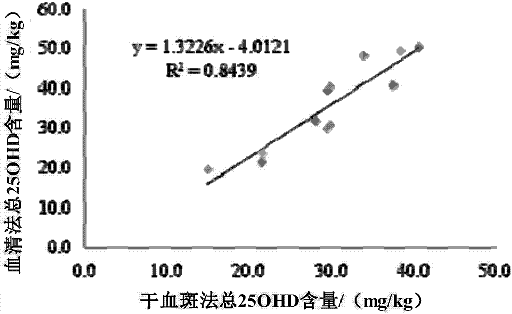
[0071] In this example, in order to evaluate the accuracy of the dried blood spot detection method in Example 1, a recovery rate test of standard addition was carried out, and the specific method is as follows:

[0072] According to the dry blood spot pretreatment method, the dried blood spot samples with known 25OHD2 and 25OHD3 spiked concentrations of 12.5, 25, and 100 ng / mL were processed. Each concentration was repeated six times in parallel, and each parallel was tested with one needle to complete the addition. Standard recovery rate test (standard addition recovery rate should meet 80%-120%), Table 3 is the average recovery rate of three concentrations of two kinds of 25OHD.

[0073] Table 3 Added recovery results

[0074]

[0075]


PUM

PropertyMeasurementUnit
Diameteraaaaaaaaaa
Concentrationaaaaaaaaaa

Abstract

The invention provides a detection method of content of vitamin D in dried blood spot. The method comprises the following steps: infiltrating blood spot by the use of ultrapure water; extracting an object to be detected by the use of acetonitrile and precipitating impurity protein, carrying out PTAD derivation for signal amplification, completing pretreatment of a sample; and completing simultaneous detection of two 25-hydroxy vitamin D by an LC-ESI-MS/MS method for 3 min. According to the pretreatment method, the system is micro, time of a hygroplasm detection method is short, and use amount of an organic solvent is greatly minimized. Thus, the method of the invention is environment-friendly and energy-saving, can be used for large-scale clinic promotion, and has high commercial value.

Description

technical field [0001] The invention belongs to the technical field of chemical analysis and relates to a method for detecting vitamin D content in dried blood spots. Background technique [0002] Vitamin D (Vitamin D (VD)) is a fat-soluble vitamin, mainly including plant-derived vitamin D2 and vitamin D3 synthesized by sunlight. The biological activities of the two are different and need to be monitored simultaneously. 25-Hydroxyvitamin D is the main storage form of vitamin D in the human body, and it is the best indicator for clinical detection and evaluation of vitamin D nutritional status (Bunch D R, Miller A Y, Wang S. Development and validation of a liquid chromatography-tandem mass spectrometry assay for serum25-hydroxyvitamin D2 / D3 using aturbulent flow online extraction technology. [J]. Clinical Chemistry & Laboratory Medicine, 2009, 47(12): 1565-72.). Vitamin D can help the body utilize calcium and phosphorus to strengthen bones (Müller DN, Kleinewietfeld M, Kvaka...

Claims


Application Information

Patent Timeline
IPC IPC(8): G01N30/06G01N30/02
CPCG01N30/06G01N30/02
Inventor 高鹏郝维维周高英周雅琼王红亮
Owner 北京爱普益医学检验中心有限公司
Why Eureka
Try Eureka