One-step adsorption method used for preparing protein microcapsules

A protein and microcapsule technology, applied in the preparation of protein microcapsules, refers to the field of protein microcapsules prepared by one-step adsorption method, which can solve the problems of cumbersome operation steps, difficult process, high loss, etc., and achieve simple preparation process, mild conditions, Good universal effect

Active Publication Date: 2018-08-28
WENZHOU INST OF BIOMATERIALS & ENG
View PDF3 Cites 7 Cited by
  • Summary
  • Abstract
  • Description
  • Claims
  • Application Information

AI Technical Summary

Problems solved by technology
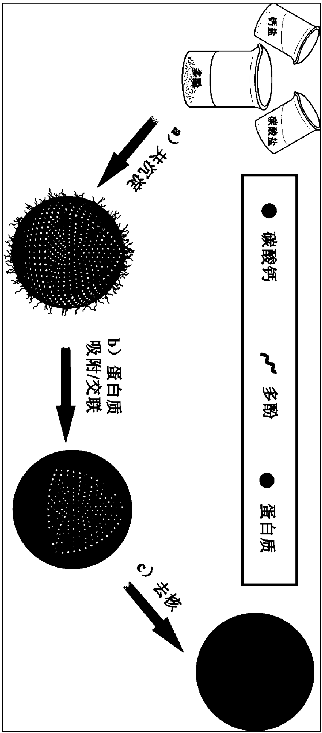
[0007] However, when the layer-by-layer self-assembly technology is used to prepare protein microcapsules, it needs to be assembled and cleaned multiple times according to the number of assembly layers. The operation steps

Method used


Image

  • One-step adsorption method used for preparing protein microcapsules
  • One-step adsorption method used for preparing protein microcapsules
  • One-step adsorption method used for preparing protein microcapsules

Examples


Embodiment 1

[0036] At 25°C, stir 1mL of 1M calcium chloride solution and 2mL of 2.5mg / mL tannic acid solution evenly, adjust the rotation speed to 1200 rpm, and quickly add 3mL of 0.33M sodium carbonate solution , stirred for 30 s, stood still for 10 min, centrifuged, and washed three times with ultrapure water to obtain tannin-doped calcium carbonate particles.

[0037] Dry the collected tannic acid-doped calcium carbonate particles in an oven at 60°C overnight, and observe the particle morphology with a scanning electron microscope (FESEM, SU8010 HITACHI, the same below), see figure 2 shown.

[0038] Put the collected particles into a centrifuge tube, add 15mL bovine serum albumin solution with a concentration of 20mg / mL, adjust the pH of the buffer solution to 9.5, incubate at 20°C for 2 hours, centrifuge, wash with the buffer solution, and redisperse in In a centrifuge tube, add 10 mL of 4-(4,6-dimethoxytriazin-2-yl)-4-methylmorpholine hydrochloride solution with a concentration of 60...

Embodiment 2

[0041] At 15°C, mix 1mL of calcium nitrate solution with a concentration of 0.1M and 2mL of a solution of epigallocatechin gallate with a concentration of 2mg / mL, stir evenly, adjust the speed to 450 rpm, and quickly add 3mL of It is a 0.033M potassium carbonate solution, stirred for 35s, allowed to stand for 10min, centrifuged, and washed three times with ultrapure water to obtain epigallocatechin gallate-doped calcium carbonate particles.

[0042] Put the particles in a centrifuge tube, add 20mL of thyroglobulin solution with a concentration of 0.1mg / mL, adjust the pH of the buffer solution to 7.0, incubate at 25°C for 6 hours, centrifuge, wash with the buffer solution, and redisperse in the centrifuge tube Add 10 mL of 4-(4,6-dimethoxytriazin-2-yl)-4-methylmorpholine hydrochloride solution with a concentration of 20 mg / mL, incubate for 6 hours, centrifuge, and wash with ultrapure water three times.

[0043] Immerse with 0.01M hydrochloric acid for 5 hours, centrifuge, and ...

Embodiment 3

[0045] At 20°C, mix 1mL of 0.33M calcium chloride and 2mL of 10mg / mL tannic acid solution, stir evenly, adjust the ultrasonic frequency to 20kHz, and quickly add 3mL of 0.33M ammonium bicarbonate solution , ultrasonically shaked for 40 s, allowed to stand for 20 min, centrifuged, and washed with ultrapure water three times to obtain tannic acid-doped calcium carbonate particles.

[0046] Put the collected particles into a centrifuge tube, add 20mL of gelatin solution with a concentration of 10mg / mL, adjust the pH of the buffer solution to 8.0, incubate at 15°C for 4 hours, centrifuge, wash with the buffer solution, and redisperse in the centrifuge tube , add 80 mL of 4-(4,6-dimethoxytriazin-2-yl)-4-methylmorpholine hydrochloride solution with a concentration of 5 mg / mL, incubate for 2 hours, centrifuge, and wash with ultrapure water three times .

[0047] Immerse with 0.05M hydrochloric acid for 5 hours, centrifuge, and wash with ultrapure water to obtain gelatin microcapsule...


PUM

PropertyMeasurementUnit
Diameteraaaaaaaaaa

Abstract

The invention relates to a one-step adsorption method used for preparing protein microcapsules, and belongs to the field of biomaterial. The one-step adsorption method is characterized in that: a polyphenol is adopted for doping of calcium carbonate micrometer particles, adsorption of protein is realized under mild conditions, crosslinking of the proteins adsorbed by the surfaces of the particlesis carried out, and at last calcium carbonate is removed so as to obtain the protein microcapsules. The advantages are that: the proteins used for preparing the protein microcapsules possess high universality; a plurality of times of assembling is not needed in preparation of the microcapsules; the one-step adsorption method is simple; preparation efficiency is high; conditions are mild; no impurity is introduced; the protein microcapsules possesses excellent biocompatibility and biodegradability, and is promising in medicine research and application prospect.

Description

technical field [0001] The invention belongs to the field of biological materials, and in particular relates to a method for preparing protein microcapsules, in particular to a method for preparing protein microcapsules by a one-step adsorption method. Background technique [0002] Layer-by-layer self-assembly (LBL) is a simple and versatile surface modification method developed rapidly in the 1990s. The technology initially uses charged substrates to be alternately deposited in oppositely charged polyelectrolyte solutions to prepare polyelectrolyte self-assembled multilayer films. With its development in membrane making, it has the following advantages: 1. A wide range of options for assembly materials, which can be polyelectrolytes or charged bioactive macromolecules such as proteins, polysaccharides, and DNA; 2. The preparation process is simple, and nanometer and submicron-scale regular structure films can be assembled on the surface of the material by simple alternate ...

Claims


Application Information

Patent Timeline
IPC IPC(8): A61K9/50A61K47/42B01J13/02B01J13/14
CPCA61K9/5052A61K9/5089B01J13/02B01J13/14
Inventor 昝兴杰史鹏忠
Owner WENZHOU INST OF BIOMATERIALS & ENG
Why Eureka
Try Eureka