Composite biology bracket material for repairing bone defects

A bio-scaffold and bone defect technology, applied in the field of bone tissue engineering, can solve the problems of side effects of degradation products, production cost, poor biocompatibility, and time for seed cell damage, degradation and absorption, etc.

Active Publication Date: 2019-06-07
JIANGNAN UNIV
View PDF5 Cites 4 Cited by
  • Summary
  • Abstract
  • Description
  • Claims
  • Application Information

AI Technical Summary

Problems solved by technology

However, this kind of scaffold material has low elastic modulus, high cost, easy deformation under stress, easy to cause damage to seed cells, and takes too long to degrade and absorb
Since the surface of these scaffold materials does not contain recognition molecules of cell adhesion molecules, such as RGD sequences, the biocompatibility with cells is relatively poor.
For example, as a scaffold material for bone tissue engineering, polylactic acid has the disadvantages of fast degradation rate, poor hydrophilicity, acidic degradation products, and prone to complic

Method used


Image

  • Composite biology bracket material for repairing bone defects
  • Composite biology bracket material for repairing bone defects
  • Composite biology bracket material for repairing bone defects

Examples


Embodiment 1

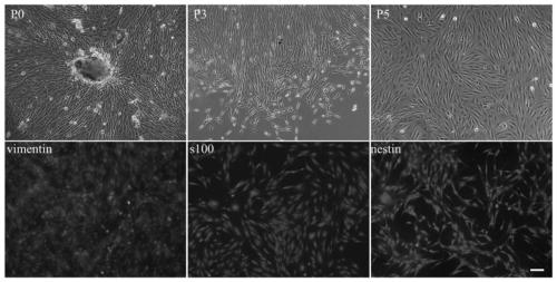
[0054] The cultivation and identification of embodiment 1 EMSCs

[0055] Deeply anesthetize SD rats (50-100g, male or female) with 10% chloral hydrate, cut the skin along the nasal cavity up to the inner canthus under sterile conditions, expose the lower nasal septum and inferior turbinate mucosa, take out the nasal septum and place it in PBS buffer solution, the full-thickness nasal mucosa was peeled off. After the nasal mucosa was taken out, it was rinsed three times with FBS medium at 4°C and shredded sufficiently, placed in a 37°C incubator and digested with trypsin for 15 minutes, centrifuged at 1000r / min to discard the supernatant, and inoculated the crushed tissue pieces in a medium containing sufficient 10% A closed culture flask of FBS DMEM / F12 was placed in a cell culture incubator. After most of the tissue pieces adhere to the wall, the medium is aspirated and new medium is added. The medium was changed every 3 days, and the cells were subcultured when the cells c...

Embodiment 2

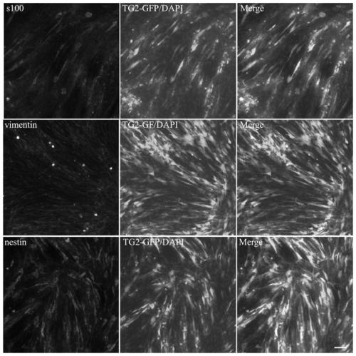
[0057] Example 2 TG2 Gene Recombinant Adenovirus Transfection EMSCs and Determination of TG2 Expression Level

[0058] The following primers were designed to synthesize the TG2 (NCBI: NP_803473.1) gene: TG2-f, CTAGCTAGCGCCACCATGGCCGAGGAGCTGAACCT and TG2-r, GGAATTCTTAGGCCGGGCCGATGATGA. The recombinant rat TG2 (rTG2) adenovirus shuttle plasmid was constructed by Nanjing Genscript Bioengineering Technology Service Co., Ltd. (Nanjing, China) and confirmed by sequencing.

[0059] The TG2 gene was inserted into the pShuttle-IRES-hrGFP2 vector to prepare the shuttle vector pShuttle-IRES-hrGFP2-TG2, and the pShuttle-IRES-hrGFP2 vector was set as a control group. 293A cells (2×10 6 cells / well) seeded in 6 cm of DMEM medium supplemented with 10% FBS 2 Petri dish and at 37 °C, 5% CO 2 Incubate overnight. Change the medium before transfection. Cells were transfected with pacAd5-9.2-100 and pShuttle-IRES-hrGFP2-TG2 shuttle plasmids at 80-90% confluence using Lipofectamine 2000. After...

Embodiment 3

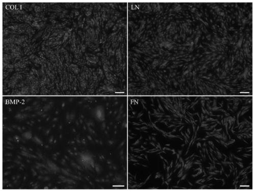
[0063] Example 3 Preparation of Fibrin Scaffold and Cell Seeding

[0064] The fibrin scaffold was made by mixing Solution A and Solution B and solidifying. Solution A was a 100 mg / mL rat fibrinogen solution dissolved in PBS, and Solution B contained 100 U rat thrombin dissolved in 5 mL PBS. Prepare fibrin scaffolds by mixing solution A and solution B in a 1:1 volume ratio so that the final concentration of fibrinogen is 50 mg / mL. Immediately and evenly drop the mixed fibrinogen-thrombin mixture onto a 96-well, 48-well or 6-well cell culture plate, and then incubate the cell culture plate at 37° C. for 30 minutes. The above-mentioned cultured TG2-EMSCs and EMSCs were digested with trypsin, centrifuged and pelleted, and the cells were collected as seed cells. Then the seeded cells are seeded on the fibrin support in the above-mentioned culture plate, and cultured by adding appropriate common complete medium. Cell-scaffolds were fixed with 4% paraformaldehyde in phosphate buff...


PUM

PropertyMeasurementUnit
Diameteraaaaaaaaaa

Abstract

The invention discloses a composite biology bracket material for repairing bone defects, and belongs to the technical field of bone tissue engineering. Reorganized TG2 gland viruses are transfected toEMSCs having multi-direction differentiation potency, in vitro assessment is performed on influence of TG2-EMSCs on osteogenesis of a fibrin bracket, and a bioactivity bracket containing TG2-EMSCs istransplanted into SD rats with skull defects, and the bone defect capacity is detected. Results prove that the fibrin bracket containing TG2-EMSCs is used for performing transplanting treatment on the skull defects, 55% of the damaged regions can be healed within two weeks, and the fibrin bracket containing natural EMSCs prove that 17% of the damaged regions are healed within the same time. The biology bracket is high and stable in biocompatibility, low in cost and simple and convenient to operate, and therefore, the fibrin bracket containing TG2-EMSCs has important clinical application valueto repair of the skull defects.

Description

technical field [0001] The invention relates to a composite biological support material for repairing bone defects, belonging to the technical field of bone tissue engineering. Background technique [0002] The repair of large bone defects in plastic surgery is a major problem that plastic surgeons often face. Common causes of bone defects include acute bone injury, benign tumors, malignant tumors, bone infection, and nonunion. The common treatment method for bone defect is autologous bone transplantation, that is, fresh bone tissue is collected from the patient's body for autologous transplantation. However, this treatment usually requires two surgeries, which not only increases the possibility of infection, but can also lead to bone deformities, pain and even functional problems. Disadvantages of autologous bone grafting prevent it from being the optimal method for repairing bone defects. At present, the use of effective genetic engineering and tissue engineering techni...

Claims


Application Information

Patent Timeline
IPC IPC(8): A61L27/22A61L27/38A61L27/50C12N5/0775
Inventor 陆乃彦张轩翁雨燕杨国锋余雪健王霁月
Owner JIANGNAN UNIV
Why Eureka
Try Eureka