
Ultrasonic nano diagnosis and treatment agent as well as preparation method and application thereof

A nano-ultrasonic technology, applied in the field of biomedicine, can solve the problems of nano-therapeutic drug loading rate and poor targeting effect of imaging ability, and achieve optimal drug loading rate and imaging ability, high drug loading rate, and compatibility good sex effect

Pending Publication Date: 2021-10-15
SHANGHAI EAST HOSPITAL EAST HOSPITAL TONGJI UNIV SCHOOL OF MEDICINE
View PDF2 Cites 0 Cited by
  • Summary
  • Abstract
  • Description
  • Claims
  • Application Information

AI Technical Summary

Problems solved by technology

[0010] Aiming at the above-mentioned technical problems in the prior art, the present invention provides an ultrasonic nano-diagnostic agent and its preparation method and application. The technical problems of poor drug loading rate, imaging ability and targeting effect of diagnostic and therapeutic agents

Method used


Image

  • Ultrasonic nano diagnosis and treatment agent as well as preparation method and application thereof
  • Ultrasonic nano diagnosis and treatment agent as well as preparation method and application thereof
  • Ultrasonic nano diagnosis and treatment agent as well as preparation method and application thereof

Examples


Embodiment 1

[0055] Lipid / PLGA-PFP nanoparticles carrying PTX and anti-miR-221 were prepared by a modified double-emulsion method.

[0056] First, 50 mg PLGA, 3 mg DSPC and 3 mg PTX were completely dissolved in 1 ml dichloromethane.

[0057] Secondly, 0.1 ml RNA aqueous solution (containing 5 nmol anti-miR-221 (anti-miR-221 was prepared by Nanjing GenScript Biotechnology Co., Ltd. according to the above sequence)) and 0.1 ml PFP (perfluoropositive Pentane) 80s (20% power; Sonic Materials, Inc.).

[0058] Third, the liquid obtained in the first two steps was mixed, and then ultrasonicated for 120 seconds in an ice bath, using pulse mode (20% power; Supersonic Materials Co.), in which mode, the ultrasonic wave was turned off for 3 seconds and turned on for 1 second, To prevent heat build-up, get a lotion.

[0059] Subsequently, 10 mL of PVA (polyvinyl alcohol) aqueous solution (4% w / v) was added, and then homogenized for 2 minutes (IKA T10basic homogenizer, gear 5, about 20000-25000 rpm). ...

Embodiment 2

[0065] Ultrasound imaging performance of the material itself: the nano-therapeutic agent in Example 1 was respectively configured into solutions with a concentration of 5ug / mL, 50ug / mL, 500ug / mL, 5mg / mL, and 50mg / mL, and placed in a solution with a mass percentage concentration of In the conical cavity in the phantom made of 2% agarose gel, the ultrasonic images of the nano-diagnostic agent in the phantom were collected under the contrast-enhanced ultrasound mode ( Figure 4 ), analyzing the time-dependent echo intensity curves before and after injection ( Figure 5 ). from Figure 5 It can be seen that as the concentration increases, the ultrasonic image tends to become brighter as the concentration increases. It shows that the ultrasonic performance is related to the drug concentration, and shows a good linear relationship with the drug concentration in the experimental concentration range, showing good performance of contrast-enhanced ultrasound, indicating that it has a ...

Embodiment 3

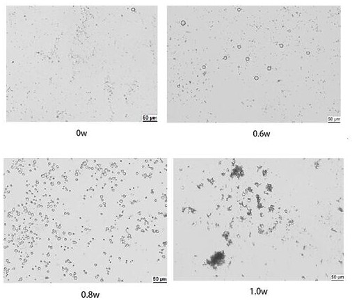
[0067] Imaging performance of the material under different ultrasonic power and time: select the nano-diagnostic agent of Example 1 with a concentration of 5 mg / mL, and place several concentrations in the phantoms made of 2% agarose gel. In the conical cavity, under the action of different ultrasonic power (0.8W / cm2, 1.0W / cm2, 1.2W / cm2, 1.4W / cm2, 1.6W / cm2) and time (20s, 40s, 60s), record the ultrasound Imaging experiment ( Image 6 ), demonstrating the time-dependent ultrasound imaging effect of nanomaterials.


PUM

PropertyMeasurementUnit
Particle sizeaaaaaaaaaa
Diameteraaaaaaaaaa

Abstract

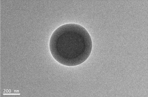
An ultrasonic nano diagnosis and treatment agent comprises an outer shell, wherein the outer shell is formed by hybridizing distearoyl phosphatidylcholine and hydroxyl-terminated poly(lactic-co-glycolic acid), paclitaxel is inlaid on the surface of the outer shell, an inner core is arranged in the outer shell, an aqueous solution mixture composed of perfluoro-n-pentane and a gene drug miR-221 inhibitor is wrapped in the inner core, and the particle size of the outer shell is 500-600 nanometers. The invention provides a preparation method of the ultrasonic nano diagnosis and treatment agent. The invention also provides application of the ultrasonic nano diagnosis and treatment agent in preparation of medicines for treating cancers. Under the stimulation of ultrasonic waves, the PFP in the inner core is stimulated to generate gas, and the appearance of the gas enhances the harmonic signal of the ultrasonic waves. Meanwhile, a large amount of gas makes the nano shell thin, external low-power ultrasonic waves are added, the shell is damaged, the paclitaxel embedded in the shell and the miR-221 inhibitor in the inner core are released, and therefore the tumor is killed.

Description

Technical field: [0001] The invention belongs to the field of biomedicine and relates to a nano-diagnosis and treatment agent, in particular to an ultrasonic nano-diagnosis and treatment agent and its preparation method and application. Background technique: [0002] Triple-negative breast cancer (TNBC) is highly aggressive and has metastatic potential. Traditional chemotherapy is currently the main treatment, but affected by side effects, the prognosis is still unsatisfactory. How to choose an appropriate treatment plan to reduce side effects and improve the effect of diagnosis and treatment has become a clinical difficulty. The development of nanotechnology in recent years has brought a glimmer of hope to solve some problems. The specific advantages are reflected in the following aspects: [0003] 1. The shell of nanoparticles can embed or encapsulate drugs to improve efficacy and reduce systemic toxicity. [0004] 2. It can carry different drugs at the same time to ach...

Claims


Application Information

Patent Timeline
IPC IPC(8): A61K49/22A61K31/337A61K45/06A61K47/24A61K47/34A61K47/69A61P35/00
CPCA61K49/225A61K47/6925A61K45/06A61K31/337A61K47/34A61K47/24A61P35/00A61K2300/00
Inventor 张丽波任真顾俊毅莫新海林杰
Owner SHANGHAI EAST HOSPITAL EAST HOSPITAL TONGJI UNIV SCHOOL OF MEDICINE
Learn More