Quantum dot micelle spherical nucleic acid sensor as well as preparation method and application thereof in Pb < 2 + > detection

A nucleic acid sensor and quantum dot technology, applied in the field of biosensing, can solve problems such as being unsuitable for on-site detection, and achieve the effects of high sensitivity and high specificity detection, good repeatability, and wide detection range

Pending Publication Date: 2022-04-15
SOUTHEAST UNIV
View PDF0 Cites 0 Cited by
  • Summary
  • Abstract
  • Description
  • Claims
  • Application Information

AI Technical Summary

Problems solved by technology

Various methods have been developed for the accurate detection of lead ions in the water environment, such as traditional atomic absorption spectroscopy, atomic emission spectroscopy and inductively coupled plasma mass spectrometry, etc., but none of the above methods are suitable for on-site detection

Method used


Image

  • Quantum dot micelle spherical nucleic acid sensor as well as preparation method and application thereof in Pb &lt; 2 + &gt; detection
  • Quantum dot micelle spherical nucleic acid sensor as well as preparation method and application thereof in Pb &lt; 2 + &gt; detection
  • Quantum dot micelle spherical nucleic acid sensor as well as preparation method and application thereof in Pb &lt; 2 + &gt; detection

Examples


Embodiment 1

[0034] Sequence design of aptamers, DNases, and substrates

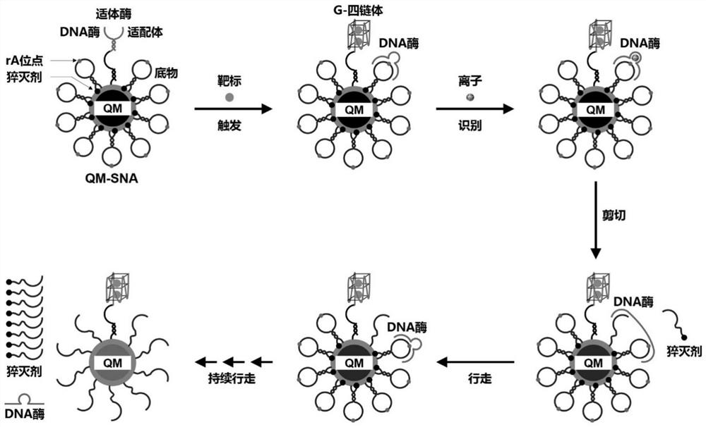
[0035] Such as figure 1 As shown, for the target Pb 2+, and designed the corresponding aptamerase DNA and substrate (Substrate). The aptamyme includes an aptamer sequence and a DNase sequence. The aptamer sequence mainly includes a target binding region and a DNase complementary region, and the amino group at the 5' end is modified for coupling to the QM surface. Among them, the target binding region is the specific recognition sequence of the target, which is used to combine with the target to form a G-quadruplex, and destroy the double-stranded structure of the aptase to release the DNase. The DNase sequence mainly includes three parts: aptamer complementary region, (substrate) orbital binding region, and catalytic center region. Wherein, the aptamer complementary region sequence is the complementary sequence of the aptamer, which is used to hybridize with the aptamer sequence to form aptazyme, immobilize and i...

Embodiment 2

[0040] Preparation of QM-SNA loaded with enzyme and substrate

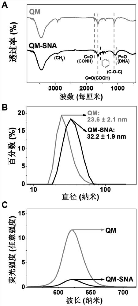
[0041] Quantum dot micelles QM are prepared by ultrasonic film hydration: the quantum dots used are CdSe / ZnS (purchased by Suzhou Xingshuo Nano Technology Co., Ltd.). 0.1 mg of quantum dots and 4 mg of dipalmitoylphosphatidylethanolamine-polyethylene glycol-carboxy (DSPE-PEG2000-COOH) were dissolved in 1 mL of chloroform. A film containing QM was obtained by reacting with a rotary evaporator at 45°C for 15 minutes; 1 mL of deionized water was added to it and dispersed by ultrasonic to prepare a QM solution.

[0042] QM-SNA was prepared by chemical cross-linking method: In a reaction system with a total volume of 500 μL, 10 μL EDC solution (1 mM) was added to 10 nM QM buffer (phosphate buffer solution: 0.01 M, pH 7.4), 10 μL Sulfo- NHS solution (0.5mM), activated at room temperature for 30min. Add 16.7 μL (1 μM) of aptamerase solution and 66.7 μL (1 μM) of enzyme substrate solution to the buffer, stir the reactio...

Embodiment 3

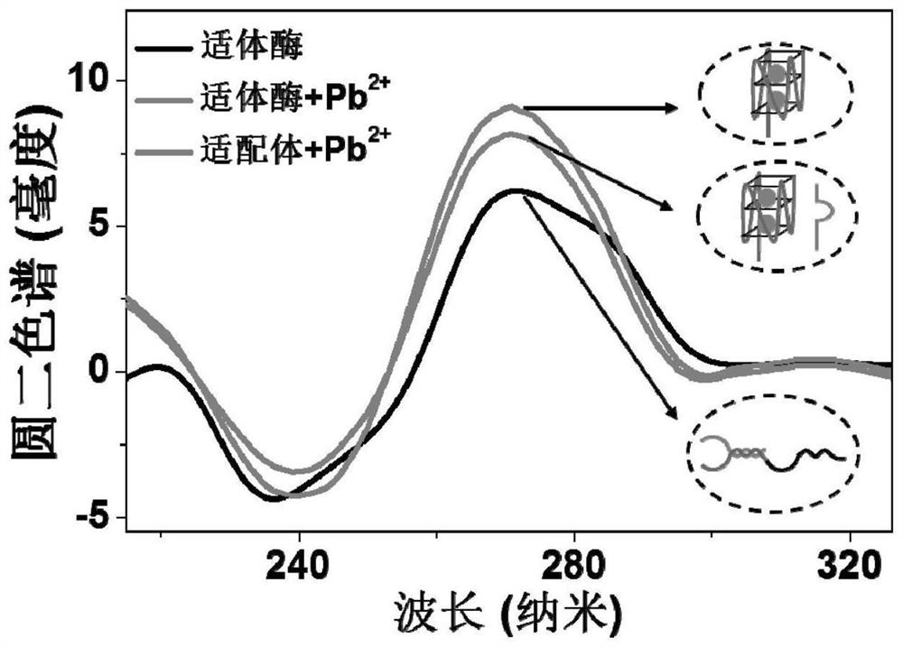
[0045] Pb 2+ triggers a conformational change in aptazymes

[0046] CD spectra of DNA were measured using a Chirascan circular dichroism spectrometer. The aptamase sample was dissolved in 0.01 M pH 7.4 phosphate buffer so that the final concentration was 5 μM, and its CD spectrum was measured. Then add 5000pM Pb to it 2+ , measured its CD spectrum after reacting at room temperature for 30 min. A quartz cell with a 2mm optical path length and an instrument scanner were used to record, the scanning range was 200 to 320nm, and the average value was obtained by scanning three times.

[0047] The result is as image 3 shown, Pb-free 2+ Under these conditions, the aptamerase has a positive peak at 275nm and a peak shoulder at 285nm, showing the characteristics of double-stranded DNA. Join Pb 2+ (5000pM), the aptamerase presents a negative peak at 242nm, and a positive peak at 270nm, which combines with the aptamer to Pb 2+ The typical G-quadruplex formed has the same charact...


PUM


Abstract

The invention discloses a quantum dot micelle spherical nucleic acid sensor as well as a preparation method and application thereof in Pb < 2 + > detection. The sensor is formed by coupling two nucleic acid molecules on the surface of a quantum dot micelle, wherein the two nucleic acid molecules are aptamer and an enzyme substrate modified by a quencher; the aptamer enzyme is formed by hybridizing a 5 '-amino modified lead ion aptamer and a DNA enzyme sequence; the enzyme substrate is hairpin structure DNA containing rA site, the 5'end of the DNA is modified with amino, and the 3 'end of the DNA is modified with a quencher. Aptamer enzyme in the sensor is specifically combined with Pb < 2 + > to form a G-quadruplex, DNA enzyme is released, and the DNA enzyme automatically walks by taking an enzyme substrate as a track; and detecting and analyzing Pb < 2 + > by measuring a fluorescence signal of the quantum dot micelle after walking is finished. According to the method for fluorescence amplification detection of Pb < 2 + > based on QM-SNA DNA enzyme walking, high-sensitivity and high-specificity detection of Pb < 2 + > is achieved, and a new detection method is provided for detection of heavy metal ions.

Description

technical field [0001] The invention belongs to the technical field of biosensing, in particular to a quantum dot micellar spherical nucleic acid sensor, a preparation method thereof and a Pb 2+ application in detection. Background technique [0002] Lead ion (Pb 2+ ) is a Class I environmental pollutant, which is commonly found in industrial production such as batteries, electric welding, and lead-zinc smelting. It can be ingested by the human body through the digestive tract and respiratory tract, and accumulate in the body, causing various health problems. After entering the human body, lead can interact with biomolecules in the body to cause chronic damage to the body and destroy the functions of the digestive system, nervous system, respiratory system, and reproductive system. Major environmental organizations have formulated relevant standards for lead ions. For example, the U.S. Environmental Protection Agency (EPA) sets the upper limit of lead ions in drinking wate...

Claims


Application Information

Patent Timeline
Patent Type & Authority Applications(China)
IPC IPC(8): G01N33/53G01N33/58C12Q1/6825C12Q1/6818
Inventor 孙清江牛亚楠沈鹏于真真王婕妤
Owner SOUTHEAST UNIV
Why Eureka
Try Eureka