Method and material for enhanced tissue-biomaterial integration

a biomaterial and tissue technology, applied in the direction of skeletal/connective tissue cells, drug compositions, prostheses, etc., can solve the problems of biomaterials not being properly integrated with the body, sacrificing implant longevity and function, and hard tissues such as cartilage and bone present particular challenges to integration

Active Publication Date: 2004-09-02
SYNTHASOME
View PDF42 Cites 27 Cited by
  • Summary
  • Abstract
  • Description
  • Claims
  • Application Information

AI Technical Summary

Problems solved by technology

Integration of biomaterials with the body is a longstanding problem in medicine.
Lack of proper integration with the body sacrifices implant longevity and function.
Hard tissues such as cartilage and bone present particular challenges to integration.
Cartilage lacks the ability to repair itself, and has a dense extracellular matrix that provides a smooth surface with weight bearing function, making cartilage extremely challenging to integrate with other materials.
This profoundly limits the strength and durability of these inventions, as they do not integrate well as possible with the tissue.
Integration of biomaterials with hard tissues such as cartilage and bone is particularly challenging due to the nature of the dense extracellular matrix and strong mechanical forces that the tissue must withstand.
Furthermore, cartilage tissue lacks the ability to heal and has difficulty integrating with natural or engineered cartilage, much less a biomaterial, and was therefore chosen as our model system.
Heretofore, true integration of biomaterials by covalent binding of the materials to an extracellular matrix has not been achieved.
Intermediate oligomeric products resulting from such degradation may have different toxicological properties, however, or biodegradation may involve oxidation or other biochemical reactions that generate molecules other than monomeric subunits of the polymer.

Method used


Image

  • Method and material for enhanced tissue-biomaterial integration
  • Method and material for enhanced tissue-biomaterial integration
  • Method and material for enhanced tissue-biomaterial integration

Examples


example i

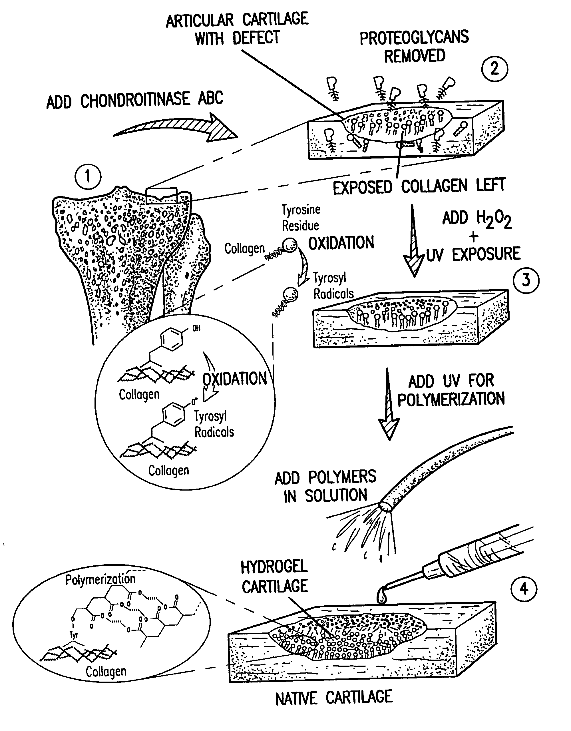
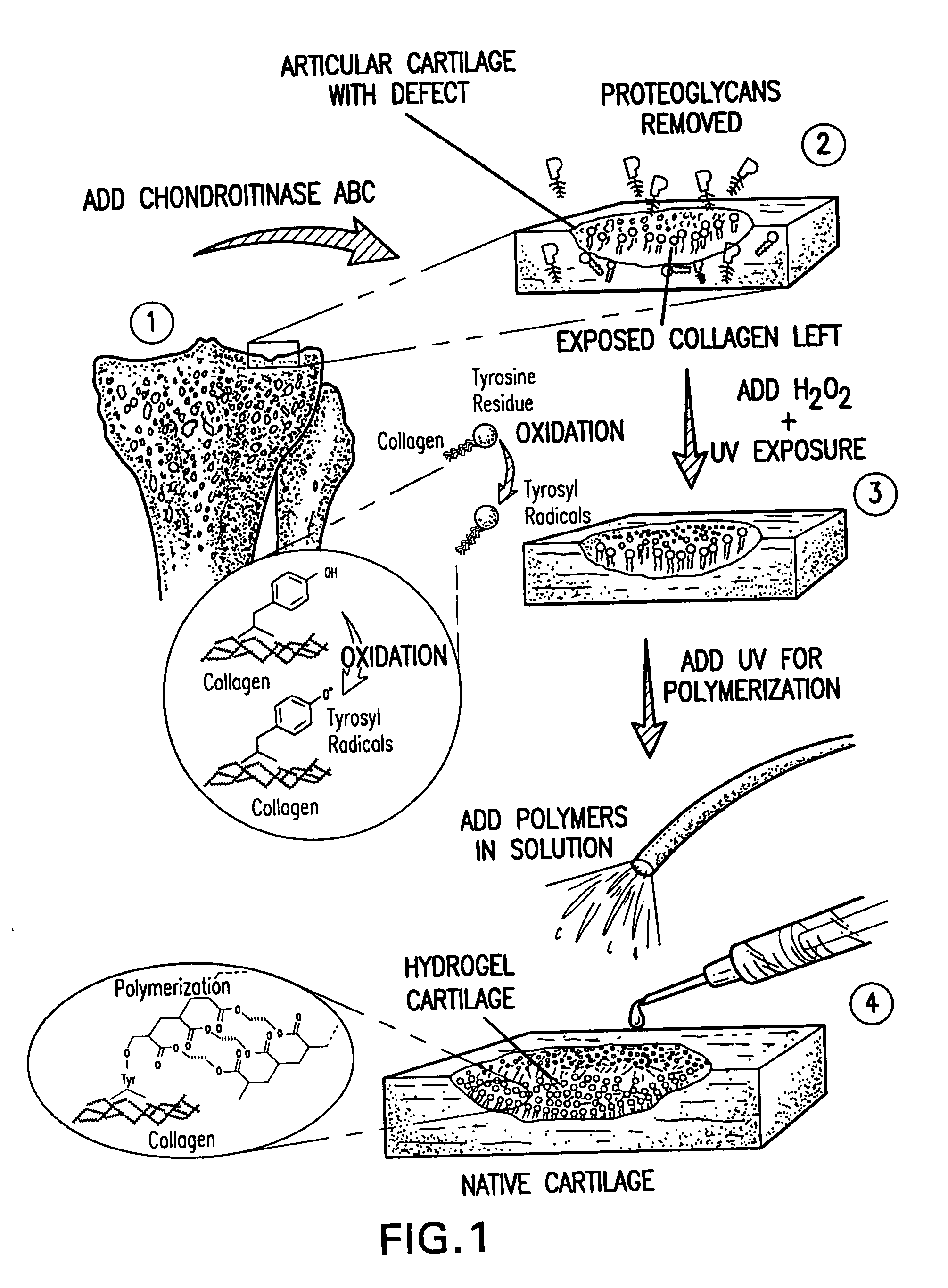
[0123] An exemplary in vitro protocol is discussed below. Surfaces of fresh fetal bovine cartilage chips were treated with chondroitinase ABC (5 unit / ml, in Tris pH 8.1) at 37.degree. C. for 1 hr. Photo-oxidation of these surfaces was performed for 5 min with H.sub.2O.sub.2 (5%) and UV-irradiation (365 nm; 3 mW / cm.sup.2). Excess H.sub.2O.sub.2 was removed. Pre-argon-bubbled PEODM (poly[ethylene glycol] dimethacrylate, 15% and 20%, w / v) were added to the cartilage surfaces without photo-initiators. The reactants underwent the UV-irradiation (365 nm; 8 mW / cm.sup.2) for 30 min.

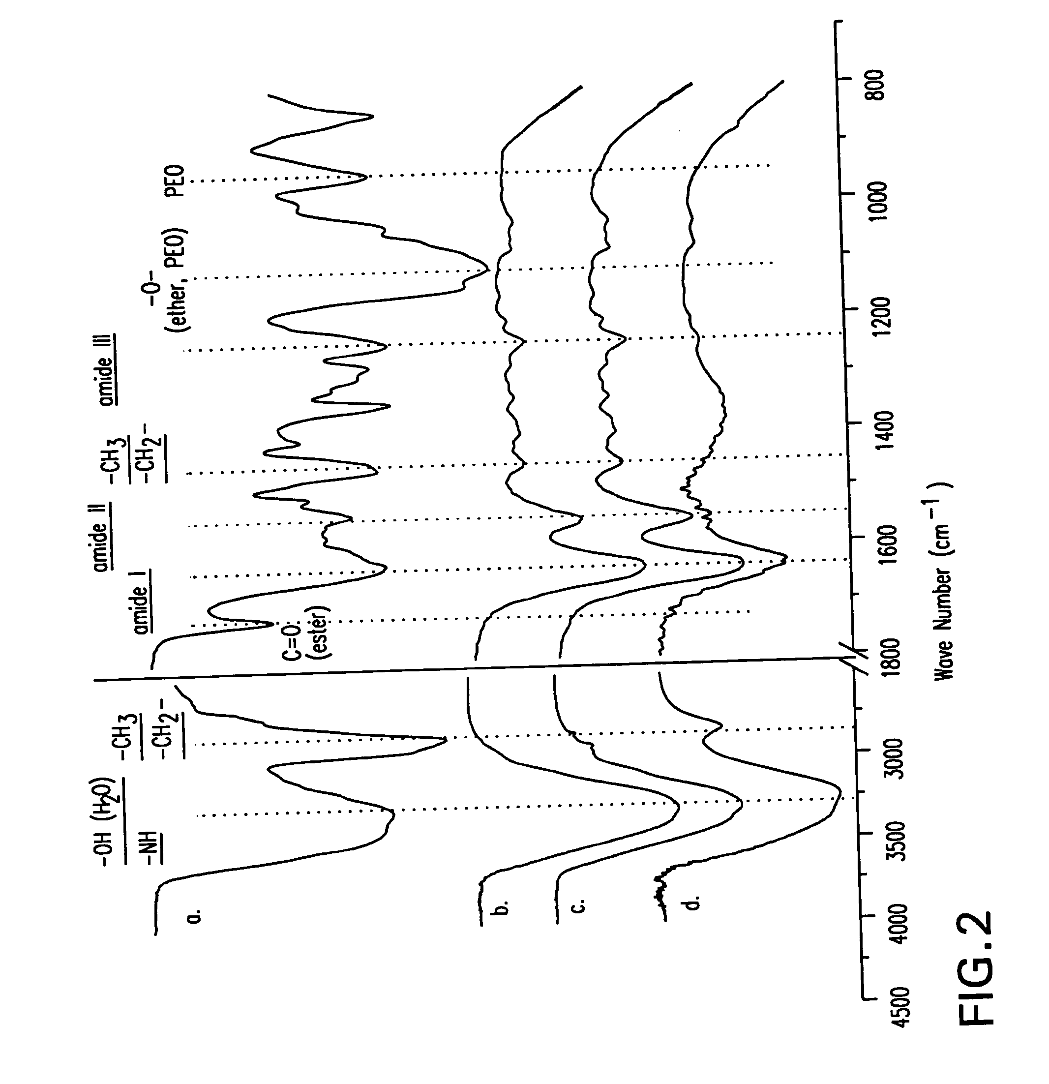
[0124] Chemical and morphological analysis was performed on the cartilage-biomaterial construct to confirm tissue-initiated polymerization and gel formation (FIG. 2). The superficial layer of native cartilage is a mixture of proteins, proteoglycans, and bonded water as seen chemically by Fourier transform infrared spectroscopy (ATR-FTIR) spectrum d. After enzymatic treatment of the cartilage surface with chondroi...

example ii

[0128] Analysis of the mechanism of the tissue-initiated polymerization and direct covalent integration according to the present invention was carried out in a simplified system using pure collagen protein and hydroxyethyl methacrylate (HEMA). Collagen was chosen as the target for reaction due to its universal presence and mechanical stability. Collagen contains tyrosine residues which may be oxidized to produce the initiating radical for polymerization. While tyrosine makes up only 0.6% of human Type II collagen, 90% of these residues are located at the protein C-terminus, providing a concentrated source of radicals for polymerization.

[0129] Type II collagen was reacted with H.sub.2O.sub.2 (1%) for 15 min. After the un-reacted H.sub.2O.sub.2 was removed, HEMA (250 mM) was added and the UV-irradiation (365 nm; 3 mW / cm.sup.2) was performed for 5 min. The product solution was loaded into the Sephadex.RTM. (Amersham) G-25 size exclusion column to remove the excess HEMA monomer and the ...

example iii

[0137] FIG. 7 illustrates the aldehyde priming embodiment of the invention. The method described below may be found in more detail in Li et al., Macromolecules vol. 36, pp. 2556-2562 (2003), the entire disclosure of which is hereby incorporated by reference.

[0138] Chondroitin sulfate A sodium salt (CS, 10 g, .about.20 mmol disaccharide repeating unit) was dissolved in 100 ml phosphate buffer saline (PBS, pH 7.4), followed by addition of glycidyl methacrylate (GMA, 3 g, .about.20 mmol) while vigorously stirring at room temperature for 1 to 15 days. The purification was performed by anhydrous acetone extraction twice to remove all the compounds that failed to covalently graft onto CS chains. The purified products were lyophilized for 48 hrs. The final yield of chondroitin sulfate methacrylate (CS-MA) product was all 7 to 8 g.

[0139] Six hundred mg of CS-MA (0.8.about.1.2 mmol of adjacent diol, 70% CS-A, Sigma) and 616 mg of sodium periodate (.about.2.88 mmol, NaIO.sub.4, Sigma) were di...


PUM

PropertyMeasurementUnit
volume %aaaaaaaaaa
molecular weightaaaaaaaaaa
pHaaaaaaaaaa

Abstract

The present invention relates to the covalent binding of a hydrogel to an extracellular matrix (ECM). The integration of the hydrogel with the tissue is superior to that in previous techniques. Moreover, unlike previous techniques, the present invention does not require a photoinitiator. Potential therapeutic applications include tissue repair and delivery of drugs or cells. The ECM is first exposed, then treated with a priming agent. Then a polymerizable agent is added and crosslinked to the ECM. Two primary embodiments of methods are disclosed. In the first, the priming agent is an oxidizer which creates tyrosyl radicals in the ECM, which are then bound by acrylate groups in the polymerizable agent. In the second, the priming agent contains aldehydes which bind amino groups in the ECM.

Description

[0001] This application claims benefit under 35 U.S.C. 119(e) of U.S. Provisional Application Nos. 60 / 416,881 and 60 / 416,882, filed Oct. 9, 2002, the entire disclosures of which is hereby incorporated by reference.[0002] This application is related to a utility patent application claiming priority to U.S. Provisional Application No. 60 / 413,152 (filed Sep. 25, 2002), entitled "Cross-linked polymer matrices, and methods of making and using same," and filed on Sep. 25, 2003, the entire disclosure of which is hereby incorporated by reference.[0003] This application is also related to a utility patent application claiming priority to U.S. Provisional Application No. 60 / 416,882 (filed Oct. 9, 2002), entitled "Multilayered polymerizing hydrogels for tissue regeneration," and filed on Oct. 9, 2003, the entire disclosure of which is hereby incorporated by reference.[0004] 1. Field of the Invention[0005] The present invention pertains to a method of creating a hydrogel covalently bound to the...

Claims


Application Information

Patent Timeline
Patent Type & Authority Applications(United States)
IPC IPC(8): A61K35/32A61K45/06A61L27/36A61L27/38A61L27/50A61L27/52C12N5/00C12N5/071C12N5/077
CPCA61K35/32C12N2533/30A61L24/0005A61L24/0031A61L27/3604A61L27/3616A61L27/3633A61L27/3683A61L27/3687A61L27/3691A61L27/38A61L27/50A61L27/52C12N5/0062C12N5/0655C12N5/0697A61K45/06A61P19/00
Inventor WANG, DONGANRATCLIFFE, ANTHONYELISSEEFF, JENNIFER H.
Owner SYNTHASOME
Why Eureka
Try Eureka