
Implantable Biosensor and Methods of Use Thereof

a biosensor and implantable technology, applied in the field of glucose monitoring, can solve the problems of lack of normal sensitivity, no magic pill to treat diabetes, failure of the health regulation system, etc., and achieve the effect of rapid oxygen transport and oxygen transpor

Active Publication Date: 2006-10-26
METRONOM HEALTH
View PDF11 Cites 23 Cited by
  • Summary
  • Abstract
  • Description
  • Claims
  • Application Information

AI Technical Summary

Benefits of technology

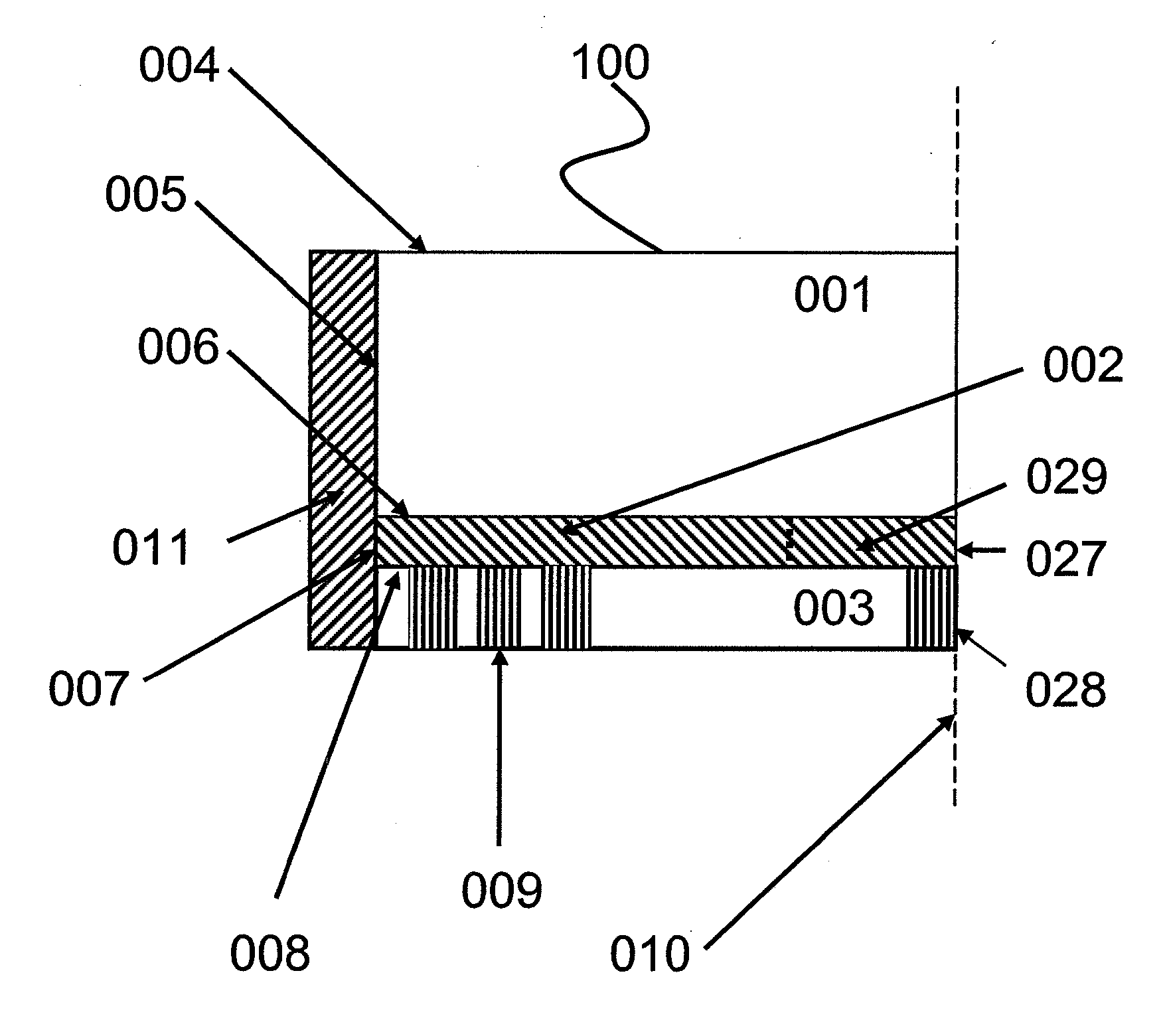
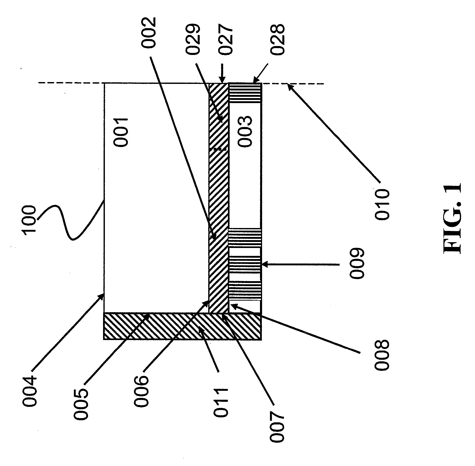
The present invention is based on the discovery of a stabilized oxygen transport matrix that includes a reversible oxygen binding protein, such as hemoglobin or myoglobin, immobilized throughout the matrix. This matrix can rapidly transport oxygen from one area to another, overcoming challenges of implantable sensors. The stabilized oxygen transport matrix can be used in an implantable glucose sensor to selectively and sensitively monitor glucose in the subcutaneous tissues under low oxygen tension. The sensor includes an oxygen transport region, a target analyte reaction region, and a sensing region. The target analyte can be glucose, and the sensor can be an implantable glucose sensor. The invention provides a solution for selectively and sensitively monitoring glucose in the subcutaneous tissues under low oxygen tension.

Problems solved by technology

In diabetics this healthy regulation system primarily fails due to the following two factors, either alone or in combination: 1) insufficient insulin production and secretion, 2) a lack of normal sensitivity to insulin by the tissues of the body.
There is still no magic pill to treat diabetes.
However, current blood glucose tests are painful, requiring finger sticking to obtain a blood sample.
They are inconvenient due to disruption of daily life and difficult to perform in long-term diabetic patients due to calluses on the fingers and poor circulation.
Further, even the recommended testing schedule is far from sufficient to allow blood glucose normalization.
Thus with present technology, the necessary monitoring is frequently unachieved chore.
The required sampling schedule cannot realistically be expected of even the most committed patients during the day and is not feasible at night.
Present blood glucose monitoring methods are not automatic, chronically requiring user initiative.
This system cannot therefore be relied upon to detect spontaneous hypoglycemia or other glycemic excursions.
Consequently, even the most diligent patients fail to avoid severe complications.
However, this approach is not directly applicable to the subcutaneous tissue.
Many sensing methods will perform well under in vitro or carefully controlled conditions, but will then fail to perform well in the body.
Their failure has been attributed to inadequate selectivity, electrode poisoning, and insufficient glucose sensitivity.
The tissue matrix contains many constituents that are constantly changing and which may interfere with varying types of measurement approaches.
However, while appealing, non-invasive optical measurements are generally not sufficiently selective for glucose without a detailed knowledge of the matrix being probed.
This poses a severe challenge for purely optical measurements to be highly selective for glucose.
However, problems exist in the direct application of both of these approaches.
Hydrogen peroxide probes often suffer from electrochemical interference by oxidizable species in a complex matrix such as encountered in the body.
The electrode oxidizes these other electroactive constituents as well as hydrogen peroxide, which results in measurements with a net positive and variable error.
The hydrogen peroxide if not eliminated may also have an undesirable reaction with the surrounding tissue as well as degrade the oxidase enzyme necessary for the operation of the sensor over the course of sensor operation.
Additionally, unless oxygen is available in excess to the glucose being reduced by the reaction, variations in bulk oxygen will also change how much glucose is oxidized, resulting in erroneous measurements if the oxygen concentration influencing the reaction is not directly accounted for in a calibration, or prevented from impacting reaction dynamics.
The problem of sensitivity to varying oxygen concentration is also present in approaches based on measuring oxygen consumption.
Unfortunately, the subcutaneous tissue environment has been shown to have both a variable and heterogeneous oxygen concentration.
However, problems of stability, drift, selectivity, and sensitivity must be overcome if using an electrode system.
In the subcutaneous environment, oxygen is scarce, making an optical method that measures the result of the glucose oxidation reaction while not consuming oxygen even more desirable.
In coupling glucose oxidase with an optical transducer, an additional problem of selective sensing of oxygen is posed.

Method used


Image

  • Implantable Biosensor and Methods of Use Thereof
  • Implantable Biosensor and Methods of Use Thereof
  • Implantable Biosensor and Methods of Use Thereof

Examples


example 1

Analysis of the Oxygen Conducting Capability of Hemogloblin

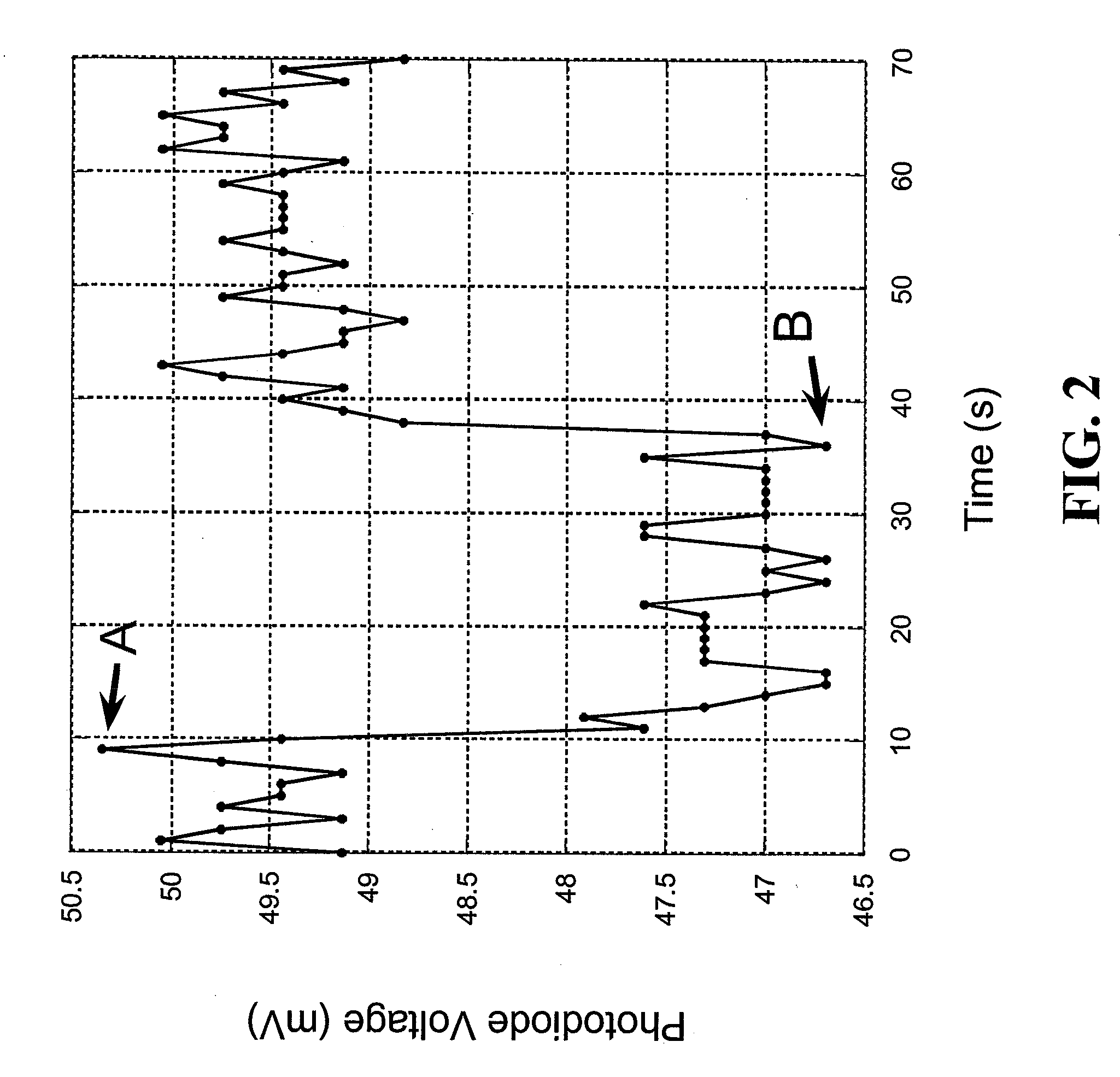
[0111] The following example illustrates the oxygen conducting capability of hemoglobin and the intended functionality of the reversible oxygen binding protein. A film of hemoglobin was prepared as follows. Human blood was extracted with a finger-stick device. A large drop of the blood was deposited on one end of a rectangular microscope glass slide. A blood smear ⅛ inches by 1½ inches in dimension was created by sliding a microscope cover glass from the blood drop, across the glass slide to create a thin film of blood across the glass slide. The glass slide was then covered in cellophane leaving approximately ¼ inch of the blood smear exposed in the long axis. A glass coverslip was placed on the cellophane to ensure a good seal. This setup ensures no-flux oxygen boundaries on all surfaces and edges of the blood smear, except for at the exposed ¼ inch at the end. A diode laser's beam was directed through the glass slide, bl...

example 2

Illustrative Implantable Glucose Sensor

[0113] This Example discloses certain features of an illustrative glucose sensor 400 of the present invention, as illustrated in FIG. 3 and FIG.4, and exemplifies a method for manufacturing the illustrative glucose sensor 400. In the illustrative glucose sensor 400 provided in this example and illustrated in FIG. 3, the electronic light source 490 and the photodetector 495 communicate through a series of fiber optic bundles 410, 415 with the illustrative glucose sensor 400. In the method used to create the glucose sensor 400, an array of individual optical fiber emitters 410 was bent into a hook so that light emanating from the emitter array 410 traveled through the reaction zone 402 and was collected by a detection fiber optic bundle 415 (i.e. an array of optical fiber receivers). In this illustrative glucose sensor 400 the entire back end of the detection bundle 415 was coupled to a photodiode type photodetector 495.

[0114] During use of the...

example 3

Glucose Measurements Using an Illustrative Sensor

[0137] This Example demonstrates detection of glucose in a relatively simple glucose sensor that includes two stabilized oxygen transport matrices in communication with one another. One of the illustrative stabilized oxygen transport matrices was an oxygen transport region and the other was a glucose reaction region that included a stabilized glucose oxidase-hemoglobin thin gel.

[0138] A contiguous oxygen transport region and glucose reaction region were deposited on a glass slide, coated with silicon, and a glucose inlet was present on a surface of the glucose reaction region. The glucose reaction region was made with 20 mg glucose oxidase and 0.1 ml of human blood crosslinked with a dilute formaldehyde solution. The steps for constructing the glucose reaction region are outlined below in more detail.

[0139] 1. 0.1 ml of whole human blood was combined with 0.3 ml of a solution containing a 1:3 ratio of 70% isopropyl alcohol to disti...


PUM

PropertyMeasurementUnit
pore sizesaaaaaaaaaa
concentrationsaaaaaaaaaa

Abstract

Provided herein is a stabilized oxygen transport matrix that includes a reversible oxygen binding protein, such as hemoglobin, immobilized throughout the stabilized oxygen transport matrix. The stabilized oxygen transport matrix is used to transport oxygen and can be used as an oxygen transport region and a reaction region of an analyte sensor, such as an implantable glucose sensor. The reversible binding protein can also function as an oxygen probe within the analyte sensor.

Description

RELATED APPLICATION DATA [0001] This application claims the benefit of priority under 35 U.S.C. §119(e)(1) of U.S. Ser. No. 60 / 531,447, filed Dec. 18, 2003, the entire content of which is incorporated herein by reference.BACKGROUND OF THE INVENTION [0002] 1. Field of the Invention [0003] The invention relates generally to glucose monitoring and more specifically to implantable glucose sensors. [0004] 2. Background Information [0005] Diabetes is a disease of insufficient blood glucose regulation. In non-diabetic people, the body's beta cells monitor glucose and deliver just the right amount of insulin on a minute-by-minute basis for tissues in the body to uptake the right amount of glucose, keeping blood glucose at healthy levels. In diabetics this healthy regulation system primarily fails due to the following two factors, either alone or in combination: 1) insufficient insulin production and secretion, 2) a lack of normal sensitivity to insulin by the tissues of the body. [0006] The...

Claims


Application Information

Patent Timeline
Patent Type & Authority Applications(United States)
IPC IPC(8): A61B5/05C12Q1/00C12Q1/54A61B5/00G01F1/64G01N27/26
CPCA61B5/14532A61B5/1473A61B5/1459A61B5/14865A61B5/1455A61B5/1495
Inventor BOTVINICK, ELLIOTBREMER, TROY M.
Owner METRONOM HEALTH
Learn More