Eureka AIR delivers breakthrough ideas for toughest innovation challenges, trusted by R&D personnel around the world.

Multichamber device for processing of biological samples using high pressure

a multi-chamber device and biological sample technology, applied in the field of methods and multi-chamber devices, can solve the problems of limited current methods for extracting biological molecules from such samples, the difficulty of molecular techniques being applied in plant and animal tissues, and the extensive enzymatic digestion of samples

Inactive Publication Date: 2007-01-18
BBI BIOSEQ A MASSACHUSETTS CORP
View PDF20 Cites 36 Cited by
  • Summary
  • Abstract
  • Description
  • Claims
  • Application Information

AI Technical Summary

Benefits of technology

[0009] The new devices and methods provide a simple format for loading and unloading samples and the methods generally can be automated while still effectively fragmenting, homogenizing, and processing tissue samples. The methods and devices reduce the necessity to transfer samples and reagents as the samples are generally processed sequentially. In one embodiment of the invention, large chunks of tissue are first fragmented into smaller pieces and then homogenized to completion, while avoiding unacceptable degradation of targeted biomolecules, before the sample is further processed. Release of biomolecules such as nucleic acids or proteins is efficient, and the function of those molecules (e.g., in the lysate) is well preserved. After the sample has been homogenized and the biomolecules have been released, the sample undergoes further processing, typically after being forced into a subsequent chamber of the multichamber device. Further processing of the sample can include, but is not limited to, one or a combination of the following processes: purification by chromatography, solid phase capture, or gel electrophoresis; enzymatic processing; amplification of biomolecules (e.g., PCR); processing and / or detection of protein interactions; chemical modification; substrate labeling; solublization of substrate; and substrate detection.
[0010] The homogenization aspect of the method is readily used in conjunction with high hydrostatic pressure cycling technology (“PCT”). The use of PCT technology to analyze biological samples is generally described elsewhere, for example in the following documents which are incorporated by reference in their entirety: Laugharn, Jr. et al., U.S. Pat. No. 6,111,096; Laugharn, Jr. et al., U.S. Pat. No. 6,120,985; Hess, R. and Laugharn, Jr., U.S. Pat. No. 6,127,534; Hess, R. and Laugharn, Jr., U.S. Pat. No. 6,245,506; Laugharn, Jr. et al., U.S. Pat. No. 6,258,534; Laugharn, Jr. et al., U.S. Pat. No. 6,270,723; Laugharn, Jr. et al., U.S. Pat. No. 6,274,726 and PCT Application No. WO99 / 22868. Physical changes effected on the sample can include pressure-driven phase changes, volume changes under high pressures and homogenization, for example, as solids or other matter pass through screens. Such processes are readily adaptable to automation.

Problems solved by technology

Often, sample preparation also entails extensive enzymatic digestion, the use of harsh chemical reagents, and / or mechanical disruption.
Particular difficulties can be encountered in the application of molecular techniques to plant and animal tissues and to bacteria with rigid cell walls such as certain mycobacteria.
Current methods for extracting biological molecules from such samples are limited by the requirement for complex processing using multiple steps and can be very time-consuming, labor intensive, and costly.
For some cells and tissues, additional mechanical disruption is also often necessary, requiring equipment such as a mortar and pestle, bead mills, a rotor-stator homogenizer, a blade blender, an ultrasonicator, a pulverizer, a pestle and tube grinder, a meat mincer, a Polytron®, or a French Press™.

Method used


Image

  • Multichamber device for processing of biological samples using high pressure
  • Multichamber device for processing of biological samples using high pressure
  • Multichamber device for processing of biological samples using high pressure

Examples


example 1

Lysis of Rat Liver Sample using Cycled Pressure at Low Temperature

[0122] Whole pieces of fresh frozen rat liver, which were immediately frozen on dry ice, and kept either on dry ice or at −70° C., were obtained from Pel-Freez (Rogers, Ariz.). The frozen tissue was cut on a block of dry ice using a razor blade to approximately 0.20 g, as determined by weighing on an electronic scale. The sample was kept frozen during the cutting procedure and stored in microcentrifuge tubes at −70° C. until use.

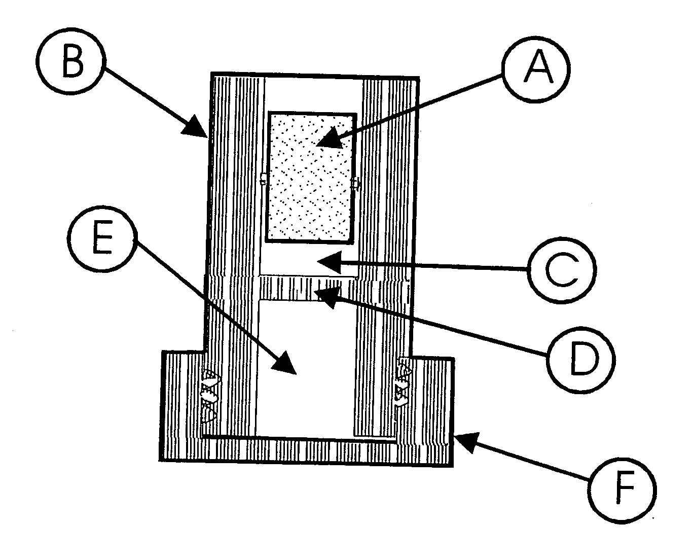
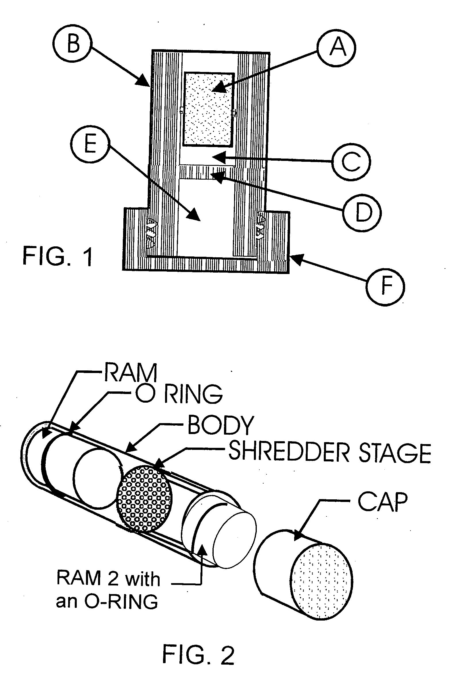
[0123] The device used in this experiment was comprised of a body, two rams, and a shredder stage made of polypropylene (see FIG. 2). The body was a cylindrical tube, 38.1 mm long, with an outside diameter of 13.8 mm, and an inside diameter of 11.6 mm. One end of the tube was threaded to accept a cap. The cap was used to seal and hold the ram at the end of the tube. The rams were cylindrical shaped parts that were 12.7 mm long and 11.5 mm diameter. Each of the rams was equipped with an O-rin...

example 2

Study of Tissue Lysis Efficiency and Improvements of the Two Chamber Homogenization Module Design

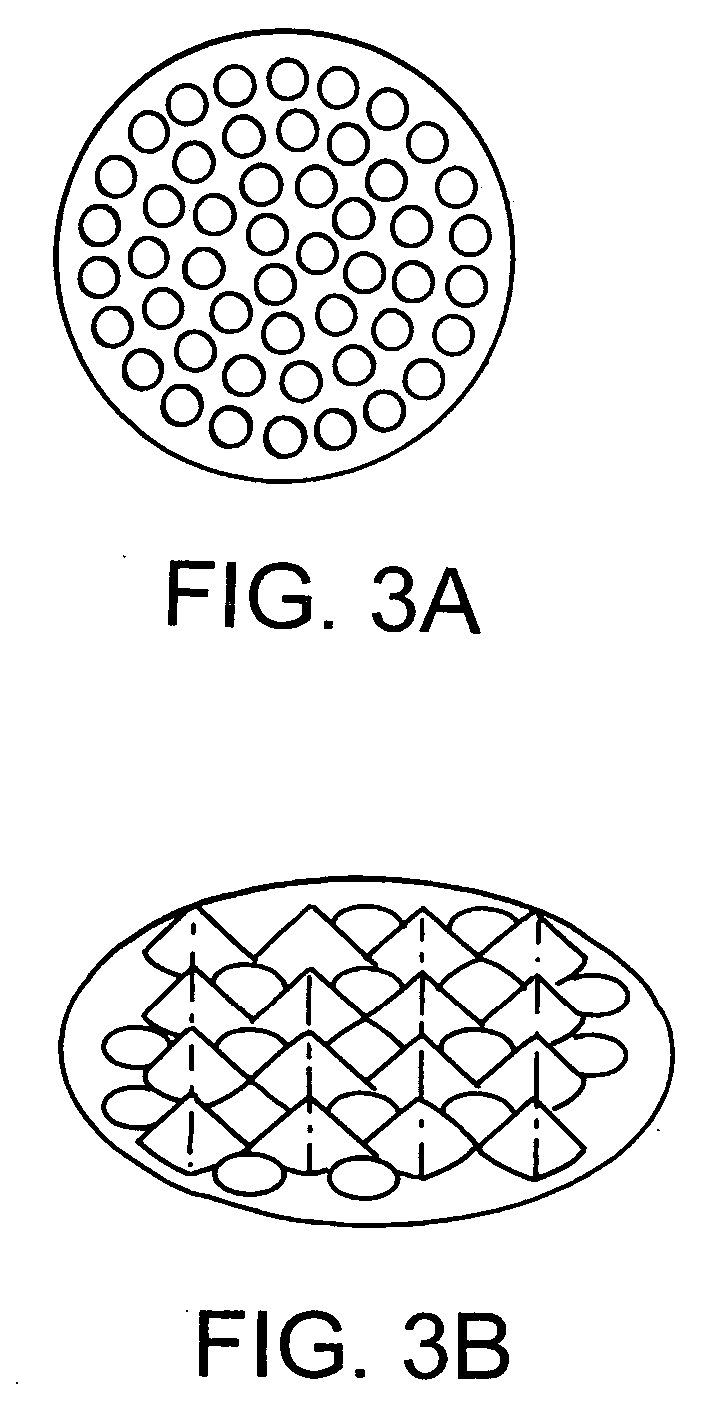
[0130] Using the rat liver as a model, tests were performed on pressure profile (from 15 to 50 kpsi), cycle number profile (from 2 to 45 cycles), temperature profile (from −30 to 0° C.), number of shredder orifices (from 10 to 89 holes on identical area) and a variety of capture fluids (Gdn / 1% CHAPS; phosphate buffered saline, GTC / 1% NP40). Following each test condition, the capture fluid was transferred to a new microcentrifuge tube and briefly centrifuged at 7,600×g. The supernatant was collected and stored in new microcentrifuge tubes. Nucleic acids were extracted from the supernatant using commercially available kits. For analysis of DNA yields, purified nucleic acids were subjected to RNase treatment followed by analysis by agarose gel electrophoresis. The recovered material was treated with RNase prior to electrophoresis in a 0.8% agarose gel in 0.5× TBE buffer, running at constan...

example 3

PCT Homogenization of Rat Brain, Pancreas Large Intestine and Skeleton Muscle

[0133] The effectiveness of PCT homogenization was also evaluated on several additional tissues, which present distinctive difficulties in extractions. Brain tissue is particularly rich in proteins and lipids, which usually present white, flocculent material in the aqueous phase during an aqueous-organic liquid phase extraction. Pancreas is extremely high in nuclease concentration, such that RNA is usually degraded and difficult to recover by conventional methods. Large intestine and skeleton muscle are fibrous tissues with connective tissue components, making them difficult to homogenize.

[0134] These tissues were PCT processed under the same experimental procedure in the liver used for FIG. 12, except 100 μl, instead of 200 μl lysate was extracted using the Roche High Pure PCR template kit. The final elution volume was 60 μl. The gDNA and rRNA were determined by an agarose gel. FIG. 5 shows that the homo...


PUM

PropertyMeasurementUnit
volumeaaaaaaaaaa
volumeaaaaaaaaaa
volumeaaaaaaaaaa

Abstract

Devices and methods are described for homogenization, processing, detection, and analysis of biological samples such as insects, fungi, bacteria, and plant and animal tissues. Multiple chambers in these devices permit different processing functions to be carried out at each stage, such that the resulting homogenized product can be further processed, purified, analyzed, and / or biomolecules such as metabolites, proteins and nucleic acids, or pharmaceutical products can be detected. The device can be used in a hydrostatic pressure apparatus, in which different activities, i.e. incubations, addition or renewal of reagent, and generation and detection of signal can be carried out in the appropriate chamber. The method improves the preservation of biomolecules from chemical and enzymatic degradation relative to conventional means. Additionally, this method enables automated sample preparation and analytical processes.

Description

FIELD OF THE INVENTION [0001] The invention is in the general field of methods and multichamber devices for preparation (for example, homogenizing) and processing of biological samples, optionally in connection with analysis and / or detection of materials from a sample. Particular embodiments have applications in biotechnology, medical diagnostics, agriculture, food, forensic science, pharmaceutical, environmental and veterinary science. BACKGROUND OF THE INVENTION [0002] Biological samples are frequently subjected to processing and analysis after they are isolated. Processing of such samples typically involves one or more of the following: homogenization of biological tissues, lysis of cells, suspension or dissolution of solid particulates, and liquefaction of solid material. Often, sample preparation also entails extensive enzymatic digestion, the use of harsh chemical reagents, and / or mechanical disruption. Following this initial preparation, the sample can be further processed us...

Claims


Application Information

Patent Timeline
Patent Type & Authority Applications(United States)
IPC IPC(8): G01N31/22G01N33/53B01L3/00C12M1/33C12N15/00C12Q1/68G01N1/00G01N1/28G01N33/566
CPCB01L3/502B01L2200/026B01L2300/044B01L2300/0663B01L2300/0832B01L2300/087Y10T436/25375B01L2400/0677B01L2400/0683G01N1/286G01N1/4077G01N2001/2866B01L2400/0481
Inventor LAWRENCE, NATHAN P.TAO, FENGKAKITA, ALLANMANAK, MARK M.SCHUMACHER, RICHARD T.LAUGHARN, JAMES A. JR.
Owner BBI BIOSEQ A MASSACHUSETTS CORP
Why Eureka
Eureka Blog
Learn More