Systems and methods for making noninvasive assessments of cardiac tissue and parameters

a cardiac tissue and non-invasive technology, applied in the field of systems and methods for assessing cardiac tissue and cardiac parameters non-invasively, can solve the problems of difficult characterization of internal tissues using non-invasive and non-traumatic techniques, unreliability, and difficulty in non-invasive detection of various cancers, and achieve the effect of maximizing the displacement of tissu

Inactive Publication Date: 2007-01-18
PHYSIOSONICS +1
View PDF23 Cites 93 Cited by
  • Summary
  • Abstract
  • Description
  • Claims
  • Application Information

AI Technical Summary

Benefits of technology

[0047] The clinical diagnosis of diastolic dysfunction is relatively easy if a pulmonary artery catheter is placed. If the patient has normal left ventricular function, yet the pulmonary artery catheter reveals a high left atrial pressure, then the diagnosis is confirmed. However, most clinicians are unwilling to place a pulmonary artery catheter, so a great deal of effort has been made to estimate left atrial pressure using echocardiography. At present, all the techniques that have been proposed have met with very limited success. Certainly there is no consensus on how to diagnose diastolic dysfunction with echocardiography alone. The ability to make determinations of cardiac properties and parameters noninvasively, such as cardiac tissue stiffness and contractility, however, makes the diagnosis of diastolic dysfunction trivially easy (and non-invasive) because wall stiffness in late diastole reflects left atrial pressure.
[0054] Peak strain rate may thus provide the best clinical estimation of myocardial contractility, particularly if strain rates can be measured in a completely non-invasive fashion. Currently, measurement of strain and strain rates require a high quality echocardiogram machine, or rely on predictions made from tissue velocity measurements. Predictions of strain based on tissue velocity measurements, though they can be made using non-invasive ultrasound techniques, are not consistently accurate. Myocardial velocity measurements, furthermore, conventionally relate to net, or bulk, tissue movement. Continuous measurement of contractility over a prolonged period of time using echocardiogram techniques is not practical or cost effective, especially in an intensive care unit or operating room, where it may be desirable to monitor many patients simultaneously. Methods and systems of the present invention provide determinations of strain rate as the rate of change of strain, measured directly using non-invasive ultrasound techniques over time. Strain rate is measured, not as bulk movement of myocardial tissue but, rather, as relative movements of selected target sites within myocardial tissue. And, because the time of peak strain rate is evanescent, improvements in accuracy of peak strain measurements provided using methods and systems of the present invention, reduce the amount of necessary time averaging of the signal, and improve the cycling rate of the measurement. Both passive and active modes of the present invention may be implemented to determine strain in myocardial tissue. Moreover, the improved accuracy, non-invasiveness and cost-effective attributes of methods and systems of the present invention permit use of strain and strain rate measurements for monitoring myocardial contractility and tissue properties, as well as diagnosis of myocardial dysfunction.
[0067] The concept that the tension wave can be accurately determined over time, especially during isovolumic relaxation and early diastole, has the potential to characterize uniquely a wide range of disorders. Constrictive and restrictive pericariditis and cardiomyopathies have distinctive patterns of pressure in the ventricular chamber. If tension follows the same pattern as pressure, then tension measurements could easily accomplish what currently requires invasive measurement. Graded myocardial ischemia, as opposed to abrupt total occlusion of coronary artery blood flow, may first present as diastolic dysfunction and therefore precede ECG changes and even changes in systolic function. Thus, if a system can monitor both diastolic and systolic function, that system has the most chance of detecting ischemia early and provide the physician a greater opportunity for intervention before the condition worsens. As with all applications, assessment of diastolic function may be accomplished using either the passive or active ultrasound modes of the present invention, or both modes simultaneously or alternately.
[0071] Single or multiple interrogation signals administered from different places and / or at different times may insonify single or multiple target tissue sites. Intrinsic and / or induced acoustic properties of the insonated target tissue may be assessed, by acquiring scatter or emission data, simultaneously and / or sequentially. One of the advantages of the methods and systems of the present invention is that target tissue sites may be volumetrically small, and spatially resolved, to provide data from localized tissue sites with a high degree of spatial resolution. In this way, localized differences in tissue properties may be identified and associated with a spatial location within the interrogated tissue. According to one embodiment, tissue sites of varying size and / or location are assessed simultaneously or sequentially. For most applications, the use of acoustic source(s) and / or transducer(s) capable of interrogating and detecting target tissue sites having a volume of from 1 mm3 to 100 cm3 are suitable.
[0084] Following the (optional) environmental assessment, an acoustic force is applied by an acoustic transducer, at a predetermined frequency, to displace targeted cardiac tissue at a targeted location. The deformation may be produced at any desired location within cardiac tissue, depending on the focus (foci) of the ultrasonic transducer(s) producing the acoustic radiation force. In some systems, variable foci ultrasonic transducers are provided, and a diagnostic procedure is carried out using a plurality of target tissue sites. According to one embodiment for assessment of cardiac output, the focus (foci) of the ultrasonic transducer(s) is preferably provided in proximity to the surface or a small distance below the surface of a ventricle wall, to maximize the tissue displacement induced by the radiation pressure that arises from the impedance mismatch between cardiac tissue and fluid.

Problems solved by technology

Characterization of internal tissues using non-invasive and non-traumatic techniques is challenging in many areas.
Non-invasive detection of various cancers remains problematic and unreliable.
Similarly, non-invasive assessment and monitoring of important internal clinical parameters, such as intracranial pressure and cardiac output, are also practical challenges, despite the efforts devoted to developing such techniques.
The major limitation in the delivery of nutrients to the tissues of the body is the delivery of oxygen.
An inadequate cardiac output translates into some tissues of the body receiving too little oxygen and leads to dysfunction of the affected organ or even tissue damage or cell death of the deprived tissue.
The risks to the patient from using the pulmonary artery catheter preclude routine use.
This technique is safer to the patient, but it is technically more difficult, less accurate, and impractical to use for longer than a few minutes at a time.
Other techniques exist, but none have gained universal acceptance.
The objective of providing a non-invasive measurement of cardiac output thus becomes a problem of how to measure stroke volume in a non-invasive fashion.
Consequently, the difficult aspect in the clinical manipulation of cardiac output is generally reduced to a problem of how to manipulate stroke volume.
First, the ventricles cannot pump to the lungs or body (right and left ventricles, respectively) what the ventricles don't have in them at the start of contraction.
Second, as more blood is put in the ventricle, the muscle cells of the heart become more stretched.
However, a heart with poorly functioning muscle will have a low EF at baseline and will not demonstrate much of an improvement in its contraction when EDV is increased (See FIG. 1).
In fact, more volume may worsen the status of the patient if the heart does not improve its performance in response to the volume.
If performance does not improve, the heart may become distended, which results in impaired function.
In the case of the right ventricle, high venous pressures cause congestion in the abdominal organs and legs that can lead to liver and intestinal dysfunction and to peripheral edema.
If these pressures get too high, fluid leaks out into the lungs and causes symptoms of heart failure (shortness of breath, inability to lay flat) or even pulmonary edema, a life-threatening event where the air sacs in the lung fill with fluid and limit the ability to get oxygen into the blood.
Unfortunately, interpretation of pressures provided by the pulmonary artery catheter can be difficult, making optimal fluid management problematic.
The difficulty, in part, is that the relationship between the filling pressure (end-diastolic pressure) and volume (end-diastolic volume) is not linear. FIG. 2 illustrates this relationship between end-diastolic pressure and volume for heart tissue that is stiff and compliant.
Furthermore, as the condition of the heart changes, the curve can shift around making it harder to interpret the pressure measurements as a measure of end-diastolic volume.
An echocardiogram may provide a volume measurement, but this measurement does not tell the clinician whether that volume is too high, too low or just right.
The accuracy and clinical usefulness of tissue strain predictions based on the estimation of strain rate from Doppler tissue velocities is problematic.
Existing methods of measuring ventricular filling and cardiac contractility using intra-arterial lines or echocardiograms have limited application because of the risk to the patient, high expense and difficulty in interpretation of the information provided.
Lack of direct, non-invasive and inexpensive methods to measure ventricular filling and cardiac contractility means that optimal management of stroke volume is missing from the care of many patients who would benefit from such optimization.
While this mode of measurement is simple and inexpensive to perform, it does not provide the most accurate measure of ABP, and it is susceptible to artifacts resulting from the condition of arterial wall, the size of the patient, the hemodynamic status of the patient, and autonomic tone of the vascular smooth muscle.
Additionally, repeated cuff measurements of ABP result in falsely elevated readings of ABP, due to vasoconstriction of the arterial wall.
While such catheters are very reliable and provide the most accurate measure of ABP, they require placement by trained medical personnel, usually physicians, and they require bulky, sophisticated, fragile, sterile instrumentation.
Additionally, there is a risk of permanent arterial injury causing ischemic events when these catheters are placed.
As a result, these invasive monitors are only used in hospital settings and for patients who are critically ill or are undergoing operative procedures.

Method used


Image

  • Systems and methods for making noninvasive assessments of cardiac tissue and parameters
  • Systems and methods for making noninvasive assessments of cardiac tissue and parameters
  • Systems and methods for making noninvasive assessments of cardiac tissue and parameters

Examples


example 1

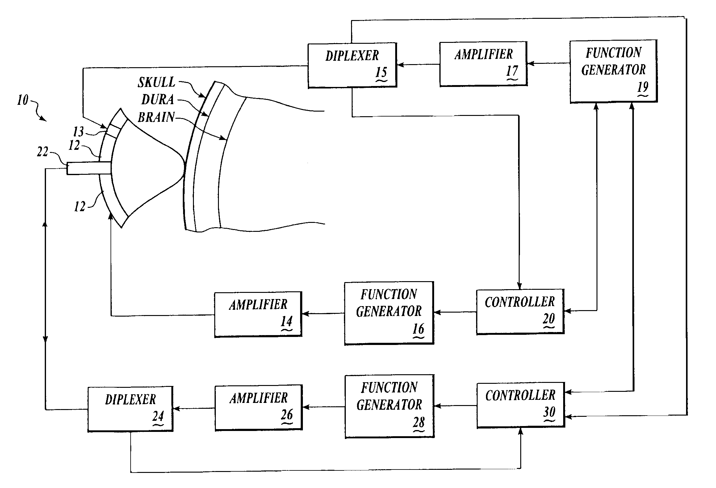
[0155] Brain tissue was used as a model experimental system. We have shown in vitro (FIG. 10A) and in vivo (FIG. 10B-D) and describe in detail below, that intrinsic displacements of brain tissue (e.g. compressions and distensions), and their various acoustic scatter properties, can be directly measured using a standard transcranial Doppler (TCD) transducer, off-the-shelf data acquisition systems, and novel analysis of the acoustic backscatter signal from brain. Myocardial tissue displacement may be measured in the same fashion and related to tissue strain, tension, and the like, as described above, to make noninvasive assessment of cardiac tissue and parameters.

[0156] An in vitro model for examining changes in ICP using acoustic techniques was constructed using fresh bovine brain immersed in fluid in a water-tight, visually and acoustically transparent bottle attached to a hand-pump for changing the pressure on the brain. An acoustic transducer (ATL / Philips Medical Systems, Bothell...

example 2

[0164] We have shown, in vitro, using a beef brain model similar to that described above, that a palpation pulse of ultrasound across a range of acoustic intensities can cause increasing displacements of brain without causing gross tissue damage. Palpation of myocardial tissue using ultrasound pulses may be achieved in a similar fashion.

[0165] Fresh bovine brain was immersed in fluid in a water-tight, visually and acoustically transparent bottle attached to a hand-pump for changing the pressure on the brain. ATL acoustic transducers (ATL-Philips Medical Systems, Bothell, Wash.), and the bottle, were placed in water so that the focus of the acoustic palpation and interrogation transducers were near the edge of the brain, but within the brain. Using LeCroy Waverunner oscilloscope, we collected acoustic interrogation waveforms backscattered from brain. For palpating and interrogating beef brain, in vitro, the interrogation pulses were administered as described with respect to FIG. 10A...


PUM


Abstract

Systems and methods for noninvasive assessment of cardiac tissue properties and cardiac parameters using ultrasound techniques are disclosed. Determinations of myocardial tissue stiffness, tension, strain, strain rate, and the like, may be used to assess myocardial contractility, myocardial ischemia and infarction, ventricular filling and atrial pressures, and diastolic functions. Non-invasive systems in which acoustic techniques, such as ultrasound, are employed to acquire data relating to intrinsic tissue displacements are disclosed. Non-invasive systems in which ultrasound techniques are used to acoustically stimulate or palpate target cardiac tissue, or induce a response at a cardiac tissue site that relates to cardiac tissue properties and / or cardiac parameters are also disclosed.

Description

REFERENCE TO PRIORITY APPLICATION [0001] This application is a continuation of U.S. patent application Ser. No. 10 / 612,483 filed Jul. 1, 2003, which claims priority to U.S. Provisional Application No. 60 / 393,293 filed Jul. 1, 2002 and U.S. Provisional Application No. 60 / 475,803 filed Jun. 3, 2003, and is also a continuation-in-part of U.S. patent application Ser. No. 09 / 995,897 filed Nov. 28, 2001, which claims priority to U.S. Provisional Application No. 60 / 253,959, filed Nov. 28, 2000.TECHNICAL FIELD OF THE INVENTION [0002] This invention relates to systems and methods for assessing cardiac tissue and cardiac parameters noninvasively using ultrasound techniques. BACKGROUND OF THE INVENTION [0003] Methods and systems for determining and characterizing various systems and tissue properties are known. Characterization of internal tissues using non-invasive and non-traumatic techniques is challenging in many areas. Non-invasive detection of various cancers remains problematic and unre...

Claims


Application Information

Patent Timeline
Patent Type & Authority Applications(United States)
IPC IPC(8): A61B8/00A61B5/03A61B8/04A61B8/06A61B8/08
CPCA61B5/0048A61B8/0883A61B5/4519A61B5/4824A61B5/4869A61B6/503A61B6/504A61B8/00A61B8/04A61B8/065A61B8/08A61B8/0891A61B8/4472A61B8/485A61B8/488A61B8/0808A61B8/4236A61B5/031A61B8/4444
Inventor MOURAD, PIERRE D.KLIOT, MICHELPATTERSON, REXROOKE, ALEC
Owner PHYSIOSONICS
Why Eureka
Try Eureka