Radiolabeled-pegylation of ligands for use as imaging agents
a technology of radiolabeled and ligands, which is applied in the field of bioactive compounds and diagnostic imaging methods using radiolabeled compounds, can solve the problems of difficult direct imaging of amyloid deposits in vivo, poor image quality, and inability to obtain much information by other means,
- Summary
- Abstract
- Description
- Claims
- Application Information
AI Technical Summary
Benefits of technology
Problems solved by technology
Method used
Image
Examples
example 1
Synthesis of 2-phenylbenzothiozole (PE3) derivatives
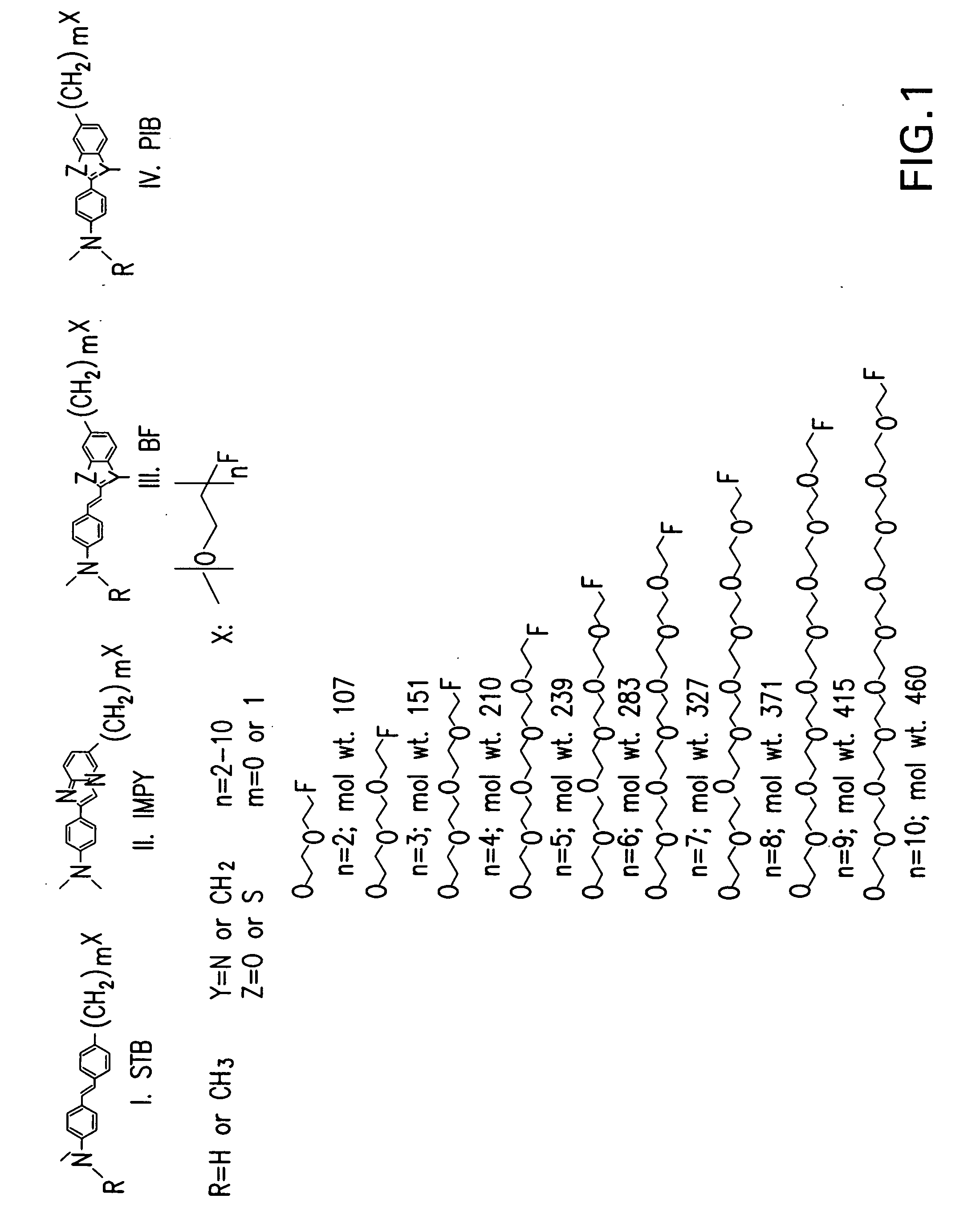
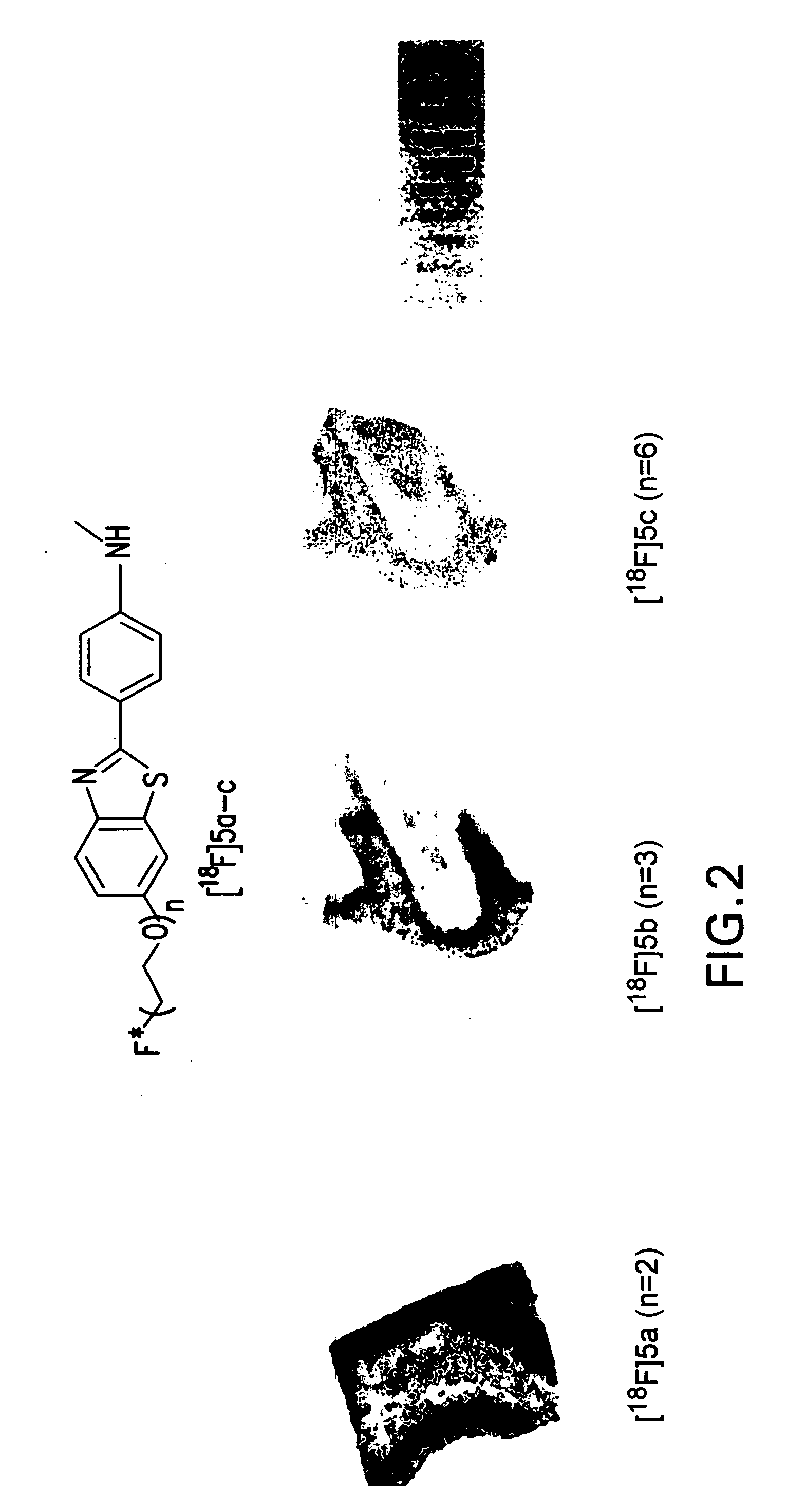
[0185] Compound 4 (2-phenylbenzothiozole (PIB) core) was prepared using Mathis and co-workers approach (26). Monomethylation was accomplished via standard reported procedures (27) to yield 4 that was used in subsequent steps.
1. General Procedure for the O-alkylation of 4
[0186] To a solution of 4 (1 eq) in anhyd. N′,N″-dimethylformamide (2 mL / 0.1 mmol of 4) in a microwavable vial (from Biotage) was added anhyd. cesium carbonate (2.5 eq) and the mixture stirred at room temperature under argon for 30 min. Alkylating agent (1.2 eq) followed by sodium iodide (1.5 eq) were then added, the vial was sealed and subjected to microwave irradiation (Biotage Initiator system). The microwave conditions were, 200° C. for 10 min. with 10 sec. pre-stirring and with fixed hold time “on”. After cooling the reaction mixture to room temperature the vial was opened, the contents were transferred to a round-bottom flask and the volatiles were removed...
example 2
1. Preparation of [2-(4-dimethylaminophenyl)-vinyl]-benzoxazol derivatives
[0209] 2-(2-(4-dimethylaminophenyl)vinyl)-benzooxazol-6-ol (3′): 2-methyl-benzoxazol-6-ol (prepared following Schreiner and coworkers method (28)) (1.7 mmol) was dissolved in anhydrous tetrahydrofuran (8 mL) and cooled to 0° C. Trimethylsilyl chloride (1.8 mmol) and diisopropylethylamine (1.84 mmol) were then added and the resultant solution stirred for 2 hours at room temperature. After cooling to −78° C., sodium hexamethyldisilazane (11.7 mmol, 1.0 M solution in tetrahydrofuran) was added slowly over 1.5 hours and then stirred at −78° C. for an additional hour. 4-(dimethylamino)-benzaldehyde was then added and the reaction allowed to warm to room temperature overnight. The reaction was then poured into a 1M solution of sodium hydrogen sulfate and extracted with ethyl acetate. The organic layers were then washed with brine, dried over magnesium sulfate and concentrated to yield a yellow solid that was purifi...
example 3
1. Synthesis of 6-iodo-2-(4′-dimethylamino)phenyl-imidazo[1,2-a]pyridine (IMPY) (2) derivatives
[0217] Preparation of 2 (IMPY core) has been described elsewhere (29). The general procedure for the synthesis of 6-FPEG substituted-SPY conjugates was accomplished using the following procedure:
[0218] Conventional synthesis: The mixture of 2 (prepared as reported previously reported (29)), fluoro-polyglycols (2-5 eq.), CuI (10% mol), Cs2CO3 (2 eq.), 1,10-phenanthroline (20% mol) in Toluene (1 mL / 0.1 mmol 2) was stirred in a sealed tube for 48 h. Solvent was removed and PTLC [Ethyl Acetate or dichloromethane-methanol (95:5) as developing solvent] gave the desired product (Yield: 17-60% depending on the glycol used).
[0219] Microwave synthesis: The mixture of reactants and reagents described above in a sealed tube was put in the microwave oven—condition: 170° C., 60 min, normal absorption level. (Yields were similar to those used the conventional synthesis).
[0220] 6-(2-fluoroethoxy)-2-(4...
PUM
| Property | Measurement | Unit |
|---|---|---|
| temperature | aaaaa | aaaaa |
| half life | aaaaa | aaaaa |
| half life | aaaaa | aaaaa |
Abstract
Description
Claims
Application Information
 - R&D
- Intellectual Property
- Life Sciences
- Materials
- Tech Scout
- Unparalleled Data Quality
- Higher Quality Content
- 60% Fewer Hallucinations
Browse by: Latest US Patents, China's latest patents, Technical Efficacy Thesaurus, Application Domain, Technology Topic, Popular Technical Reports.
© 2025 PatSnap. All rights reserved.Legal|Privacy policy|Modern Slavery Act Transparency Statement|Sitemap|About US| Contact US: help@patsnap.com


