Use of Keratinocyte Composition for the Treatment of Restenosis

Inactive Publication Date: 2008-12-18
INTERSTITIAL THERAPEUTICS
View PDF4 Cites 4 Cited by
  • Summary
  • Abstract
  • Description
  • Claims
  • Application Information

AI Technical Summary

Problems solved by technology

Restenosis constitutes one of the main limitations in the success of percutaneous interventions, such as, for example, angioplasty for opening up a coronary artery or other vessel which has become blocked due to build up of deposits on the vessel wall.
However, the damage caused to the wall of the vessel during angioplasty causes restenosis i.e. proliferation of cells in the vessel wall which o

Method used


Image

  • Use of Keratinocyte Composition for the Treatment of Restenosis
  • Use of Keratinocyte Composition for the Treatment of Restenosis

Examples


example 1

Preparing Cultured Keratinocytes In Vitro

[0059]Skin biopsies were harvested from burn patients, cleaned with 70% ethanol, and processed according to Rheinwald and Green's keratinocyte isolation method (Rheinwald J. G., Green H.: Cell serial cultivation of strains of human epidermal keratinocytes: the formation of keratinizing colonies from single cells. Cell, 6: 331-44, 1975). Skin biopsies were trypsinized overnight at 4 deg C. in a 0.05% trypsin / 0.02% EDTA solution. The cells were isolated from the epidermal / dermal junction and plated onto a feeder monolayer of lethally irradiated 3T3 cells. The medium was made up of three parts Dulbecco's modified with Eagle's medium plus one part Ham's F12 medium. The following were added to the mixture: cholera toxin (0.1 nM), hydrocortisone (0.4 pg / ml), triiodo-I-thyronine (20 pM), insulin (5 pg / ml), transferrin (5 pg / ml), fungizone (250 pg / ml), penicillin (1000 IU / ml), streptomycin (1000 pg / ml), and 10% foetal calf serum. Epidermal growth fac...

example 2

Treatment of a Wound

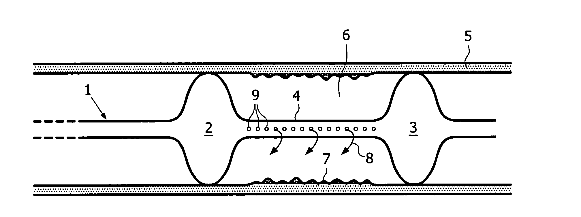
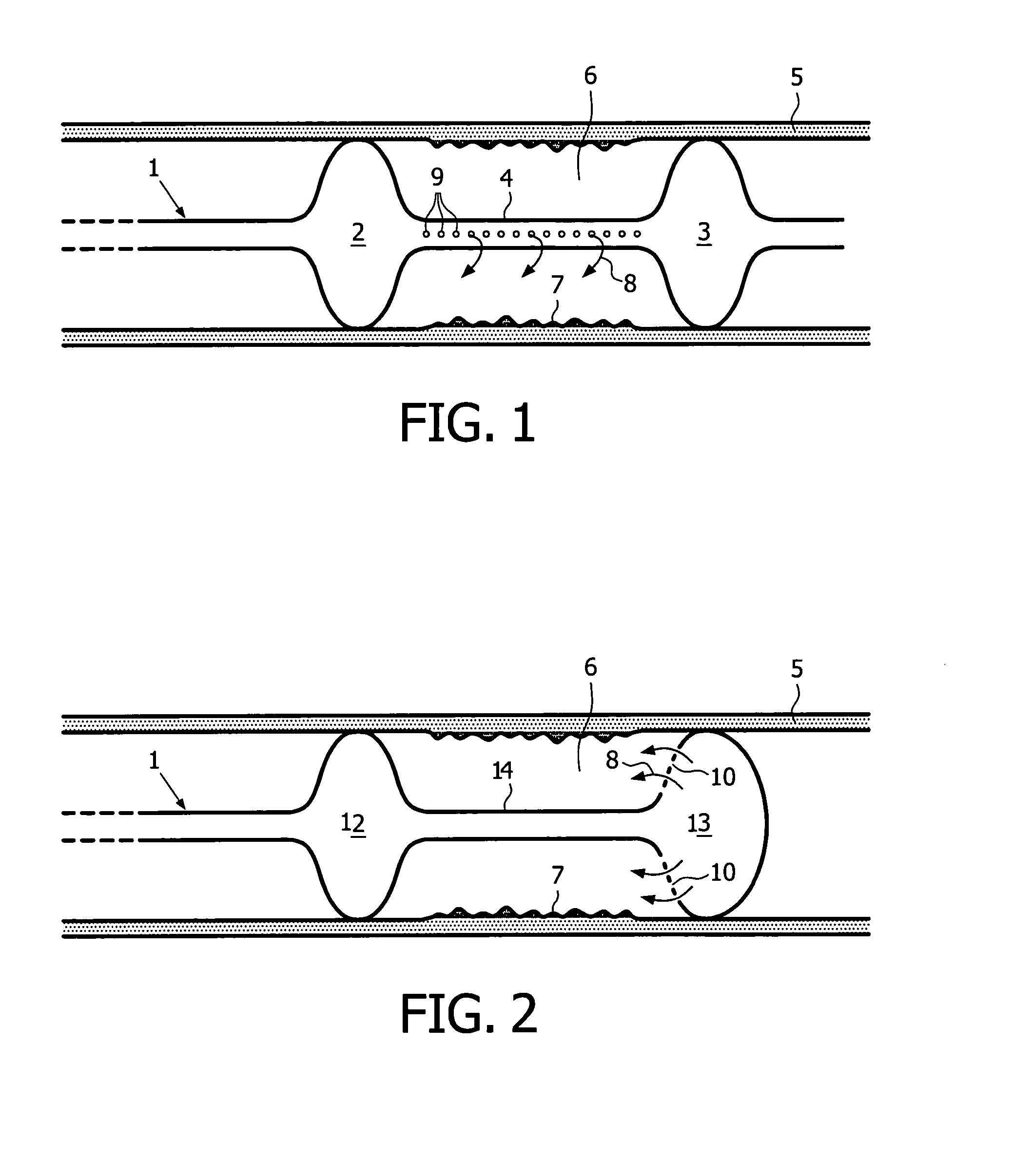
[0060]A 51 year old patient presents a 8 mm long stenotic lesion in the proximal part of the right coronary artery. A 3.5 mm diameter balloon is used to dilate the artery and a 16 mm long magnesium alloy stent is inserted and positioned at the level of the former vessel narrowing. A deflated Genie dumbbell balloon is introduced and accurately positioned at the level of the magnesium alloy stent. Ten ml of a solution containing 100000 keratinocytes / ml with 0.1 mM of Mn2+ is used to inflate the Genie balloon. The inflation of the Genie balloon is followed by the flow of the composition through small holes located on the inner wall of the distal balloon enlargement. This contributes to the filling of the space located between both larger balloon parts and the tight contact of the solution of keratinocytes with the vessel wall. A pressure of 2 Atm is necessary to keep the Genie balloon expanded and to let the keratinocyte solution stay in contact with the vessel wall...

example 3

Treatment of a Wound

[0061]A 76 y old patient presents with a narrowing of his popliteal artery with some pain and claudication after a walk of 300 m. An angiographic examination shows a narrowing of the artery on a length of 5 cm. A percutaneous transcatheter angioplasty (PTA) is performed at the level of the vessel narrowing and a wallstent is inserted at the level of the lesion. A 8 cm long Genie balloon is introduced inside the artery and is accurately placed at the level of the dilated lesion. Thirty ml of a keratinocyte suspension stimulated by nanomolar suspension of fibronectin and by 0.1 mM of Mn2+ is infused in the Genie balloon and in the intra-vascular space. The patient does not show any sign of recurrence 8 months later. A Doppler evaluation shows a regular, non interrupted blood flow in the vessel.


PUM


Abstract

Methods for treating and/or preventing restenosis in a subject by local administration to the subject of a composition that includes keratinocytes is disclosed. Administration is preferably by way of a catheter having an inflatable balloon, such as a double or dumbbell-shaped balloon.

Description

BACKGROUND TO THE INVENTION[0001]Restenosis constitutes one of the main limitations in the success of percutaneous interventions, such as, for example, angioplasty for opening up a coronary artery or other vessel which has become blocked due to build up of deposits on the vessel wall. During angioplasty, a balloon catheter is introduced into the region of the blockage, and is hydraulically inflated in order to open the obstructed vessel lumen by compressing the closed wall. Commonly, a stent—an expandable wire mesh—is employed on the balloon and is implanted to support the new stretched open position of the vessel. However, the damage caused to the wall of the vessel during angioplasty causes restenosis i.e. proliferation of cells in the vessel wall which over a period of time contributes to reblocking of the vessel.[0002]Where stents are deployed during angioplasty, they may be coated with drugs to prevent restenosis, such drugs being, for example, sirolimus, everolimus or paclitax...

Claims


Application Information

Patent Timeline
IPC IPC(8): A61K35/12C12N5/08A61P43/00A61K35/36
CPCA61K35/12A61K35/36A61L27/3813A61L27/3886A61M25/1011A61M2025/105A61L27/3808A61P43/00
Inventor POPOWSKI, YOURI
Owner INTERSTITIAL THERAPEUTICS
Why Eureka
Try Eureka