
Optical imaging agent

an optical imaging agent and agent technology, applied in the field of optical imaging agents, can solve the problems of difficult in vivo fluorescence signal quantification, and achieve the effect of high molecular weigh

Inactive Publication Date: 2011-02-03
INVICRO
View PDF3 Cites 4 Cited by
  • Summary
  • Abstract
  • Description
  • Claims
  • Application Information

AI Technical Summary

Benefits of technology

[0003]The present invention provides a new class of macromolecular optical contrast agents, compositions incorporating these agents and methods of using these agents in optical imaging modalities. Of interest in the present invention are contrast agents used for imaging the blood pool and monitoring its movement. Optical imaging assisted by such agents is useful for such procedures as assessments of relative tissue blood volume, estimation of tissue perfusion, and detection of abnormal capillary permeability. Clinical applications include assessments of myocardial and cerebral ischemia, pulmonary embolism, transplants, and neoplasia. To be useful as blood-pool markers, the contrast agents must remain in the pool rather than leaving it through such means as diffusion into extravascular compartments or glomerular filtration. A requisite property of contrast agents is therefore a relatively high molecular weight, which prevents the agents from diffusing through normal capillaries and glomerular endothelium. A further advantage of the agents of the invention is that the prolonged intravascular retention of these agents permits imaging of the blood pool in multiple body regions without repeated dosing, thereby eliminating the need for critical timing of the imaging.
[0005]In an exemplary embodiment, the contrast agent of the invention comprises a fluorophore linked, through a linker, to a carrier molecule. The fluorophore can be selected based on the optimization of one or more of its characteristics, e.g., water-solubility, quantum yield, absorbance maximum, emission maximum, stability, and the like. Similarly, the linker is generally selected for a desired property, e.g., the ease of converting its precursor(s) into a linker joining the carrier molecule and the fluorophore, water-solubility, stability (or lack thereof under selected biological conditions), lack of quenching of the fluorophore, and the like. The carrier molecule can be a species that simply increases the molecular size or weight of the fluorophore or it can have properties that enhance the delivery of the contrast agent of the invention to a particular tissue (or prevent or retard its delivery to a particular tissue). Natural and synthetic polymers are exemplary choices for the carrier. In an exemplary embodiment, the carrier molecule is a polysaccharide.

Problems solved by technology

Due to this background signal, the quantification of fluorescence signals for in vivo imaging can be a difficult task when working in this range.

Method used


Image

  • Optical imaging agent
  • Optical imaging agent
  • Optical imaging agent

Examples


examples

Materials and Methods

Contrast Agent

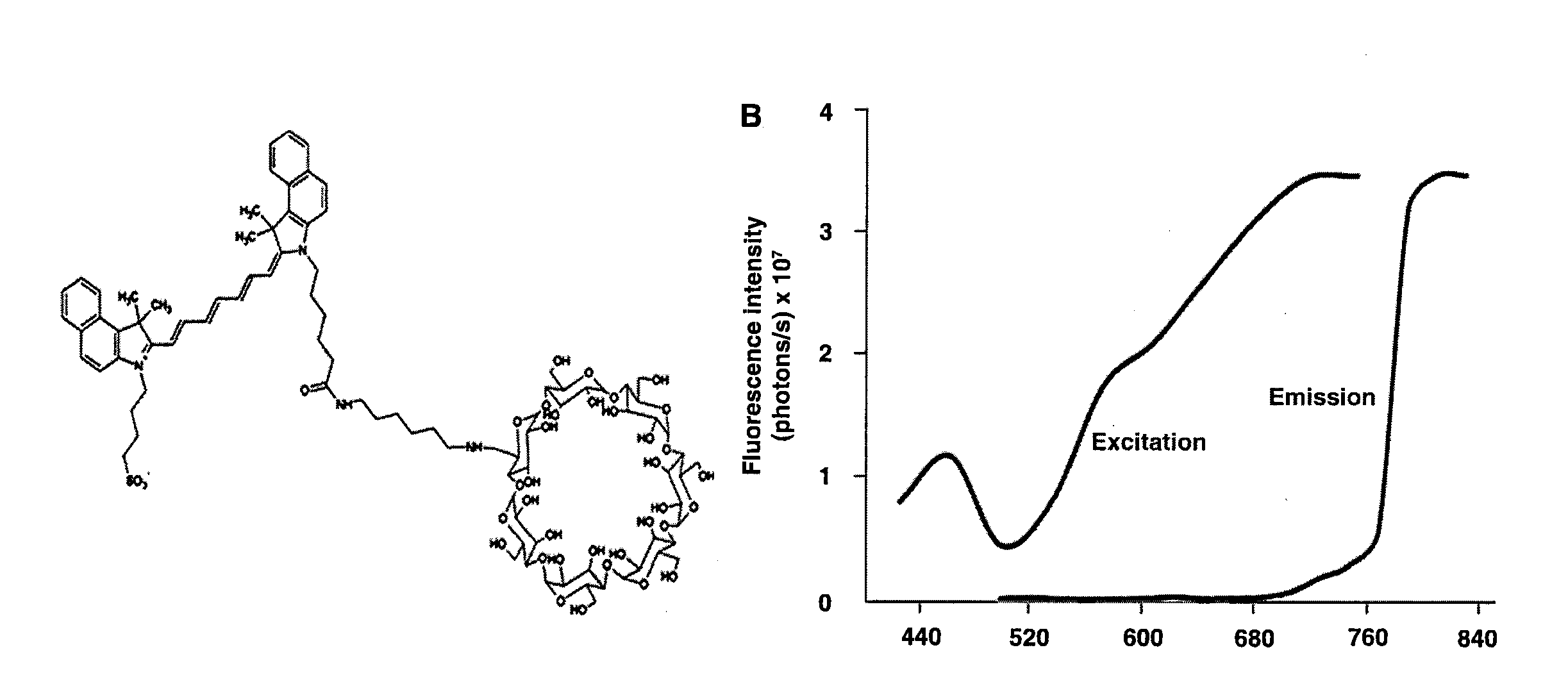
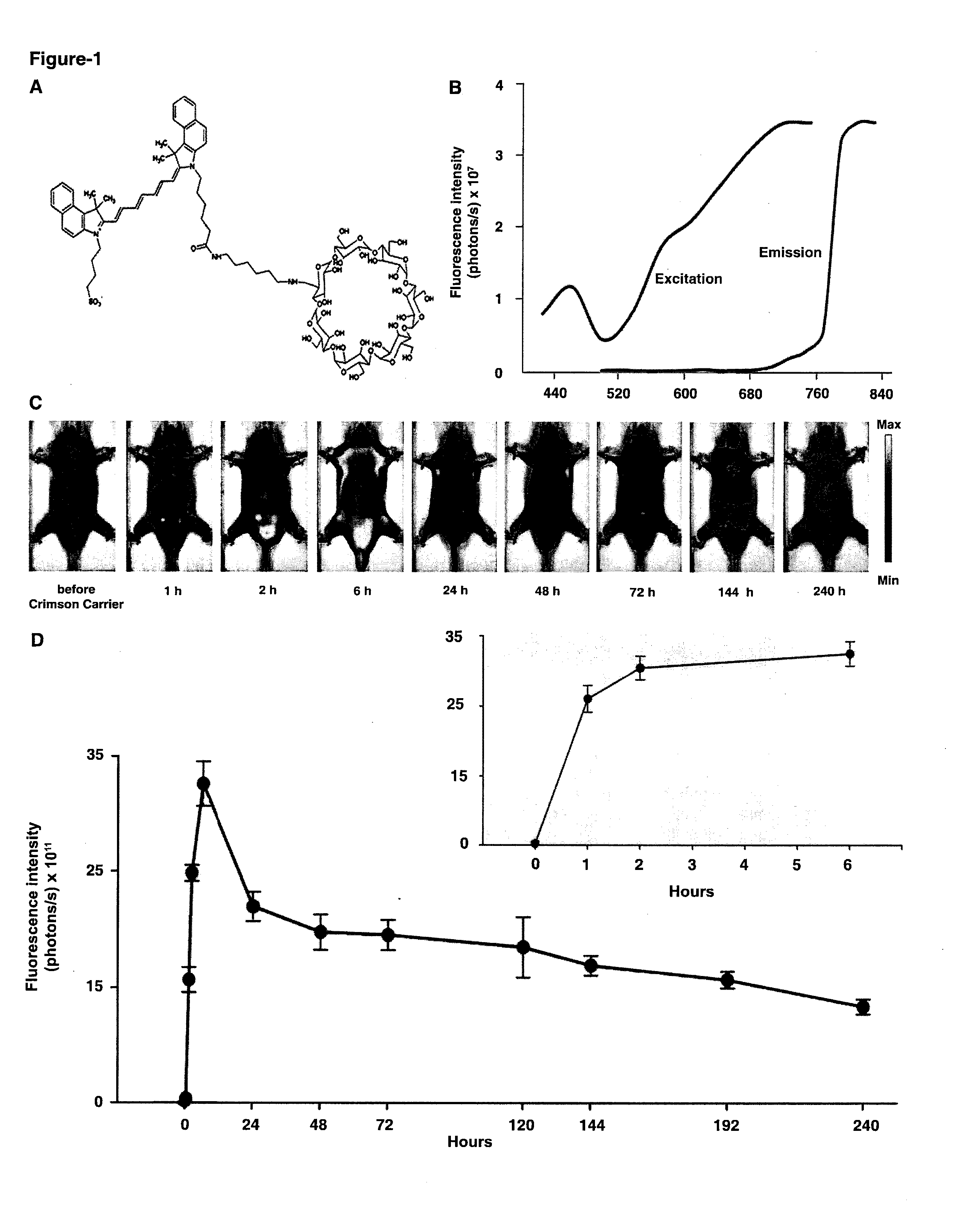
[0075]Compound 1 (C93H132N4O38S) is composed of a β-cyclodextrin ring conjugated to Indocyanine Green (ICG) via a short linker (CH2)6. 1 has a molecular weight of 1946.11 atomic mass unit (amu). The powdered 1 was freshly constituted with phosphate buffer saline (PBS) to a final concentration of 0.2 mM.

Live Animal Imaging

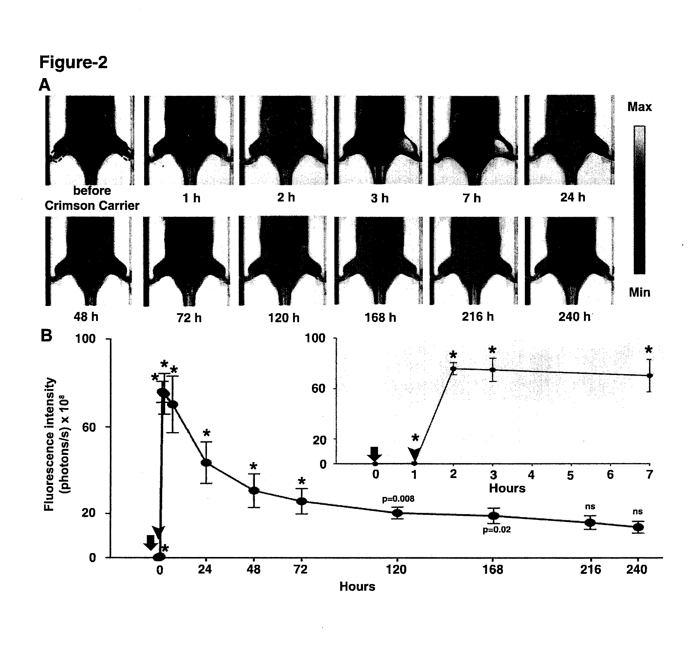
[0076]Optical imaging for all experiments was performed using Xenogen IVIS® Spectrum system (Caliper-Xenogen, Alameda, Calif., USA). All animal procedures were conducted in accordance with the Guidelines for the Care and Use of Laboratory Animals and were approved by the Institutional Animal Care and Use Committee (IACUC) at University of Texas Health Science Center at San Antonio. All images were acquired using epiillumination at excitation wavelength of 745 nm and emission wavelength of 820 nm unless otherwise stated. The camera settings were kept constant at 1 sec exposure time, 4×4 binning, 12.6 cm or 6.5 cm field of view, and ...


PUM

PropertyMeasurementUnit
Nanoscale particle sizeaaaaaaaaaa
Structureaaaaaaaaaa

Abstract

The near-infrared wavelengths (700 nm-900 nm) are a suitable optical window for light penetration and deep tissue imaging. The present invention provides a near-infrared fluorescent blood pool contrast agent. The agent is of use to detect and quantify pathological capillary leak in live animals, e.g., serially imaging of traumatized tissue (muscle) as well as tumors.

Description

CROSS-REFERENCE TO RELATED APPLICATIONS[0001]This application claims benefit of priority to U.S. Provisional Patent Application Ser. No. 61 / 230,574, filed Jul. 31, 2009, which is hereby incorporated by reference in its entirety for all purposes.BACKGROUND OF THE INVENTION[0002]Preclinical and clinical fluorescence optical imaging instruments are increasingly utilized to obtain biological information on the path from gene to clinic (1). Skin and red blood cell autofluorescence occurs to some extent at all excitation wavelengths in visible wavelength range of light (400-700 nm). Due to this background signal, the quantification of fluorescence signals for in vivo imaging can be a difficult task when working in this range. To achieve better signal: background and increasingly accurate quantitative imaging in vivo, working within the near-infrared (NIR) wavelength range (700-900 nm) is desirable. In this spectral region, light absorption and scattering by physiologically abundant molecu...

Claims


Application Information

Patent Timeline
IPC IPC(8): A61B5/00C08B37/16A61P27/02
CPCC08B37/0012A61K49/0054A61K49/0034A61K49/0032A61P27/02
Inventor DOW, WILLIAMDOW, KAREN B.
Owner INVICRO
Learn More