Biocompatible materials for mammalian stem cell growth and differentiation

a technology of stem cell growth and biocompatibility, applied in the field of biocompatibility materials, can solve the problems of increasing public health problems, increasing the cost of culture systems, and not always giving rise to es cells suitable for all forms of therapy, and achieve the effect of promoting the above-mentioned cell function

Inactive Publication Date: 2011-06-30
AARHUS UNIV
View PDF2 Cites 33 Cited by
  • Summary
  • Abstract
  • Description
  • Claims
  • Application Information

AI Technical Summary

Benefits of technology

[0018]The manufacture of a structure having the desired surface topography (that may be entirely artificial or may mimic a surface architecture observed in nature) requires techniques capable of defining features that have micrometer scale or nanometer scale dimensions. The present invention exploits the tools and techniques presently developed within micro- and nano-technology, which allow the design and construction of structures whose surface architecture may have a lateral feature size on a micro- or nano-meter scale. This feature size can be achieved e.g. by colloidal lithography of ferritin followed by removal of the organic phase leaving behind ion dots. In particular, the use of e-beam lithography and photolithography allows the manufacture of a surface topography which is precisely defined and which can be precisely reproduced in relevant applications.
[0032]In another aspect, the invention relates to the use of a cell or tissue culture container to promote uniform differentiated growth of mammalian embryonic stem cells and thereby facilitate the production of a population of differentiated cells wherein the number of undifferentiated cells is substantially reduced, such that the use of the differentiated cells in therapy (e.g. an implant) carries a reduced risk that the cells can give rise to teratomas in the patient, when compared to current differentiation protocols. Accordingly, the invention provides a container, the container having a surface for exposure to a culture during use, where at least a part of the surface is defined by a biocompatible material, wherein at least a part of an exposed face of the biocompatible material has a nano- and / or micrometer scale topographical structure comprising a plurality of protrusions arranged on grid points of a regular two-dimensional grid, characterized in that the protrusions have a cross section and a cross-sectional diameter of between 1.0 μm-8.0 μm and wherein the lateral dimension of the maximum gap between any protrusion and its nearest neighbor (d;Y) is between 1.0 μm-2.0 μm, and wherein each of the protrusions of said topographical structure has a vertical height / depth dimension that is equal to or less than 1.0 μm, preferably between 0.6 μm-1.0 μm, more preferably about 0.6 μm. The use of a cell or tissue culture container on the invention allows the production of a population of differentiated cells wherein the number of undifferentiated cells is substantially reduced, such that the use of the so produced cells in an implant carries a reduced risk that the implanted cells give rise to teratomas in the patient, when compared to current cell differentiation protocols.
[0042]The manufacture of a structure having the desired surface topography (that may be entirely artificial or may mimic a surface architecture observed in nature) requires techniques capable of defining features that have micrometer scale or nanometer scale dimensions. In particular, it has turned out that when at least a part of a surface of such a biocompatible material is characterized by a micrometer scale topographical structure, a number of cell functions of at least one of a variety of different cell types are significantly improved. In particular, regular patterns of spaced-apart protrusions that extend out of the surface have been found to be particularly efficient for promoting the above-mentioned cell functions.

Problems solved by technology

Medical conditions whose treatment might benefit from such materials include degenerative disorders, cancer and trauma of the musculoskeletal apparatus, each of which constitutes an increasing problem in public health.
Thirdly, in order to facilitate the use of mammalian ES cells for medical purposes it is essential that the cells at no point come into contact with biological materials derived from another species, since xeno-contamination is most likely to cause immunogenic problems upon transplantation into a patient.
Feeder- and serum-free culture conditions have been described for murine ES cells, for example using gelatin-coated dishes combined with Leukemia Inhibitory Factor (LIF)-supplemented media, but these culture systems are expensive and do not always give rise to ES cells suitable for all forms of therapy.
Degenerative disorders, cancer and trauma of the musculoskeletal apparatus constitute an increasing problem in public health.
Major advances and results have been achieved in this area during the last decades, but implant loosening over time continues to be a significant problem for successful long-term joint replacements.
The current implant surfaces, alone, are not able to bridge larger bone defects and maintain long-term stability.
Furthermore, as the near-future patient population will include a significant number of younger patients, the problem concerning long-term aseptic implant loosening is predicted to increase dramatically.
The biocompatibility / biointegration of an implant in the body is extremely complicated, involving processes traditionally belonging to medical science, surface science, materials science, and molecular biotechnology.
All these mechanisms contribute to the response of the tissue to the implant and influence whether the implant is successfully anchored with sufficient mechanical strength in the bone of the patient or whether an inflammatory reaction against the implant occurs, which finally results in aseptic loosening and operative failure.
Achieving a successful outcome of such treatment presents a formidable challenge, since an implant needs to allow tissue regeneration at the implant site, while avoiding becoming a target for the body's own powerful rejection mechanisms.

Method used


Image

  • Biocompatible materials for mammalian stem cell growth and differentiation
  • Biocompatible materials for mammalian stem cell growth and differentiation
  • Biocompatible materials for mammalian stem cell growth and differentiation

Examples


example 1

Manufacture of a BSSA Wafer Comprising 13×3 Tantalum Array of Tester Areas

[0150]A single-sided polished silicon wafer with a thickness of 525±25 μm provided a substratum for the manufacture of a biocompatible material. The wafer was an n-type wafer with a resistivity of 1-20 ohm cm. A micrometer-sized pattern was printed onto the polished side of the silicon wafer by standard photolithography and reactive ion etching in a SF6 / 02 discharge according to the following protocol:[0151]1. The wafers were pre-etched with buffered hydrofluoric acid (BHF, BHF is a solution of concentrated HF (49%), water, and a buffering salt, NH4F, in about the ratio 1:6:4) for 30 seconds and then dried under N2 flow, and[0152]2. the wafer was then spin-coated with a 1.5 μm thick layer of photoresist AZ5214, Hoechst Celanese Corporation, NJ, US (the chemical composition can be found at the Material Safety Data Sheet (MSDS) supplied by Hoechst Celanese Corporation). and pre-baked at around 90° C. for 120 sec...

example 2

Identification of Biocompatible Surfaces for Promoting Growth of Pluripotent Mouse Embryonic Stem (ES) Cells

[0164]Undifferentiated murine ES cells grow in compact, well-defined colonies (FIG. 10). When colonies flatten and spread out, the ES cells most likely have started to differentiate. Thus, colony morphology is an obvious and generally accepted marker of pluripotency. Examples of well established intracellular pluripotency markers are the transcription factors Oct4 and Nanog and the enzyme Alkaline Phosphatase (AP).

[0165]ES cells (KH2 cells and CJ7 cells) were seeded at a density from 1.3-5×10⁶ cells / p10 Petri dish on tantalum BSSA wafers (FIG. 11), produced as in Example 1. The cells cultured for cultured for 3 days under conditions of 5% CO2, 90% air humidity and 37° C. The cells were grown in DMEM supplemented with 15% FCS, 2 mM glutamine, 50 U / ml penicillin, 50 μg / ml streptomycin, non-essential amino acids, 100 μM β-mercaptoethanol and nucleosides, but in the absence of a f...

example 3

Pluripotency and Growth Morphology of Mouse Embryonic Stem (ES) Cells is Maintained During Serial Passaging on Biocompatible Surfaces

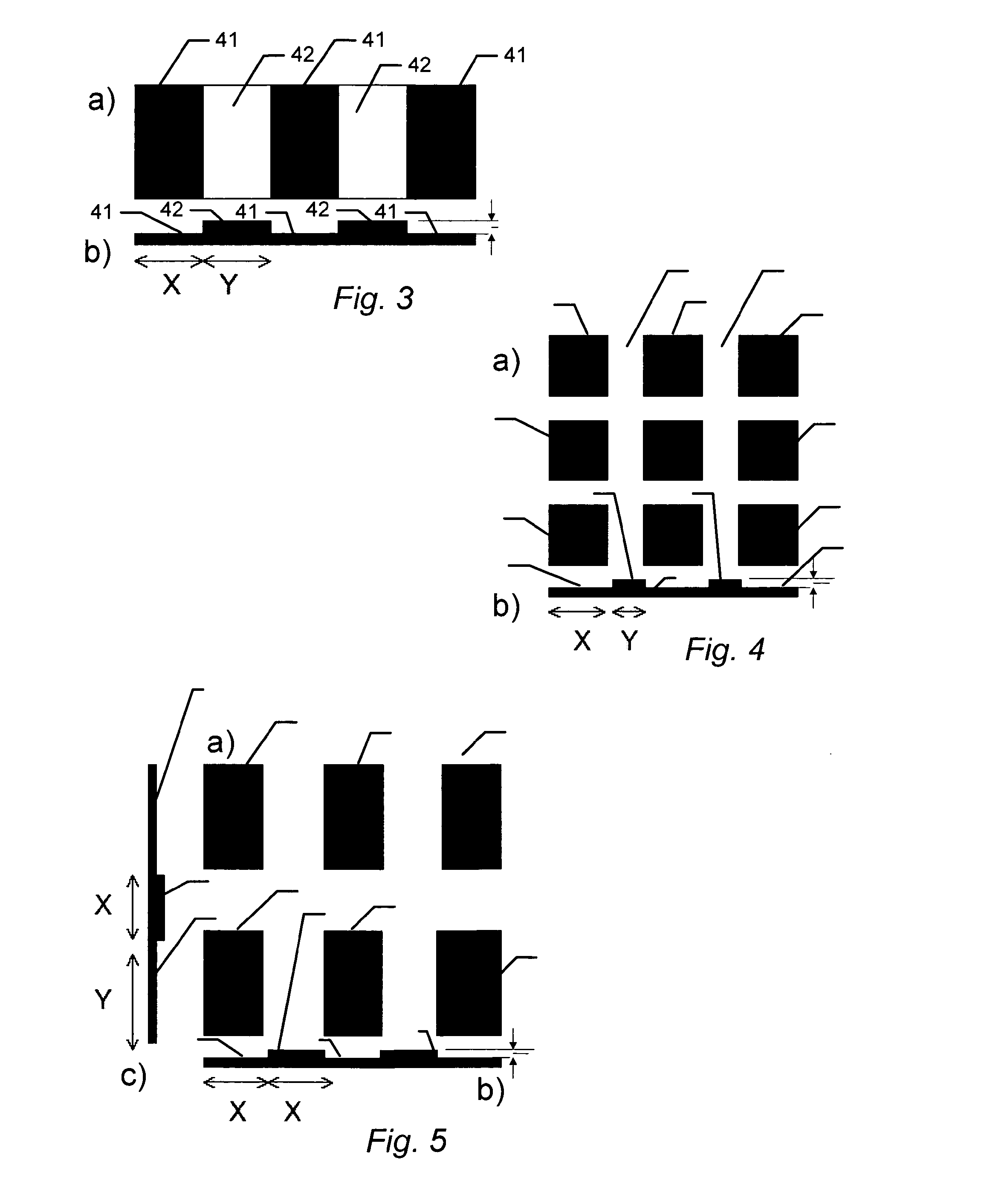
[0174]The previous examples demonstrate that biocompatible surfaces having different surface structures can be used to promote a high ES colony number (for example structure: F1.2) or to promote low ES colony number (for example structure: F4.1). The sustained effect of these structures on the growth of ES cells was examined during serial passaging of CJ7 and KH2 ES cells. After each passage, the ES cells were fixed and analysed for Oct4 and Nanog expression using antibodies specific for Oct4 and Nanog. The ES cells CJ7 and KH2 still express the pluripotency markers after 6 and 10 passages respectively on F1.2 (F1.2 has dimensions X=1;Y=2) or F4.1. (F4.1 has dimensions X=4;Y=1). The CJ7 and KH2 cells also expressed alkaline phosphatase in the last passage. ES colonies generated on F1.2 structures retained a compact and well defined morphology during pa...


PUM

PropertyMeasurementUnit
diameteraaaaaaaaaa
diameteraaaaaaaaaa
diameteraaaaaaaaaa

Abstract

A biocompatible material, wherein at least a part of a surface of the biocompatible material is characterized by a micro or nano-meter scale topographical structure comprising a plurality of features where the structure is selected to promote the growth of undifferentiated pluripotent stem cells or serve to promote the uniform differentiated growth of stem cells. Furthermore, a biocompatible material is provided having a surface structure and composition that affects a cellular function, in particular cellular functions related to gene induction, cell differentiation and the formation of bone tissue in vivo and ex-vivo.

Description

TECHNICAL FIELD[0001]The present invention provides biocompatible materials having a surface structure and composition that affects the cellular function and growth of embryonic stem cells. In a first aspect the present invention provides a biocompatible material having a composition and surface structure that either serves to promote the growth of undifferentiated pluripotent stem cells or serves to promote the uniform differentiated growth of stem cells. In second aspect the present invention provides a biocompatible material having a surface structure and composition that affects a cellular function, in particular cellular functions related to gene induction, cell differentiation and the formation of bone tissue in vivo and ex-vivo.BACKGROUND OF THE INVENTION[0002]With regards to the first aspect of the invention, the promotion of selected cellular functions is an important task in a variety of applications, such as the production of undifferentiated mammalian embryonic stem cell...

Claims


Application Information

Patent Timeline
Patent Type & Authority Applications(United States)
IPC IPC(8): A61F2/28C12N5/02C12M3/00B29C59/02
CPCC12M25/06C12M25/00C12N2535/10C12N5/0606
Inventor DUCH, MOGENS RYTTERGARDMARKERT, LOTTELOVMAND, JETTEFUCHTBAUER, ANNETTE CHRISTINEFUCHTBAUER, ERNST MARTINFOSS, MORTENBESENBACHER, FLEMMINGPEDERSEN, FINN SKOU
Owner AARHUS UNIV
Why Eureka
Try Eureka