Therapeutic encapsulated embryonic stem cells and mesenchymal stromal cells

a technology of embryonic stem cells and mesenchymal stromal cells, which is applied in the direction of biocide, drug composition, skeletal/connective tissue cells, etc., can solve the problems of msc tissue persistence, potential msc differentiation and/or msc migration away from the injury site, and is difficult to resolve, control and quantify. , to achieve the effect of reducing the inflammatory aspects of trauma-mediated tissue damage, promoting tissue repair, and reducing the inflammatory aspects

Inactive Publication Date: 2012-01-26
RUTGERS THE STATE UNIV
View PDF6 Cites 14 Cited by
  • Summary
  • Abstract
  • Description
  • Claims
  • Application Information

AI Technical Summary

Benefits of technology

[0017]In another aspect the present application discloses that by incorporating the soluble inducer, e.g., retinoic acid (RA), into the permeable microcapsule system, cell aggregation was decreased and neuronal lineage differentiation enhanced.
[0020]In another aspect the present invention provides that differentiation approaches to efficiently generate large homogenous neural cell populations offer the potential to investigate and treat a variety of neurological disease processes mediated both by traumatic and naturally occurring events. The inventors have developed a method, using alginate microencapsulation, to generate all neuronal lineage cell types from stem cells. This process is scalable and may be used for both clinical treatment and drug discovery protocols. In addition, the differentiation process of the present invention can be augmented by culture supplementation of specific differentiation pathway regulators (e.g., PPAR agonists) which yield, for example, more than 80% myelin basic protein+ (protein positive) cells. Other pathways may also be targeted for cell specific enrichment.
[0023]In another aspect the present invention sought to determine if alginate encapsulated MSCs could attenuate inflammation and promote tissue repair. Through evaluation of an immobilization platform combining several different alginate concentrations and cell seeding densities, among others, the results indicate that this system can sustain MSC proliferation and viability for at least 3 months in vitro. In addition, depending on the alginate concentration, MSCs could either be maintained as undifferentiated cells (vital to sustaining tissue protective properties), or induced to differentiate into chondrocyte lineage cells. The data demonstrate that the microencapsulation platform can support constitutive MSC secretion patterns and that this function is also dependent upon alginate concentration. Furthermore, MSCs in the presence of pro-inflammatory stimuli, can be induced to secrete these factors at increased rates. Finally, via an in vitro model of macrophage inflammation attenuation, we have demonstrated that MSCs can mitigate inflammatory aspects of trauma mediated tissue damage by inducing a macrophage phenotype which promotes tissue remodeling rather than degeneration.

Problems solved by technology

Although studies have indicated that the microenvironment as well as the developmental status of MSCs can alter neural stem cell inductive signals (Croft, A. P. and Przyborski, S. A., Exp. Neurol., 216, 329-341 (2009)), MSC tissue persistence, potential MSC differentiation and / or MSC migration away from the injury site are very complex and present dynamic problems, which are difficult to resolve, control and quantify.
In particular, several drawbacks in current MSC implantation approaches limit safe and controlled clinical trial implementation.
These include, 1) directly transplanted MSCs exposed to the complex injury environment may be adversely affected early in the treatment process, 2) MSCs may migrate to undesired tissue locations, and 3) MSCs may differentiate into undesired end stage cells.
These issues severely limit the development of controlled feasibility studies and ultimately translatability of MSC treatments into clinical settings.
Moreover, although studies have established techniques to successfully differentiate stem cells into different mature cell lineages using growth factors or extracellular matrix protein supplementation in both two and three-dimensional configurations, their practicality is limited by lack of control and low yields of differentiated cells.

Method used


Image

  • Therapeutic encapsulated embryonic stem cells and mesenchymal stromal cells
  • Therapeutic encapsulated embryonic stem cells and mesenchymal stromal cells
  • Therapeutic encapsulated embryonic stem cells and mesenchymal stromal cells

Examples


example 1

ES Cell Culture

[0132]All cell cultures were incubated in a humidified 37° C., 5% CO2 environment. The ES cell line D3 (ATCC, Manassas, Va.) was maintained in an undifferentiated state in T-75 gelatin-coated flasks (Biocoat, BD-Biosciences, Bedford, Mass.) in Knockout Dulbecco's modified Eagles medium (Gibco, Grand Island, N.Y.) containing 15% knockout serum (Gibco), 4 mM L-glutamine (Gibco), 100 U / ml penicillin (Gibco), 100 U / ml streptomycin (Gibco), 10 mg / ml gentamicin (Gibco), 1,000 U / ml ESGRO™ (Chemicon, Temecula, Calif.) and 0.1 mM 2-mercaptoethanol (Sigma-Aldrich, St. Louis, Mo.). Media was changed every two days until plates were confluent. ES cultures were split and passaged every 6 days. Following media aspiration, cells were washed with 10 mL of phosphate buffered solution (PBS) (Gibco), detached using 3 mL of trypsin EDTA (Gibco) for 3 minutes, and subsequently 12 mL of Knockout DMEM was added. Cells were then replated in gelatin-coated T-75 flasks at a density of 1 millio...

example 2

Alginate Encapsulation

[0133]Alginate solution was prepared by dissolving 2.2 g of alginic acid sodium salt (MW: 100,000-200,000 g / mol, G Content: 65%-70%, Sigma-Aldrich) in 100 mL of Ca2+ free DMEM (Gibco), using a heated magnetic stir plate at a temperature of 65° C. The solution was then filtered using a 45 μm syringe filter (Fisher Scientific, Pittsburgh, Pa.). To create the cell-alginate mixture, 1 mL aliquot of cell suspension with a seeding density of 5×10⁷ cells / mL was added to 9 mL of either 1.2%, 1.7%, 2.2% or 2.5% (w / v) alginate solution to yield a final cell seeding density of 5×10⁶ cells / mL. This solution was transferred to a 10 mL syringe, and was connected to a syringe pump (KD Scientific, MA). Alginate beads were generated using an electrostatic bead generator (Nisco, Zurich, Switzerland) at a flow rate of 40 mL / h, and an applied voltage of 6.4 kV. The beads were extruded into a 200 mL bath of CaCl2 (100 mM), containing 145 mM NaCl, and 10 mM MOPS (all from Sigma-Aldr...

example 3

Assessment of Cell Proliferation and Neural Specific Protein Expressions

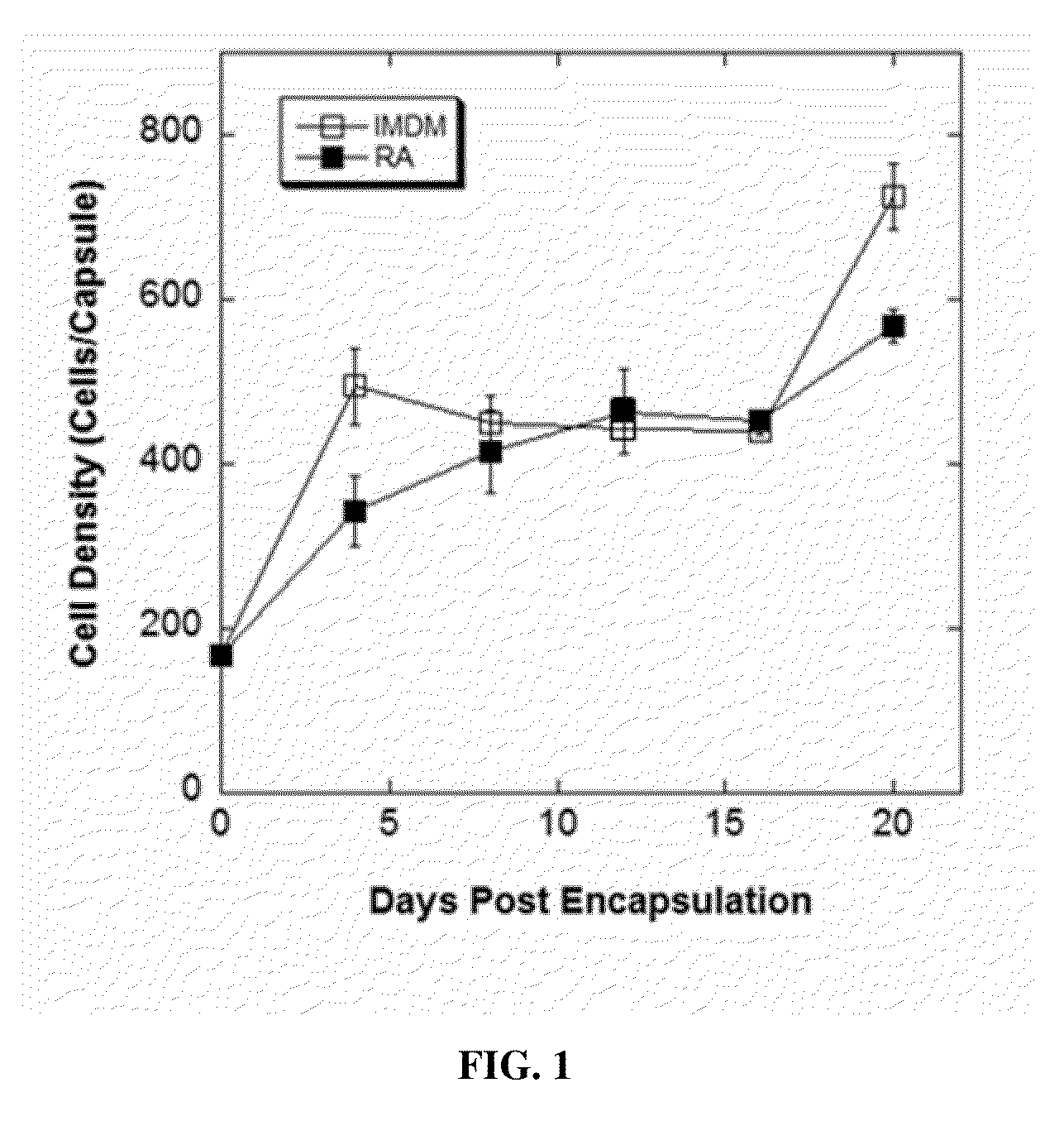
[0134]Under an optimized encapsulation condition (i.e. 2.2% w / v alginate and 5×10⁶ cells / mL), experiments were designed to assess cell proliferation and the expression of an array of neural special markers during a 20-day differentiation period. Encapsulated cells were cultured in the presence or absence of RA, recovered on days 4, 8, 12, 16 and 20 post encapsulation by depolymerizating the alginate microcapsules, and cell number and viability were determined. As indicated in FIG. 1, encapsulated cell numbers in the presence or absence of retinoic acid were similar. The cell proliferation in both conditions exhibited biphasic kinetic properties, and the cultures ultimately reached a final density 2.5 times greater than the initial seeding density. Cell viability was greater than 95% in both conditions and throughout the culture period.

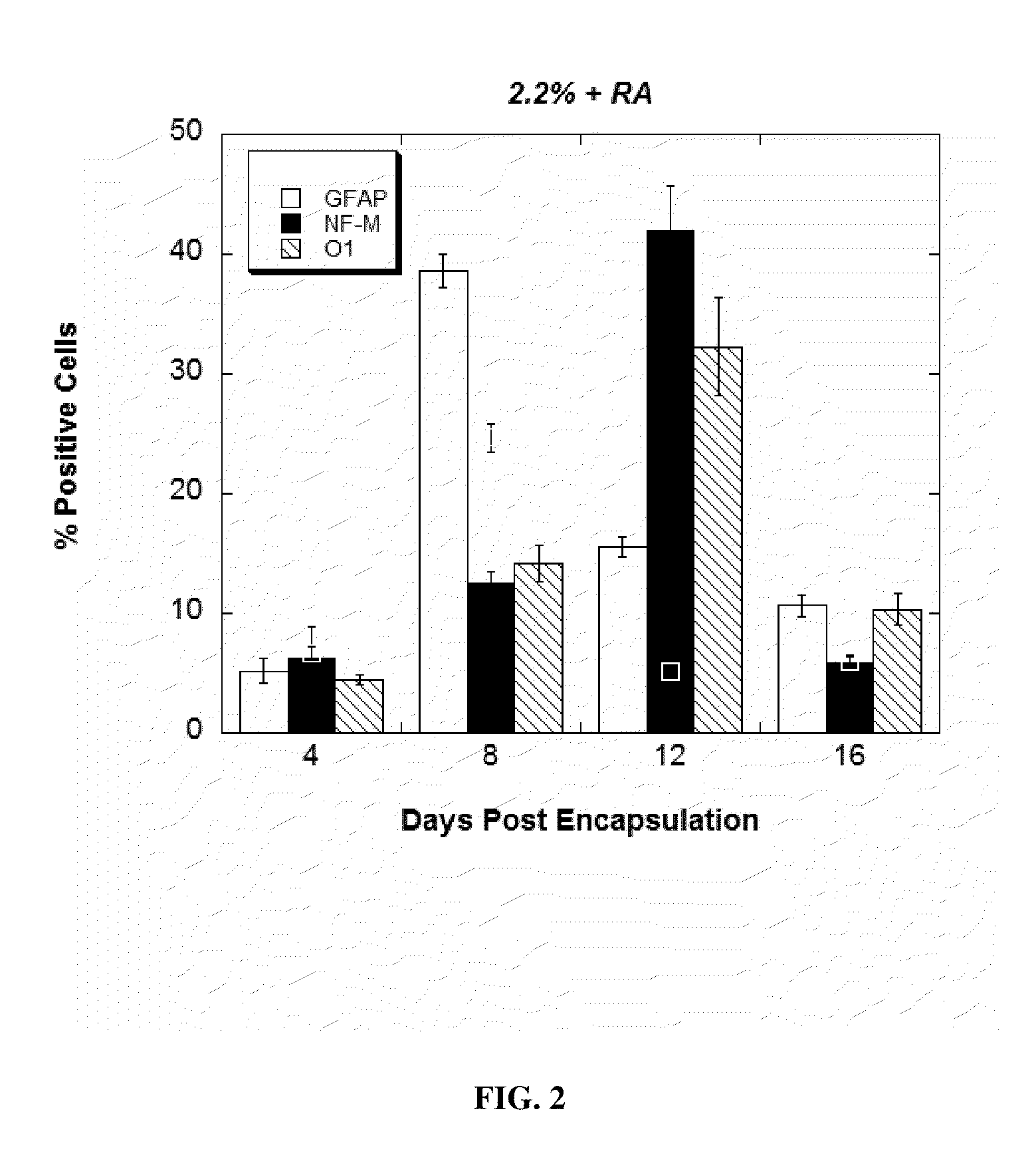
[0135]To assess the effect of RA on lineage commitment within the alginate mic...


PUM

PropertyMeasurementUnit
step sizeaaaaaaaaaa
cell seeding densityaaaaaaaaaa

Abstract

This application discloses alginate microencapsulation-mediated differentiation of embryonic stem cells and use of the stem cell differentiation method for the development of effective treatment of various diseases and disorders. The microencapsulation of embryonic stem (ES) cells results in decreased cell aggregation and enhanced neural lineage differentiation through incorporating the soluble inducer retinoic acid (RA) into the permeable microcapsule system. This application also discloses a micro-encapsulation system for immobilizing mesenchymal stromal cells (MSCs) while sustaining the molecular communication. Thus, the invention provides the use of encapsulated mesenchymal stromal cells in the cellular transplantation therapies. Moreover, the invention provides methods for delivery of encapsulated MSCs into the central nervous system and therapies derived therefrom, such as, the treatment of spinal cord injury (SCI) and other inflammatory conditions.

Description

CROSS-REFERENCE TO RELATED APPLICATIONS[0001]This application claims priority under 35 U.S.C. §119(e) to U.S. Provisional Patent Application Ser. Nos. 61 / 350,760, filed on Jun. 2, 2010, and 61 / 354,998, filed on Jun. 15, 2010, which are both hereby incorporated by reference in their entirety for all purposes.FIELD OF THE INVENTION[0002]The present invention relates to alginate microencapsulation-mediated differentiation of stem cells and use of the stem cell differentiation method for the development of effective treatment of various diseases or disorders. The invention also relates to a micro-encapsulation system for immobilizing stem cells, methods for delivery of encapsulated stem cells into the central nervous system, and use of the encapsulated stem cells as cellular transplantation therapies.BACKGROUND OF THE INVENTION[0003]Unlike adult differentiated tissue cells, pluripotent stem cells, such embryonic stem (ES) cells, can divide and self-renew indefinitely in-vitro and can al...

Claims


Application Information

Patent Timeline
Patent Type & Authority Applications(United States)
IPC IPC(8): C12N11/10A61P25/00A61K35/12A61K35/28
CPCA61K35/28C12N11/10C12N5/0606C12N5/0618C12N2533/74A61K2035/122A61K2035/128C12N2501/58C12N2506/02C12N5/0663A61P25/00A61K9/4816A61K35/545A61K47/36A61K2035/124C12N5/0622C12N2501/60
Inventor YARMUSH, MARTIN L.SCHLOSS, RENE S.GRUMET, MARTINBARMINKO, JEFFREYKIM, JAE HWANMAGUIRE, TIMDOLLE, JEAN-PIERRELI, LULU
Owner RUTGERS THE STATE UNIV
Why Eureka
Try Eureka