Global Proteomic Screening Of Random Bead Arrays Using Mass Spectrometry Imaging

a mass spectrometry and random bead technology, applied in the field of global proteomic screening of random bead arrays using mass spectrometry imaging, can solve the problems of not yet demonstrated commercial viability, unsuitable for proteomic or nucleotide analysis, and the date has not exceeded, and achieves low cost, high precision, and wide commercial availability

Active Publication Date: 2012-03-29
AMBERGEN
View PDF6 Cites 29 Cited by
  • Summary
  • Abstract
  • Description
  • Claims
  • Application Information

AI Technical Summary

Benefits of technology

[0079]Importantly, photocleavage from individual beads and collection of DNA tags (or mass tags) from positive beads can be easily accomplished almost simultaneous with fluorescent scanning by using a modified fluorescence microarray scanner. In particular, a laser normally used for scanning the image can be replaced with a laser capable of photocleavage of DNA tags from individual beads such as a pulsed Nd—Yag laser with 355 nm output which are widely commercially available at low cost. It has been demonstrated by us that such lasers can photocleave >90% of the tags on a bead sample in less than a few seconds. Since commercial fluorescent scanners operating with multiple wavelengths and different lasers are designed to perform sequential scans maintaining image registration, software image identifi...

Problems solved by technology

However, in practice, such methods to date have not exceed a few hundred codes [Mathur and Kelso (2009) Cytometry A].
(Pregibon D C, Toner M, Doyle P S. Science. 2007 Mar. 9; 315(5817):1393-6) or fluorescence of rare earth elements (Parallel Synthesis Technologies Inc; www.parallume.com) can potentially generate hundreds of thousands of unique codes but have not yet demonstrated their commercial viability.
However, in these studies the detection was performed after elution of the mass tags and not directly from individual beads or from arrays of particles using mass spectrometric imaging techniques.
The SIMS technique provides high lateral resolution down to sub-micron range, but unlike MALDI MS generates only small ions (MW below 400 Da) and is therefore not suitable for proteomic or nucleotide analysis.
Importantly, all of the above studies do not describe MALDI-MS on individual beads or arrays of individual beads.
These methods are often slow, labor intensive and heavily dependent on highly specialized robotics, such as serial microarray printers/spotters which are expensive and subject to failure; the net result is prohibitively expensive protein arrays of limited density and limited scalability for larger protein content.
While costs may come down as more efficient methods of protein production and isolation are introduced, fundamental limitations still remain—namely the need for individually cloning each gene, individually expressing each protein in cells, separate isolation of each protein, mechanical microarray printing of the proteins, stability of the protein stocks or arrays derived from them and difficulties in expressing proteins that are toxic to the host cell.
Furthermore, at ˜20,000 spots total (replicates and controls), Invitrogen's microarray capacity is nearly at it's maximum since protein arrays are not compatible with the photolithography used in DNA microarrays to create smaller and more densely packed spots.
An additional limitation of the current generation of proteomic arrays is the intrinsic low sensitivity and high noise which impedes biomarker discovery from clinical samples.
Part of this problem derives from the low replicate number (duplicate) for each protein represented and the variability of spot printing and subsequent readout.
For these reasons, the assay conditions are far from the ideal solution-phase functional assays.
However, t...

Method used


Image

  • Global Proteomic Screening Of Random Bead Arrays Using Mass Spectrometry Imaging
  • Global Proteomic Screening Of Random Bead Arrays Using Mass Spectrometry Imaging
  • Global Proteomic Screening Of Random Bead Arrays Using Mass Spectrometry Imaging

Examples


example 1

Affinity Purification of Cell-Free Expressed Peptides onto an Agarose Bead Affinity Resin Followed by Mass Spectrometry Detection from Single Beads

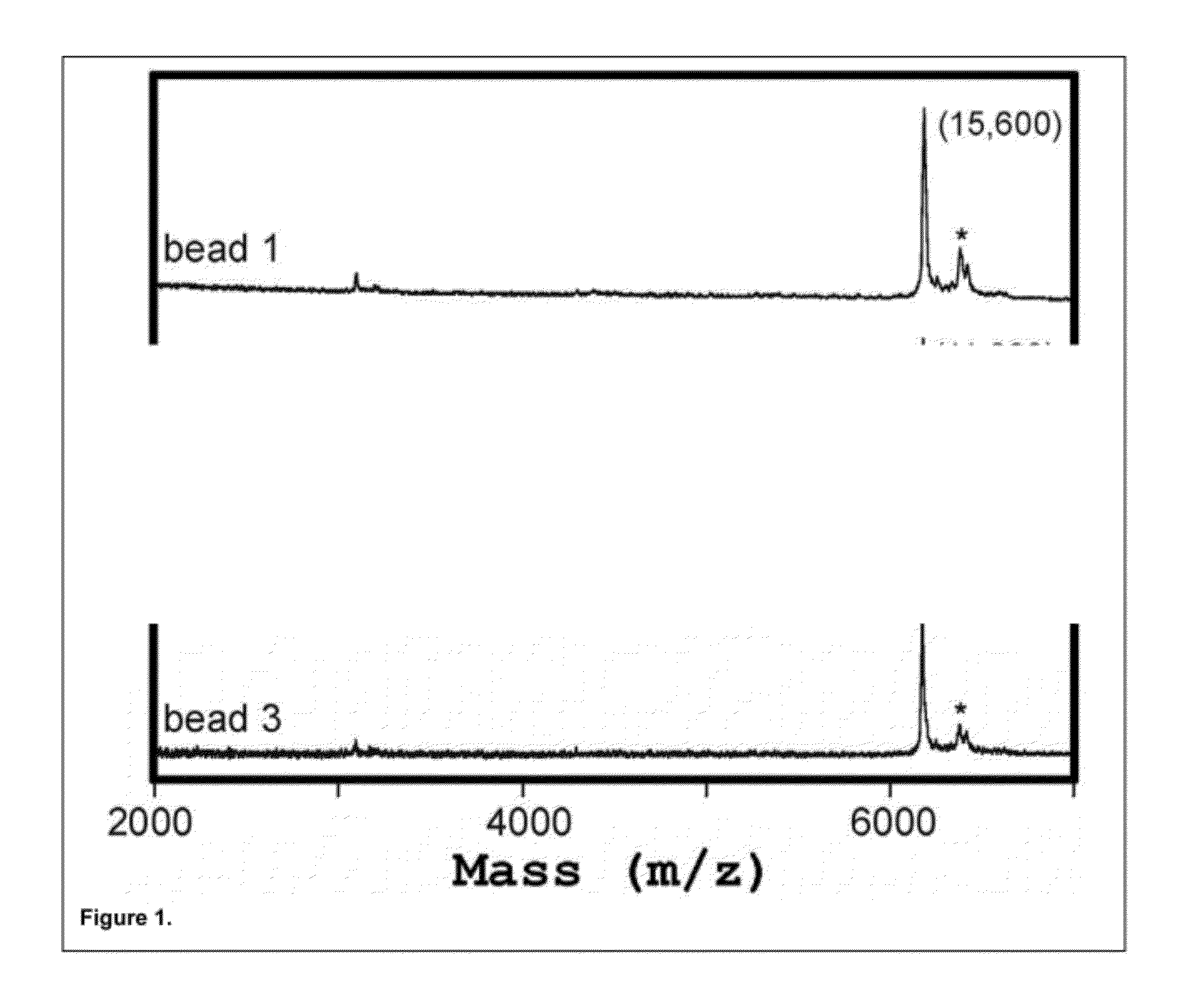
[0164]In this Example a test peptide corresponding to a segment of the APC gene, with an expected molecular weight of 6,203 Da, containing an N-terminal FLAG® epitope tag (Sigma-Aldrich, St. Louis, Mo.), was synthesized in a recombinant cell-free transcription / translation reaction according to the manufacturer's instructions (PureSystem; Post Genome Institute Co., LTD., Japan).

[0165]The nascent peptide from the reaction was then purified on mouse anti-FLAG antibody-coated agarose affinity beads (˜75-150 micron diameter). For this, the crude cell-free expression mixtures were diluted with 50 μL of AB-T [100 mM ammonium bicarbonate with 0.1% Triton X-100 (v / v)]. Mouse anti-FLAG antibody coated agarose affinity beads were used in batch mode to purify the cell-free expressed peptides (EZview™ Red Anti-FLAG® M2 Affinity Gel, Sigma-Aldrich, St....

example 2

Mass Spectrometry Readout and Mass-Imaging from Individually Resolved Beads

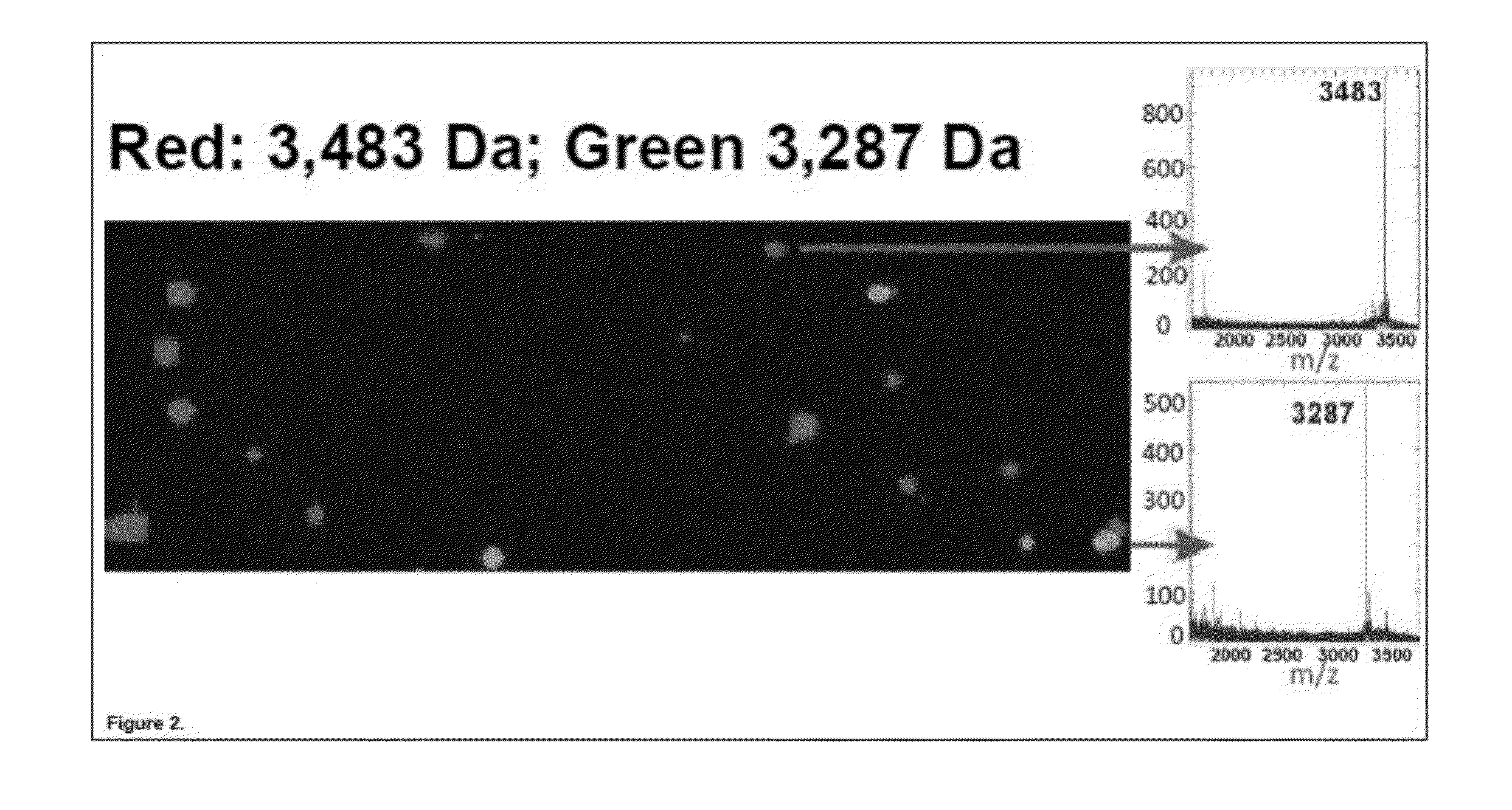
[0167]In this Example, two test peptides with molecular weights of 3,483 Da and 3,287 Da, each containing an N-terminal FLAG® epitope tag (Sigma-Aldrich, St. Louis, Mo.), were separately synthesized in a recombinant cell-free transcription / translation reaction according to the manufacturer's instructions (PureSystem; Post Genome Institute Co., LTD., Japan). The nascent peptides from each reaction were then separately purified on mouse anti-FLAG antibody-coated agarose affinity beads (EZview™ Red Anti-FLAG® M2 Affinity Gel, Sigma-Aldrich, St. Louis, Mo.) (˜75-150 micron diameter). The beads were then mixed in a 9:1 ratio and manually deposited in a random pattern on a suitable substrate for MALDI-TOF mass spectrometry. The MALDI-TOF matrix (CHCA) was sprayed on in a thin and uniform film and allowed to dry. MALDI-TOF imaging of the surface was performed using an ABI 4800 Plus MALDI-TOF / TOF mass spectrometer (A...

example 3

Mass Spectrometry Readout and Mass-Imaging from Individually Resolved 34 Micron Beads Deposited in Pico-Well Plates: Mass-Tagging of Analyte-Bearing Beads for Identification

[0169]In this Example, 34 micron agarose beads were conjugated to an antibody directed against the HSV epitope tag. The beads were then loaded with different recombinant proteins bearing this HSV epitope tag. The beads were additionally bound to one of three different peptide “mass tags” of unique mass, corresponding to the HSV peptide epitope itself, conjugated on the N-terminus to different fluorophores. The beads were then deposited into a special pico-well plate and mass-imaged by scanning MALDITOF mass spectrometry. Separately, the presence of the recombinant proteins was detected on the same batch of beads by probing with a fluorescently labeled antibody directed against the common VSV-G epitope tag, also present in the recombinant proteins.

Development of Pico-Well Plates for MALDI-TOF Bead Scanning

[0170]In...


PUM


Abstract

Methods for proteomic screening on random protein-bead arrays by mass spec is described. Photocleavable mass tags are utilized to code a protein library (bait molecules) displayed on beads randomly arrayed in an array substrate. A library of probes (prey) can be mixed with the protein-bead array to query the array. Because mass spec can detect multiple mass tags, it is possible to rapidly identify all of the interactions resulting from this mixing.

Description

CROSS-REFERENCE TO RELATED APPLICATIONS[0001]This application claims priority to co-pending U.S. Provisional Application Ser. No. 61 / 359,964 filed Jun. 30, 2010, herein incorporated by reference in its entirety for all purposes.FIELD OF THE INVENTION[0002]This invention relates to biology, molecular biology, biochemistry, cell biology, biomedicine, biomarkers and clinical diagnostics; proteomics, reverse proteomics and mass spectrometry; bio-molecular arrays, microarrays, bead-arrays, and bead-displays; multiplexed assays and bio-assays; and label-free bio-molecular detection. More specifically, the invention relates to detecting or imaging molecules or compounds on individual beads or particles using mass spectrometry as applied to the aforementioned fields.BACKGROUND OF THE INVENTIONA. Multiplexing in Bio-Molecular Detection[0003]There is a continuing realization that the complexities of biological systems can neither be fully understood nor harnessed by taking single measurements...

Claims


Application Information

Patent Timeline
IPC IPC(8): C40B30/04
CPCG01N33/6845G01N33/54306G01N33/6848G01N33/5308
Inventor BERGO, VLADISLAV B.LIM, MARK J.ROTHSCHILD, KENNETH J.
Owner AMBERGEN
Why Eureka
Try Eureka