

Identity markers
a technology of identification markers and dermal cells, which is applied in the field of biomarkers and methods for the identification and/or isolation of trichogenic dermal cells, can solve the problems of hair loss or alopecia, affecting the treatment effect, so as to reduce the need for operator intervention and reduce time and labor.
- Summary
- Abstract
- Description
- Claims
- Application Information
AI Technical Summary
Benefits of technology
Problems solved by technology
Method used
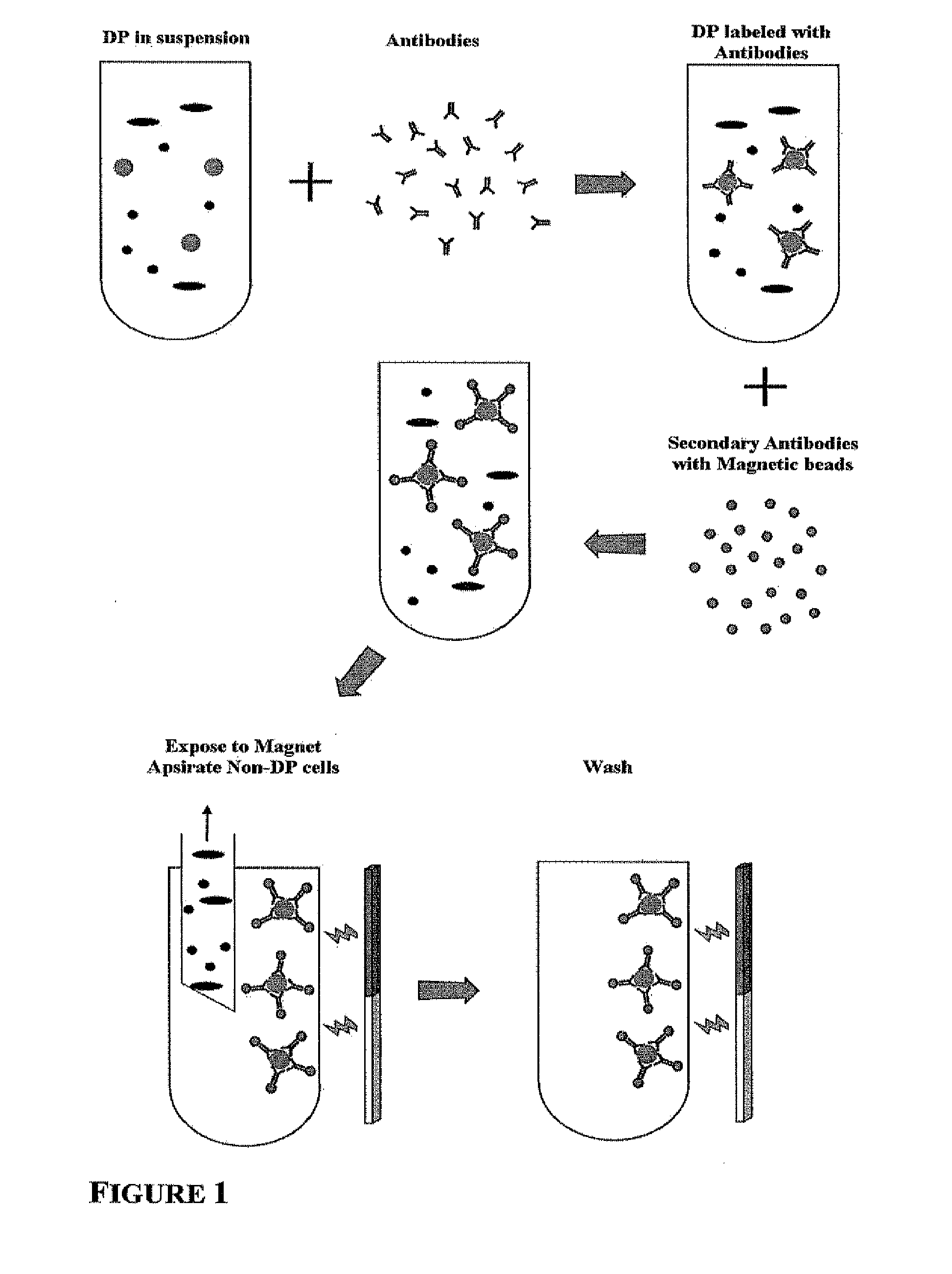
Image

Examples
example 1
Microarray Analysis of Genes Expressed in DP / DS Cells and not in Fibroblasts or Keratinocytes
[0068]In order to identify genes that are expressed in DP / DS cells and not in fibroblasts or keratinocytes, gene expression was compared in these cell types using microarray analysis.
[0069]Materials and Methods
[0070]Microarray Screening Methodology
[0071]Total RNA was prepared from 9 cell culture samples and 3 freshly isolated tissue samples. The 12 samples fell into the groups below:
[0072]Group 1: cultured human dermal fibroblasts (HDF) from 3 independent donors;
[0073]Group 2: cultured human keratinocytes (HK) from 3 independent donors;
[0074]Group 3: cultured dermal papilla cells (DP cells) from 3 independent donors; and
[0075]Group 4: freshly isolated dermal papillae (DPfr) from 3 independent pools of donors.
[0076]RNA extraction, purification, analysis, labelling, profiling on microarrays and primary microarray data analysis was performed by ALMAC Diagnostics (Durham, N.C., USA) in accordanc...
example 2
HAPLN1 is Expressed Specifically in DP / DS Cells in Hair Follicles
[0091]Materials and Methods
[0092]Human hair follicles were isolated by microdissection from a human scalp biopsy, immersed in 2M sucrose solution for 48 h, embedded in TFS compound (Triangle Biomedical Research, Cat. No. H-TFM) blocks, sectioned at 8 μm intervals and placed onto Poly-L-Lysine-coated microscope slides (Polysciences, Inc., Cat. No. 22247). Frozen sections were fixed in 100% acetone for 15 min at −20° C. After two PBS washes, sections were treated with Peroxidase Blocking reagent (Dako, Cat. No. REFS201) for 10 min at room temperature, washed with PBS three times and treated with components of Avidin / Biotin Blocking Kit (Vector Laboratories, Cat. No. SP2001) according to manufacturer's protocol. Immunostaining was perfumed using Peroxidase Goat IgG Kit (VECTASTAIN ABC kit, Vector Laboratories, Cat. No. PK-4005) as described in manufacturer's protocol. Primary antibody used was polyclonal goat anti-human H...
example 3
HAPLN1 is Preferentially Expressed in Cultured DP Cells and / or DS Cells but not in Cultured HDF or in Cultured HK
[0095]Materials and Methods
[0096]HK and HDF were obtained from infant foreskin. DP and DS cells were obtained from adult human donors.
[0097]HDF were cultured in DMEM supplemented with 10% FBS, 2 mM L-Glutamine and 0.05 mg / ml Gentamicin. HK were cultured in EpiLife Medium supplemented with EDGS (Invitrogen). DP cells were cultured in Dermal Papilla Growth Medium (combination of equal parts of human keratinocyte condition medium and Chang Medium. All cells were cultured in 8-well microscope chamber slides (LabTek II Chamber Slide System, Nulge, Nunc International, Cat. No. 154534).
[0098]For immunostaining of the cultures, cells were fixed in a mixture of 5% acetic acid and 95% ethanol for 5 min at room temperature. Fixed cells were washed twice with PBS and immunostained with a Mouse Monoclonal Anti-human HAPLN1 antibody (R&D Systems, Cat. No. MAB2608, 10 μg / ml), using VECT...
PUM
| Property | Measurement | Unit |
|---|---|---|
| volume | aaaaa | aaaaa |
| volume | aaaaa | aaaaa |
| temperature | aaaaa | aaaaa |
Abstract
Description
Claims
Application Information