Biomarker Detection From Fluid Samples

a technology of biomarkers and fluid samples, applied in the field of biomarker detection from fluid samples, can solve the problems of high false negative rate, high cost and time consumption, and many diagnostic tests rely on cell cultures, microscopy or immunoassays which are lacking sensitivity, etc., and achieve the effect of accurately and rapidly detecting target analytes

Active Publication Date: 2020-08-20
THE BOARD OF TRUSTEES OF THE UNIV OF ILLINOIS
View PDF1 Cites 1 Cited by
  • Summary
  • Abstract
  • Description
  • Claims
  • Application Information

AI Technical Summary

Benefits of technology

[0007]Described herein are systems and methods which utilize an array of wells to isolate pathogens directly from fluid, including liquefied solid samples, body fluids, food samples, environmental samples, with nucleic acid detection, including amplification techniques, to accurately and rapidly detect target analytes, such as pathogens or cell-indicating markers, in fluid samples, even in very low concentrations. The provided systems and methods dry the fluid sample, including a solid or semi-solid sample that has been liquefied, to deposit the total volume or a fraction of the volume in a number of wells and perform nucleic acid detection, such as by amplification, on each of the individual wells to detect the presence of even a single individual target analyte and provide a quantitative analysis of the amount of target analyte within the sample. The systems and methods are robust, and ensure that reagents required for detection, such as amplification or crispr enzyme, reach each individual well, and that reliable detection occurs, even approaching a single target analyte in a well. The technique is compatible with various bodily fluids, including blood, urine, saliva, sputum, etc. Similarly, the technique is compatible with solid and / or semi-solid samples that have been liquefied, including tissue, stool, food, environmental, and the like.

Problems solved by technology

Detection of low concentration pathogens is important in many applications, including food samples, environmental samples and patient samples, including blood, saliva and urine, remains a challenge for medical diagnostic professionals.
Currently, many diagnostic tests rely on cell cultures, microscopy or immunoassays which are lacking in sensitivity, costly and time consuming.
These constraints often force pathologists to rely on experience and guesswork, sometimes resulting in misdiagnoses or the overreliance on antibiotics when they may not actually be necessary, which in turn contributes to drug resistance.
For example, diagnosis of sepsis, a bacterial infection of the blood resulting from dysregulated inflammatory response, remains a challenge for medical professionals to quickly diagnosis.
The most common test for sepsis is a blood culture followed by nucleic acid amplification, which requires days to a week for a bacteria to grow to detectable quantities and has a high false negative rate.
The time required for testing is problematic, as within days of the onset of infection a patient may progress into advance stages leading to organ failure, causing sepsis to have high morbidity and mortality rates.
Even as technology surrounding nucleic acid amplification techniques have advanced, significant hurdles remain in rapid, low cost identification of pathogens in bodily fluids.
While tests have been developed for mRNA biomarkers and pathogen nucleic acids, current RNA / DNA based techniques often require expensive equipment and are directed towards single pathogens, as multiplexing increases costs.
The nucleic acid tests also often require costly and time consuming purification steps before the sample may be analyzed.

Method used


Image

  • Biomarker Detection From Fluid Samples
  • Biomarker Detection From Fluid Samples
  • Biomarker Detection From Fluid Samples

Examples


example 1

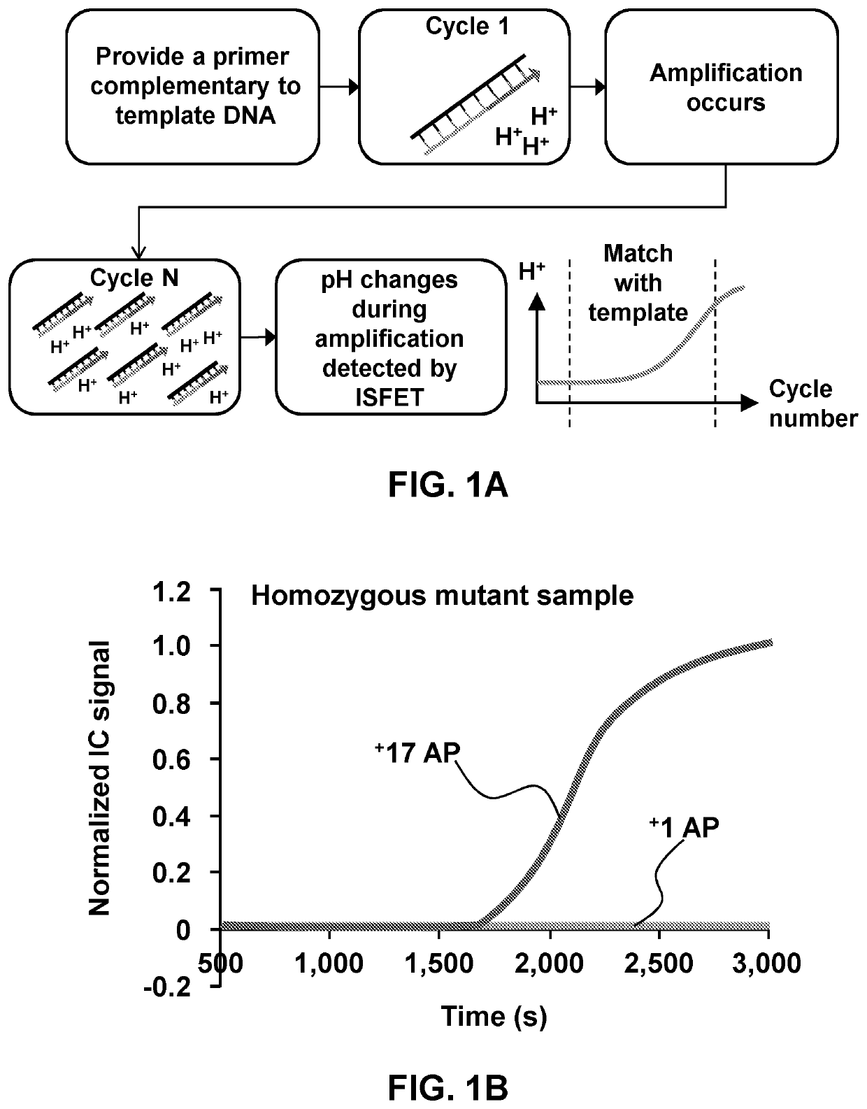
uid Sample (Blood, Urine, Saliva, Etc.) Digital Amplification

[0123]Described herein is dried sample digital amplification (LAMP, PCR etc.) on a substrate (Glass, plastic, silicon etc.) with an array of microwells and with no / minimal sample processing. The drying of the sample allows processing of larger sample volumes (up to several milliliters of blood for e.g.) and preservation of pathogen nucleic acids in the sample for room temperature transportation and storage. Additional fixation of samples can be performed of the samples using common fixatives such as Formalin or acetone for extended storage of the samples for analysis.

[0124]Digital amplification (digital lamp, digital per etc.) is performed on the array of wells or microwells with dried sample in it. The array may have large number of wells (1e5-1e6) in line with large number of partitions required for digital amplification. This technique combines higher sample volume processing capability with superior sensitivity and lim...

example 2

of Bacteria in a Dried Blood Sample

[0172]Detecting extremely low target analyte or pathogen count (1-10 CFU / ml or lower) has been a fundamental challenge in the medical diagnostics' and food testing industry2 due to extremely low signal to noise ratio. Cell culture, which usually take 1-5 days, is the only way of detecting these low pathogen levels in any sample.1,3,4 Sepsis, a clinical syndrome that results from dysregulated inflammatory response to infection leading to organ dysfunction has very high morbidity and mortality.4 It is also estimated to be the most costly inpatient diagnosis, accounting for more than $23 billion annually in US alone.5 Since sepsis usually has very low pathogen counts (1-3 CFU / m1)6 the only way for diagnosis is culture followed by nucleic acid amplification or direct molecular detection after a positive blood culture.3 Blood cultures (or other sample cultures) are very time consuming (1-5 days), have a very high false negative rate and do not work for ...

example 3

ed / Dried Biomolecule Delivery to Micro / Nano-Well Arrays and Strategies for End-Point Multiplexed Detection of Nucleic Acid Targets

[0229]To deliver material, including biomolecules, enzymes and the like, into a micro-well array in a multi-step process, the material can be lyophilized or dried on a fluid barrier, such as beads-on-a-wax paper, and pulled down into the wells using centrifugation or magnetic pull down. Since in a micro-well array, cross contamination between two adjacent wells is a challenge, loading the enzyme through beads on a wax paper allows simultaneous loading of a large array of wells (>1e6) in a single step requiring less than 1 minute. The fluid barrier may be a substrate with a hydrophobic coating, such as wax paper with its hydrophobic wax coating. Various barriers are compatible with the methdos and systems provided herein, so long as the barrier prevents any fluid exchange between adjacent wells during the process.

[0230]A schematic illustration of the beads...


PUM

PropertyMeasurementUnit
volumeaaaaaaaaaa
volumeaaaaaaaaaa
volumeaaaaaaaaaa

Abstract

Described herein are systems and methods which utilize an array of wells to isolate pathogens and nucleic acid detection techniques to accurately and rapidly detect pathogens in fluid samples, even in very low concentrations, including from solid or semi-solid samples that have been fluidized. The provided systems and methods dry the fluid sample to deposit a fraction of the total volume in a number of wells and perform nucleic acid detection on individual wells to detect even individual pathogens and provide a quantitative analysis of the amount of pathogen within the sample. Also provided are methods and systems for precise delivery of dried materials, including biomolecules that are enzymes of use in the process, to microwells.

Description

CROSS REFERENCE TO RELATED APPLICATIONS[0001]This application claims the benefit of priority to U.S. Pat. App. 62 / 569,119 filed Oct. 6, 2018, which is hereby incorporated by reference to the extent not inconsistent herewith.STATEMENT REGARDING FEDERALLY SPONSORED RESEARCH OR DEVELOPMENT[0002]This invention was made, at least in part, with United States governmental support awarded by National Science Foundation award no. SBC 5710003967, United States Department of Agriculture award nos. 8000063044, 80000069085 and 8000074077 and National Institutes of Health award no. MGH-APF 087126. The United States Government has certain rights in this invention.BACKGROUND OF INVENTION[0003]Detection of low concentration pathogens is important in many applications, including food samples, environmental samples and patient samples, including blood, saliva and urine, remains a challenge for medical diagnostic professionals. Many diagnostic techniques common today are methods which have been utilize...

Claims


Application Information

Patent Timeline
Patent Type & Authority Applications(United States)
IPC IPC(8): C12Q1/6848G01N27/414C12Q1/689B01L3/00C12Q1/6806C12Q1/6851
CPCC12Q1/6851B01L3/5027C12Q1/6806B01L2200/0689B03C1/01B01L2300/0893C12Q1/689G01N27/4145B03C2201/26B03C1/288B01L2300/0861B03C2201/18B03C1/30C12Q1/6848B01L2300/161C12N15/10G01N33/50G01N2001/4027
Inventor BASHIR, RASHIDGANGULI, ANURUP
Owner THE BOARD OF TRUSTEES OF THE UNIV OF ILLINOIS
Why Eureka
Try Eureka