

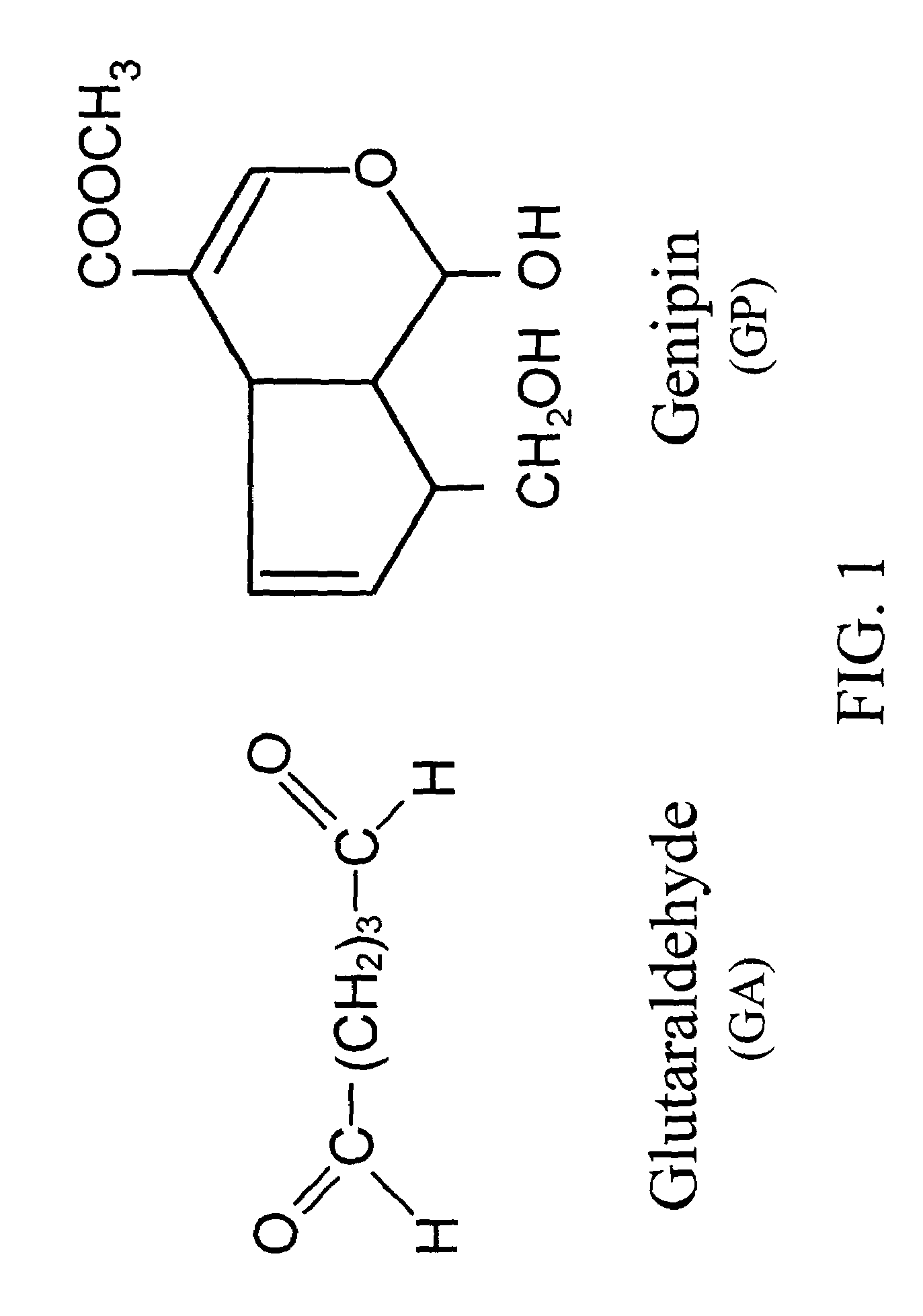
Acellular biological material chemically treated with genipin
a biological material and genipin technology, applied in the field of chemical modification of biomedical materials, can solve the problems of easy degradation of collagenase, low tensile strength, and impair the biocompatibility of biological tissue, and achieve the effects of reducing immunogenicity, reducing enzymatic degradation, and reducing antigenicity
- Summary
- Abstract
- Description
- Claims
- Application Information
AI Technical Summary
Benefits of technology
Problems solved by technology
Method used
Image

Examples
example 1
Tissue Specimen Preparation
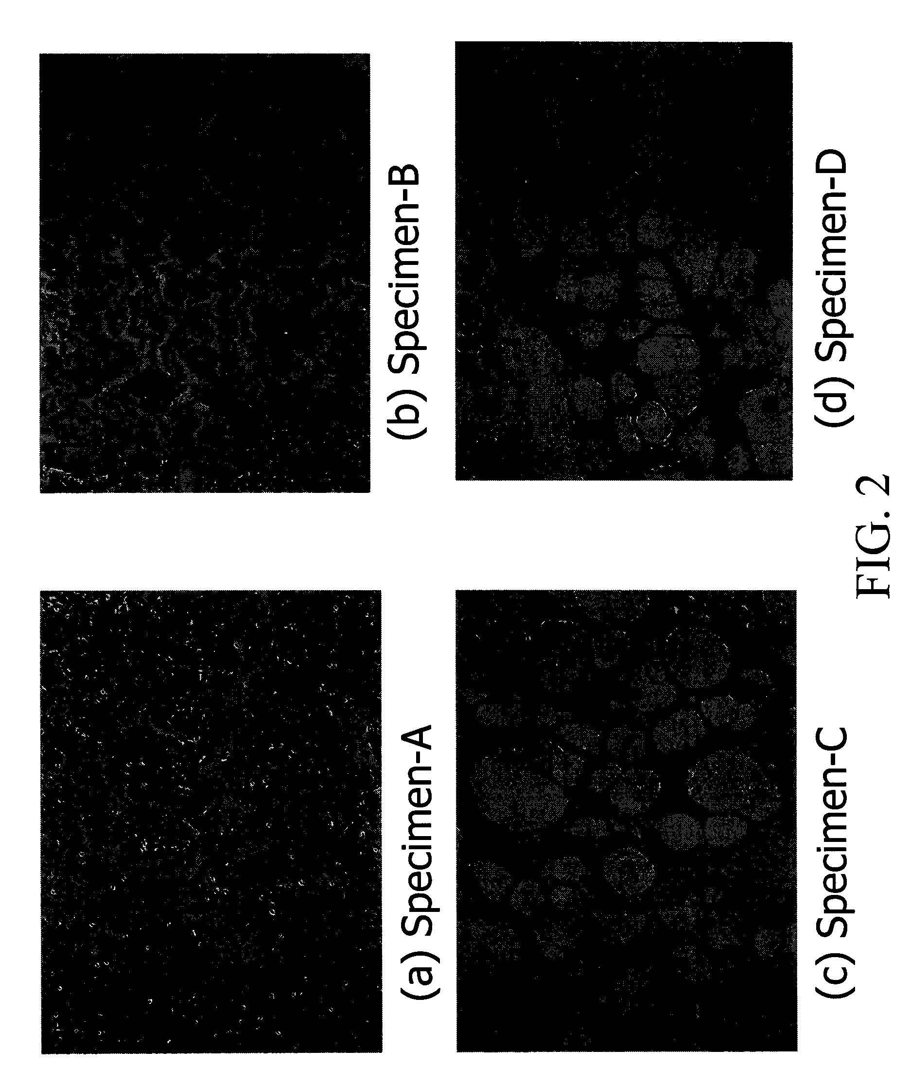
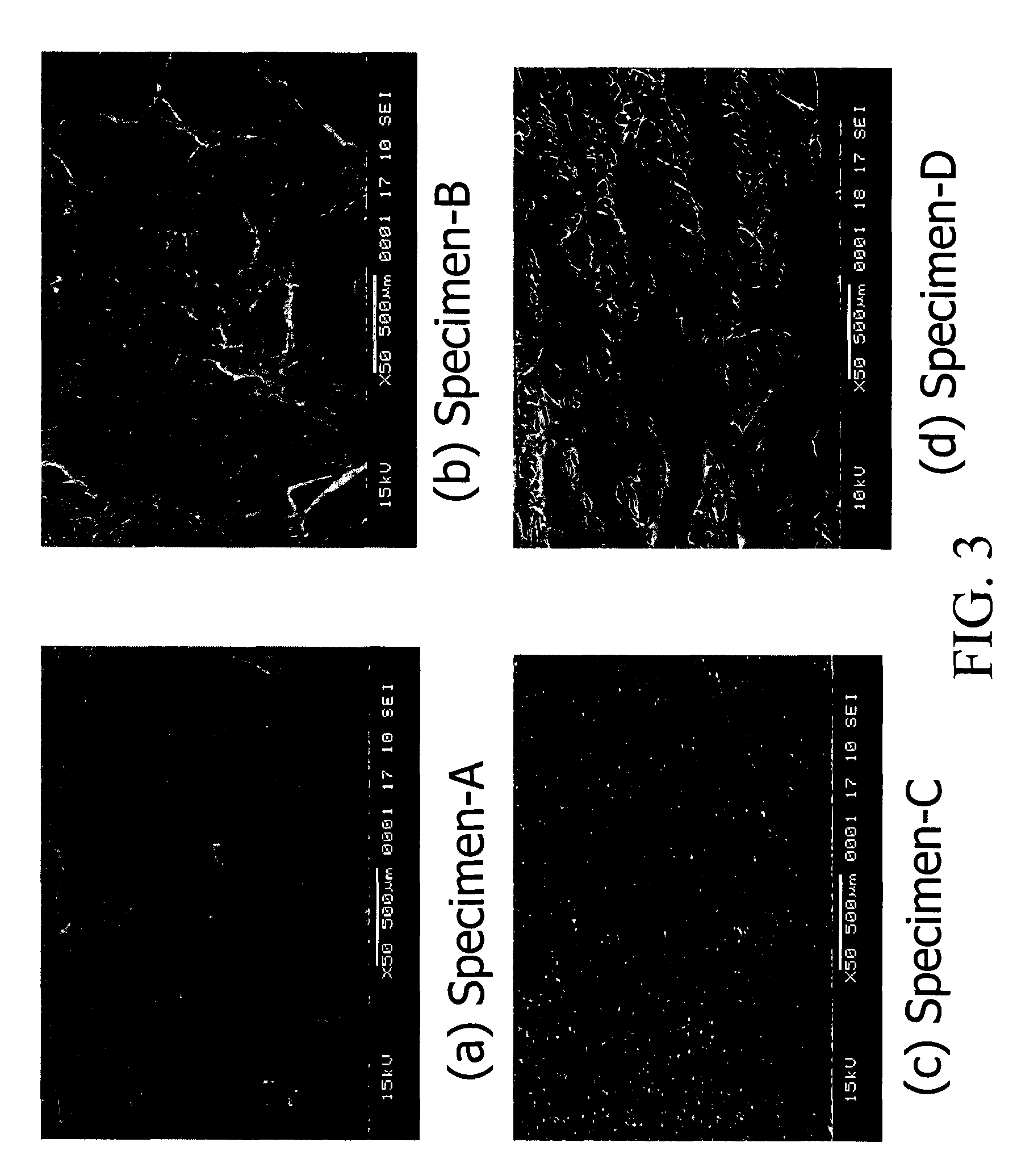
[0046]In one embodiment of the present invention, bovine pericardia procured from a slaughterhouse are used as raw materials. The procured pericardia are transported to the laboratory in a cold normal saline. In the laboratory, the pericardia are first gently rinsed with fresh saline to remove excess blood on tissue. Adherent fat is then carefully trimmed from the pericardial surface. The cleaned / trimmed pericardium before acellular process is herein coded specimen-A. The procedure used to remove the cellular components from bovine pericardia is adapted from a method developed by Courtman et al (J Biomed Mater Res 1994; 28:655–66), which is also referred to herein as “an acellularization process”. A portion of the trimmed pericardia is then immersed in a hypotonic tris buffer (pH 8.0) containing a protease inhibitor (phenylmethyl-sulfonyl fluoride, 0.35 mg / L) for 24 hours at 4° C. under constant stirring. Subsequently, they are immersed in a 1% solution of...
example 2
Tissue Specimen Crosslinking
[0050]The cellular tissue (specimen-A) and acellular tissue (specimen-B) of bovine pericardia are fixed in 0.625% aqueous glutaraldehyde (Merck KGaA, Darmstadt, Germany) and are coded as specimen-A / GA and specimen-B / GA, respectively. Furthermore, the cellular tissue (specimen-A) and acellular tissue (specimen-B, specimen-C, and specimen-D) of bovine pericardia are fixed in genipin (Challenge Bioproducts, Taiwan) solution at 37° C. for 3 days and are coded as specimen-A / GP, specimen-B / GP, specimen-C / GP, and specimen-D / GP, respectively. The aqueous glutaraldehyde and genipin solutions used are buffered with PBS. The amount of solution used in each fixation was approximately 200 mL for a 10×10 cm bovine pericardium. After fixation, the thickness of each studied group is determined using a micrometer (Digimatic Micrometer MDC-25P, Mitutoyo, Tokyo, Japan). Subsequently, the fixed cellular and acellular tissue are sterilized in a graded series of ethanol soluti...
example 3
Comparison of Glutaraldehyde and Genipin Crosslinking
[0056]Pericardia tissue chemically treated with glutaraldehyde and genipin shows different characteristics and biocompatibility. FIG. 5 shows thickness of the glutaraldehyde-fixed cellular tissue (A / GA), the glutaraldehyde-fixed acellular tissue (B / GA), the genipin-fixed cellular tissue (A / GP), and the genipin-fixed acellular tissue (B / GP) before implantation. In general, the acellular tissue shows increased tissue thickness by either type of crosslinking (with glutaraldehyde or genipin) as compared to the control cellular tissue. It is further noticed that genipin-fixed acellular tissue shows the highest tissue thickness among the samples characterized, probably due to enhanced water absorption. This high tissue thickness of genipin-fixed acellular tissue is desirable for tissue engineering in vivo or in vitro in medical devices, such as an extended-release drug delivery device, vascular or skin graft, or orthopedic prosthesis of...
PUM
| Property | Measurement | Unit |
|---|---|---|
| porosity | aaaaa | aaaaa |
| pH | aaaaa | aaaaa |
| pH | aaaaa | aaaaa |
Abstract
Description
Claims
Application Information