Waveguide immunosensor with coating chemistry and providing enhanced sensitivity

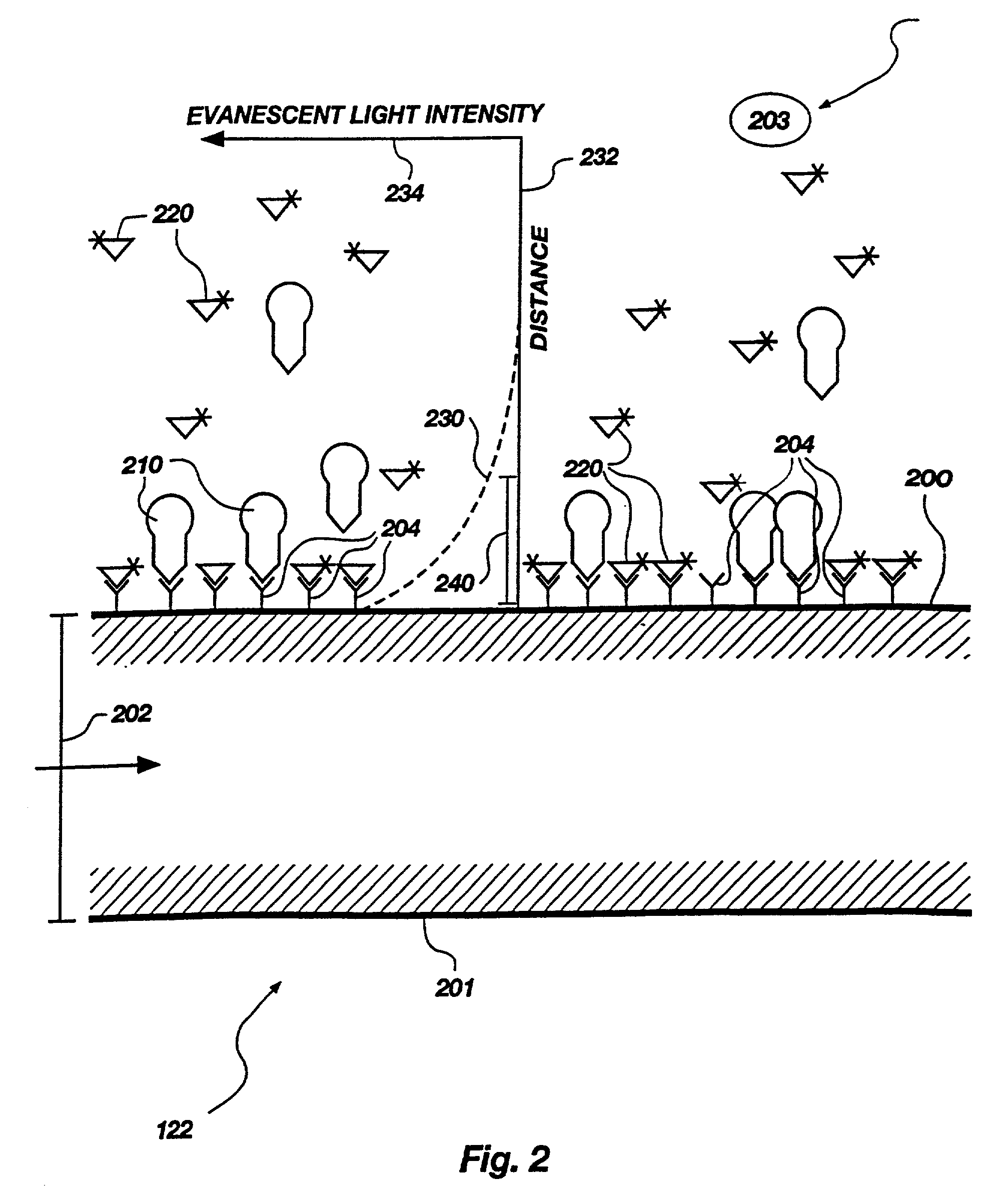
a waveguide immunosensor and coating technology, applied in the field of samples analysis, can solve the problems of difficult to build a multi-well biosensor, difficult to achieve multi-well biosensor, and ineffective internal reflection of typical rod-shaped waveguides, and achieve the effect of increasing the intensity of the evanescent field

Inactive Publication Date: 2006-04-04
UNIV OF UTAH RES FOUND
View PDF43 Cites 61 Cited by
  • Summary
  • Abstract
  • Description
  • Claims
  • Application Information

AI Technical Summary

Benefits of technology

[0017]In another preferred embodiment, a substantial portion of the surrounding edge of the waveguide is coated with a reflective coating to prevent light from escaping through the edge, thereby increasing the intensity of the evanescent field.

Problems solved by technology

However, the bulk of the unbound tracer molecules remaining in solution, will be outside the range of effective excitation and thus will not be stimulated to emit fluorescence.
However, in a typical prior art evanescent light device such as that of Block et al., U.S. Pat. No. 4,909,990 issued Mar. 20, 1990, the waveguide is a fiber optic rod whose shape makes it difficult to build a multi-well biosensor.
The typical rod-shaped waveguides are not as effective in internal reflection as a design having two parallel surfaces would be.
Previous methods of immobilizing antibodies to optical substrates in evanescent biosensors also present some problems causing reduction in sensitivity.
This approach has at least two significant disadvantages due to the fact that most proteins have multiple lysine residues.
First, the presence of multiple potential coupling sites (multiple lysine residues) results in multiple random orientations of antibodies on the substrate surface.
If the substrate-coupled lysine residue is near the N-terminal of the antibody molecule, the antibody's antigen binding site (which is near the N-terminal) may be effectively unavailable for binding of the analyte.
Second, if multiple lysines on the same antibody molecule are coupled to the substrate, the molecule may be subjected to conformational strains which distort the antigen binding site and alter its binding efficiency.
Another problem relates to the levels of non-specific binding to the antibody-coated surface of the optical substrate.
These levels are often sufficiently high to make detection of analyte at concentrations below about 10−10 molar (abbreviated M) very difficult.
However, this is time-consuming and complicates the assay procedure.
Second, non-specific binding can be a serious problem unless the surface is “passivated” with a masking agent such as bovine serum albumin or with a thin coating of hydrophilic polymer such as poly(ethylene glycol) or poly(methacrylate).

Method used


Image

  • Waveguide immunosensor with coating chemistry and providing enhanced sensitivity
  • Waveguide immunosensor with coating chemistry and providing enhanced sensitivity
  • Waveguide immunosensor with coating chemistry and providing enhanced sensitivity

Examples


example i

Preparation of Waveguide Surface—Hydrogel

[0071]A silica surface was prepared with a hydrogel coating comprised of polymethacryloyl hydrazide (abbreviated “PMahy”). Fused silica slides of CO grade and thickness about 1 mm, available from ESCO, Inc., were suitable as waveguides (optical substrates).

[0072]To graft the PMahy to the silica, the surface was derivatized with aldehyde groups. The derivatization was accomplished by silanization with 3-aminopropyltriethoxy silane (abbreviated “APS”) to add an amino functional group, followed by reaction with glutaraldehyde to produce free aldehyde groups. The PMahy was then reacted with these aldehyde groups to form the hydrogel coating.

[0073]Antibodies could be coupled to this hydrogel in at least two ways. In one method, the carbohydrate groups in the Fc antibody region are oxidized to aldehydes by treatment with sodium metaperiodate. However, few antigen-binding fragments contain carbohydrate moieties useful for this purpose. Thus, a prefe...

example ii

Preparation of Waveguide Surface—Avidin-biotin

[0082]This strategy was designed to exploit the very strong binding affinity of biotin for avidin (binding constant of around 10−15). An avidin coating was readily made by physical adsorption on a silica surface. The Fab′ fragments were then conjugated with biotin to form biotin-Fab′ conjugates, also referred to as biotinylated Fab′ fragments or b-Fab′ fragments. The biotin is coupled at specific location(s) on the Fab′ fragments. The avidin coated surface is then treated with the b-Fab′ fragments, so that the biotin binds to the avidin thereby immobilizing the Fab′ fragment to the surface in a site-specific manner.

[0083]In actual experiments, the procedure was as follows. Chromic acid-cleaned silica surfaces were immersed in a solution of 3×10⁻⁶ M (molar) avidin for about 3 hours at room temperature. The surfaces were then washed several times in PBS to remove unadsorbed avidin.

[0084]Biotinylated Fab′ conjugates were prepared from a sol...

example iii

Preparation of Waveguide Surface—Peg-type

[0087]In this method, the terminal hydroxyl groups of polyethylene glycol (abbreviated PEG) were converted to primary amine or hydrazide groups by reaction with ethylenediamine (abbreviated ED) or hydrazine, respectively. The PEG molecules so modified were then coupled to APS-glutaraldehyde activated silica surfaces.

[0088]Monofunctional (PEG M2000, M5000) or difunctional (PEG 3400, PEG 8000, PEG 18,500) of the indicated molecular weights in daltons, were reacted with p-nitrophenyl chloroformate (abbreviated p-NPC; obtained from Aldrich Chemicals) in solution in benzene. The mixture was agitated at room temperature for about 24 hours. Dry ethyl ether (less than 0.01% water, purchased from J.T. Baker Chemicals) was used to precipitate PEG-(o-NP)2 from solution. The precipitate was vacuum-dried overnight. Between about 50% and about 100% of PEG molecules were converted by this treatment to PEG-Onp, as determined by hydrolysis with 0.1N sodium hy...


PUM

PropertyMeasurementUnit
wavelengthsaaaaaaaaaa

Abstract

Methods and apparatus for evanescent light fluoroimmunoassays are disclosed. The apparatus employs a planar waveguide and optionally has multi-well features and improved evanescent field intensity. The preferred biosensor and assay method have the capture molecules immobilized to the waveguide surface by site-specific coupling chemistry. Additionally, the coatings used to immobilize the capture molecules provide reduced non-specific protein adsorption.

Description

CROSS-REFERENCE TO RELATED APPLICATIONS[0001]This application is a divisional of application Ser. No. 09 / 207,187, filed Dec. 8, 1998, now U.S. Pat. No. 6,340,598, issued Jan. 22, 2002, which is a divisional of application Ser. No. 08 / 640,141, filed Apr. 30, 1996, now U.S. Pat. No. 5,846,842, issued Dec. 8, 1998, which is a divisional of application Ser. No. 08 / 064,608, filed May 18, 1993, now U.S. Pat. No. 5,512,492, issued Apr. 30, 1996.FIELD[0002]This application relates to the art of analyzing samples for particular substances by means of fluorescent binding assays, and more particularly to apparatus, compositions and methods for such assays employing evanescent light.BACKGROUND[0003]Biosensor apparatus based on optical detection of analytes by fluorescence of tracer molecules, have attracted increasing attention in recent years. Such apparatus are useful for both diagnostic and research purposes. In particular, biosensors for a solid-phase fluoroimmunoassay, are becoming an impo...

Claims


Application Information

Patent Timeline
Patent Type & Authority Patents(United States)
IPC IPC(8): C12M3/00G01N21/25G01N21/55G01N21/64G01N21/77G01N33/543
CPCG01N21/6428G01N21/6452G01N21/648G01N33/54373G01N21/7703Y10S436/805Y10S435/808
Inventor HERRON, JAMES N.CHRISTENSEN, DOUGLAS A.CALDWELL, KARIN D.JANATOVA, VERAHUANG, SHAO-CHIEWANG, HSU-KUN
Owner UNIV OF UTAH RES FOUND
Why Eureka
Try Eureka