
Detection and treatment of prostate cancer

Active Publication Date: 2006-11-14
GLYCOMIMETICS
View PDF3 Cites 55 Cited by
  • Summary
  • Abstract
  • Description
  • Claims
  • Application Information

AI Technical Summary

Benefits of technology

[0030]Tests for prostate specific antigen (PSA) are well known in the art. As noted above, screening for PSA to detect prostate cancer is estimated to miss (i.e., false negatives) a significant proportion of the individuals tested who actually have prostate cancer. An advantage of the present invention is that the method of screening for prostate cancer may detect prostate cancer in an individual having the cancer but who tests negative in a test for PSA. The present method may be used alone or in combination with a test for PSA.
[0031]As noted above, the current approaches to screening for prostate cancer are unable to differentiate between benign prostate disease and prostate cancer. For therapy purposes, it is important to be able to determine whether a patient diagnosed or presenting with prostate dysfunction has a benign prostate disease or prostate cancer. Benign prostate disease includes benign prostatic hyperplasia (BPH). Another advantage of the present invention is that by detecting for antigen which binds antibody NUH2, prostate cancer may be distinguished from benign prostate disease in a patient diagnosed or presenting with prostate dysfunction. The presence of antigen is associated with prostate cancer. The absence of antigen is indicative of benign prostate disease. A patient diagnosed or presenting with prostate dysfunction may have been tested in a test for PSA, and may have tested negative.
[0032]It may be desirable to elicit or enhance an immune response to the antigen on prostate cancer cells that binds antibody NUH2. The immune response may be a T cell or B cell response. The immune response may be elicited for the first time in a warm-blooded animal, such as a human. Alternatively, it may be enhanced in an organism where a response is present but at insufficient levels. In one embodiment, an individual is selected based on an increased predisposition to prostate cancer. The increased predisposition may be based, for example, upon age, family history or ethnicity. The individual selected is immunized with a composition comprising the antigen and a physiologically acceptable carrier or diluent. Antigen may be coupled to an appropriate immunogenic carrier, such as keyhole limpet hemocyanin (KLH), to include in a composition to elicit or enhance an immune response. Immunizing with the antigen which binds antibody NUH2 in a composition includes immunizing with the naturally occurring antigen or truncated versions, or synthetic versions (chemical or recombinant) or variations of either. Sources of antigen for immunization include human placenta or DU145 cells. Antigen may be coated on Salmonella minnesotae (commercially available as the adjuvant Detox).
[0033]In another embodiment of immunizing, a patient is selected who is suspected to possess prostate cancer cells. For example, the patient may be selected based upon a positive result in a screening method, such as that described herein or for PSA. The individual selected is immunized with the composition described above. Immunization may be performed on the patient in combination with other procedures.
[0034]Sialyl I carbohydrate structures are also found in disialogangliosides and glycoproteins in human placenta. This is an adequate source of antigenic material for the development of humanized antibodies directed against the sialyl I carbohydrate structure for the diagnostic imaging or immunotherapy of prostate cancer patients. In addition, antibodies or other agents that bind to the antigen can be covalently or noncovalently bound to, or fusion constructs prepared from, a molecule that is directly or indirectly cytotoxic to prostate cancer cells, using methodology well known to those of ordinary skill in the art. The cytotoxic molecule may be directly cytotoxic to prostate cancer cells without the necessity for activation, or may be indirectly cytotoxic by requiring activation in vivo (e.g., by the presence or administration of an activating molecule) including as a prodrug, or itself activating in vivo another molecule(s) which is then cytotoxic to prostate cancer cells (e.g., complement mediated cell lysis). Treatment of prostate cancer may result, for example, in remission of the cancer or prolongation of survival time.
[0035]In an embodiment of the present invention, a method is provided for killing prostate cancer cells in a patient suspected to possess such cells. An agent which is directly or indirectly cytotoxic is delivered to prostate cancer cells. The agent may be delivered by attachment (e.g., covalent, noncovalent or fusion construct) to a binding partner for the antigen which binds monoclonal antibody NUH2 produced by the cell line having ATCC Deposit No. HB 9762. The binding partner may be an antibody, such as NUH2, or a nonantibody molecule that binds to the antigen (e.g., a lectin or small organic molecule). Suitable agents include therapeutic radioisotopes, chemotherapeutic compounds and cytotoxic prodrugs. Alternatively, the agent delivered to prostatic cancer cells is not attached to the binding partner for the antigen. For example, numerous pretargeting methodologies are well known in the art (e.g., U.S. Pat. No. 4,863,713 to Goodwin et al. and subsequent improvements by others) wherein the binding partner for a molecule on a cell is delivered to the cell prior to the introduction of the cytotoxic agent. Generally, a ligand / anti-ligand pair is utilized wherein the ligand is attached to the binding partner, and the anti-ligand is attached to the cytotoxic agent. A typical ligand / anti-ligand pair is avidin (or streptavidin) and biotin. Pretargeting methodologies are generally used in a two or three step process, and may additionally include a clearing agent other than anti-ligand to remove binding partner-ligand not bound to prostate cancer cells. The method of the present invention for killing prostate cancer cells may be used in combination with other cancer therapies (e.g., surgery, radiation, chemotherapy and immunotherapy).

Problems solved by technology

Chemotherapy has not proven to be successful in the past, but is being tested in combination with other treatments.
The failure to differentiate can result, for example, in exposure of patients with benign disease to treatments that are unnecessary and have side effects (e.g., impotency).
Moreover, it is estimated that PSA testing misses 20%–30% of all individuals with cancer.

Method used


Image

  • Detection and treatment of prostate cancer
  • Detection and treatment of prostate cancer
  • Detection and treatment of prostate cancer

Examples


example 1

Immunohistology of Sections of Human Tissues

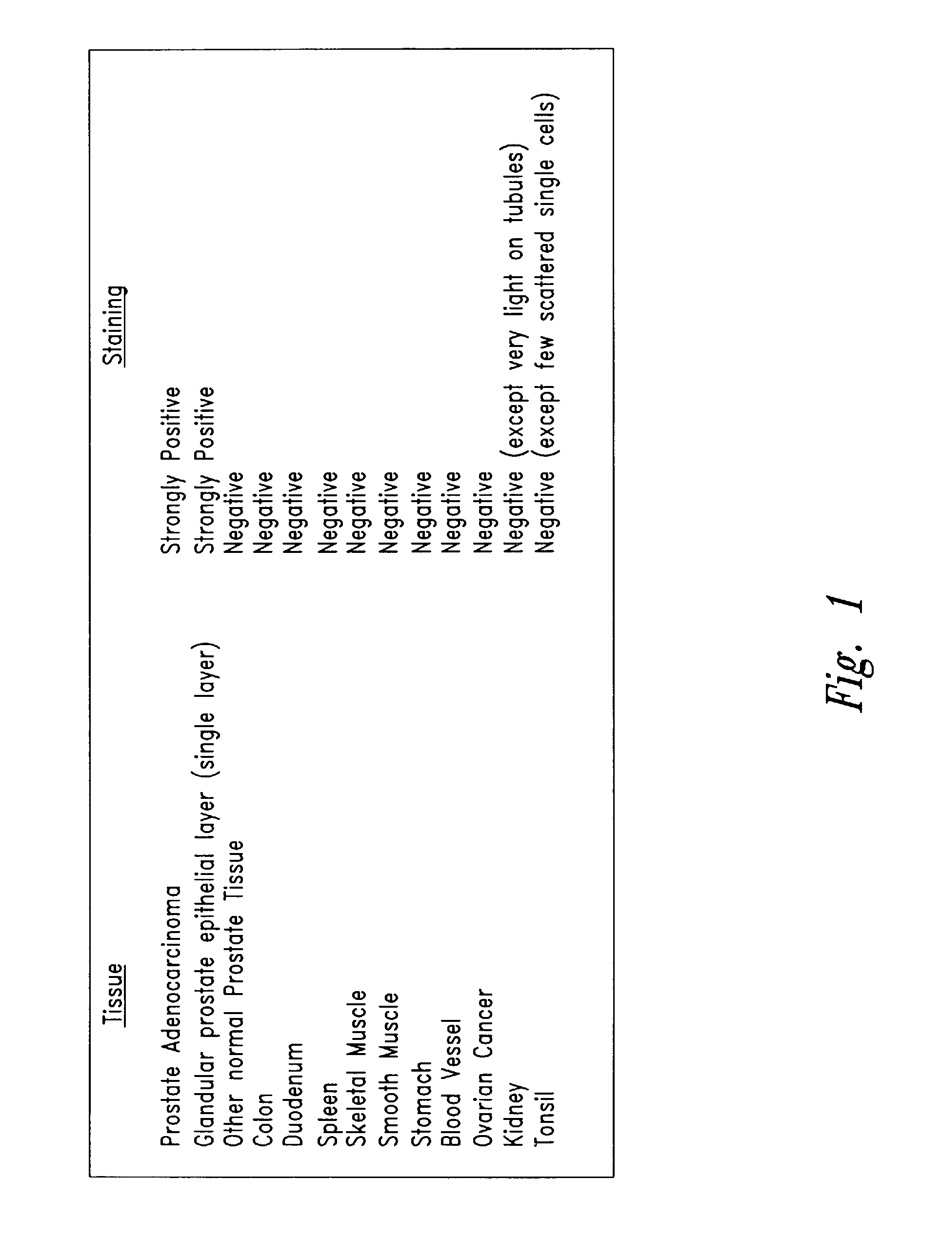
[0039]Tissue sections obtained from various human organs were directly stained with antibody NUH2. A second anti-IgM antibody chemically linked to horseradish peroxidase (HRP) further reacted with the tissue section and the location of bound antibody was observed by reaction with the FW substrate. Tissues sections were counter stained with eosin to detect location of cells in the section by a blue color. As shown in FIG. 1 most normal tissues were completely negative and did not bind antibody NUH2. Prostate adenocarcinoma, however, strongly bound NUH2 and strongly expresses the antigen, sialyl I. Normal prostate showed expression of antigen restricted to one single cell layer lining the glandular epithelium. All other cells in normal prostate were negative. This antigen is highly restricted to prostate cancer and is negative for most normal tissues tested. Over 95% of tonsil was negative, however, several scattered single cells within the ...

example 2

Expression of Prostate Cancer Antigen (Sialyl I) on Tumor Cell Lines as Determined by Cell Sorting Using Antibody NUH2

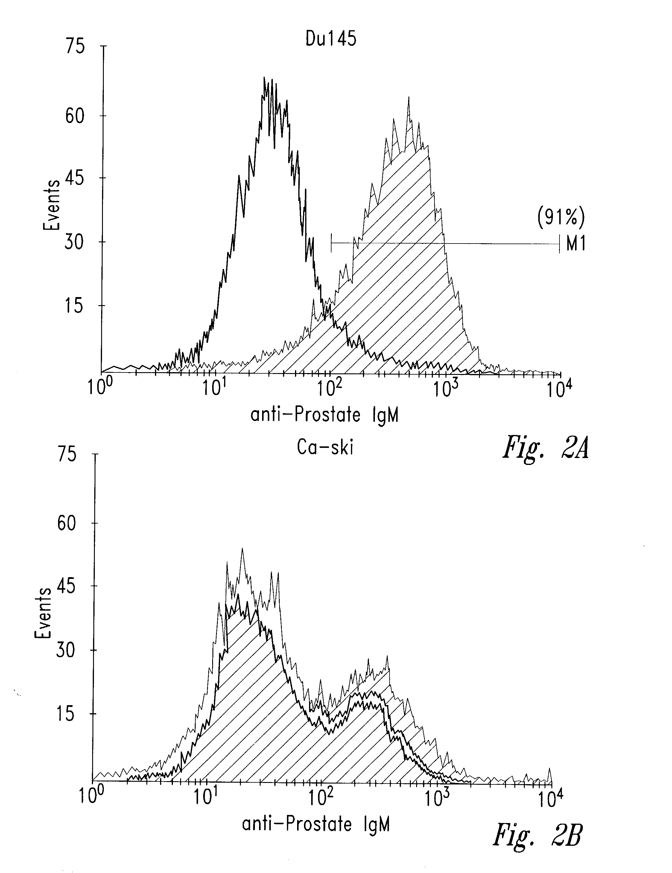
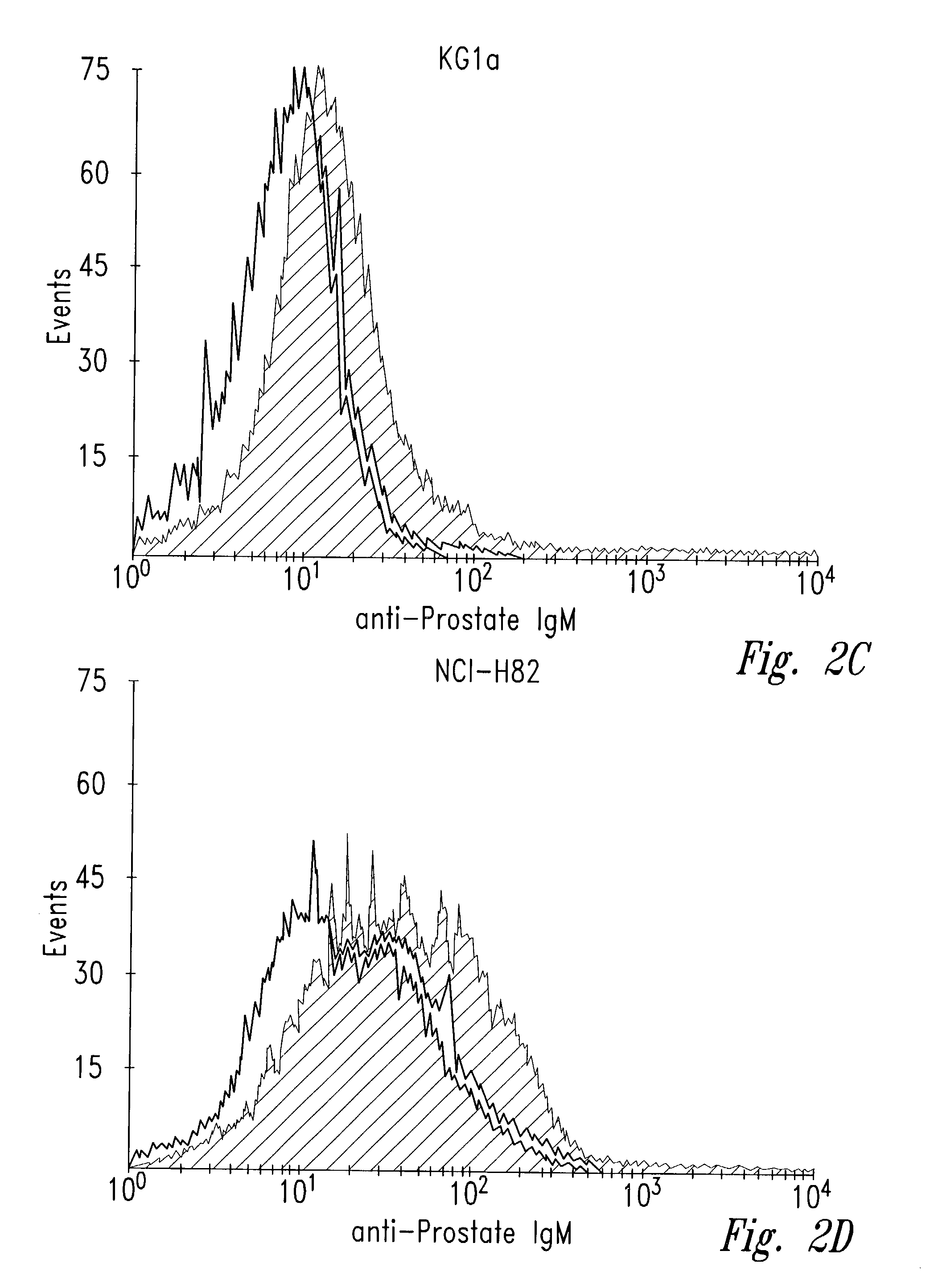
[0040]Human tumor cell lines were washed with phosphate-buffered saline (PBS) and allowed to bind to antibody NUH2 in suspension at a concentration of 10 μg / ml in PBS. A duplicate sample of the same cell line was incubated with a control IgM murine antibody in parallel as a negative control. After incubation for 1 hour at 4° C., the cells were washed with PBS and incubated with goat anti-mouse IgM coupled to FITC. The intensity of fluorescence was measured for each population by fluorescence activated cell sorter analysis (FACS). For direct visualization of the results, histograms of the same cell type reacted with either NUH2 or control antibody were superimposed (FIG. 2). Prostate cancer cell line DU145 strongly expresses sialyl I antigen, whereas most other tumor cell lines were completely negative. The lung cancer cell line NCIH446 and the colon cancer cell line ...

example 3

Immunostaining Glycolipid Antigens Directly on Thin Layer Chromatograms Using Antibody NUH2

[0043]Glycolipids were extracted from tissues using chloroform and methanol according to the Folch procedure and separated into aqueous (upper) and organic (lower) phases. The upper phase extract was dried under rotary evaporation and the glycolipids were resuspended in methanol and subjected to ion exchange chromatography using DEAE Sepharose®. Neutral glycolipids elute in the non-bound fractions while charged glycolipids were eluted with increasing concentrations of ammonium bicarbonate in methanol.

[0044]Samples from various fractions were applied to a silica gel high performance thin layer chromatography plate (HPTLC) and the chromatograms were developed in a chamber containing chloroform / methanol / 0.02%CaCl2 (5:4:1). After chromatography, the dried plate was cut into sections for chemical staining and immunostaining. Orcinol reagent was used to chemically stain a section of the plate to det...


PUM


Abstract

An antigen is shown to be associated with prostate cancer, and is useful for new methods and compositions for diagnosing or treating prostate cancer. This is particularly useful for individuals with prostate cancer who test negative for Prostate Specific Antigen. Additionally, this is useful for distinguishing between benign prostate disease and prostate cancer in a patient diagnosed or presenting with prostate dysfunction.

Description

CROSS-REFERENCE TO RELATED APPLICATION[0001]This application claims the benefit of U.S. Provisional Patent Application No. 60 / 300,250, filed Jun. 21, 2001.BACKGROUND OF THE INVENTION[0002]1. Field of the Invention[0003]The present invention relates generally to the detection or treatment of cancer, and more specifically, prostate cancer. In particular, an antigen is shown to be associated with prostate cancer, and is useful for new methods and compositions for diagnosing or treating prostate cancer including in individuals with prostate cancer who test negative for Prostate Specific Antigen (PSA).[0004]2. Description of the Related Art[0005]Each year over 300,000 men are diagnosed with prostate cancer in the U.S. alone. It is the most prevalent form of cancer for men of all races. Both the incidence of prostate cancer and its associated mortality have been increasing over the past ten years. It is estimated that about 50–65% of the prostate cancer is localized, 9–17% has spread to a...

Claims


Application Information

Patent Timeline
IPC IPC(8): G01N33/574C07K16/30A61K39/395A61K45/00A61P35/00G01N33/577
CPCC07K16/3069G01N33/57434G01N2405/10A61P35/00
Inventor MAGNANI, JOHN L.
Owner GLYCOMIMETICS
Learn More