
Method for preventing damage to or rejuvenating a cellular blood component using mitochondrial enhancer

a technology of mitochondrial enhancer and cellular blood, which is applied in the direction of biocide, other blood circulation devices, organic chemistry, etc., can solve the problems of blood supply contamination with infectious microorganisms such as malaria, west nile virus, hiv, etc., and achieves the effects of reducing the rate of lactate production, increasing oxygen consumption, and increasing the hypotonic shock respons

Inactive Publication Date: 2007-05-22
TERUMO BCT BIOTECH
View PDF331 Cites 9 Cited by
  • Summary
  • Abstract
  • Description
  • Claims
  • Application Information

AI Technical Summary

Benefits of technology

[0023]This invention also provides methods for increasing the storage life of cellular blood components, extending platelet storage life, treating a cell comprising a mitochondrion, treating a fluid comprising cells containing mitochondria to improve a quality of said fluid, said methods comprising adding an effective, substantially non-toxic amount of a mitochondrial enhancer to said fluid wherein said mitochondrial enhancer is selected from the group consisting of alloxazines, endogenous alloxazines, non-endogenous alloxazines, endogenously-based derivative alloxazines, endogenous photosensitizers, and non-endogenous photosensitizers. Fluids treatable by the methods of this invention include fluids containing living cells with mitochondria or fluids that come into contact with living cells such as peritoneal solutions, blood, and fluids comprising a blood product. Cells treatable by the methods of this invention include plant cells, animal cells, yeast cells, cellular blood components, platelets, and cells in a wound surface.

Problems solved by technology

However, it was recognized that the incidence of bacterial growth and subsequent transfusion reactions in the recipient increased to unacceptable levels with a seven-day-old platelet concentrate.
Contamination of blood supplies with infectious microorganisms such as malaria, West Nile virus, HIV, hepatitis and other viruses and bacteria presents a serious health hazard for those who must receive transfusions of whole blood or administration of various blood components such as platelets, red cells, blood plasma, Factor VIII, plasminogen, fibronectin, anti-thrombin III, cryoprecipitate, human plasma protein fraction, albumin, immune serum globulin, prothrombin complex, plasma growth hormones, and other components isolated from blood.
First, if the donor is experiencing a mild bacteremia, a condition comprising bacteria in the blood, the blood will be contaminated, regardless of the collection or storage method.
Adequate donor histories and physicals will decrease but not eliminate this problem.
Even when “sterile” methods of skin preparation are employed, it is extremely difficult to sterilize the crypts around the sweat glands and hair follicles.
Indeed, many patients requiring platelet transfusions lack host-defense mechanisms for normal clearing and destruction of bacteria because of either chemotherapy or basic hematologic disease.
The growth of even seemingly innocuous organisms in stored platelets can, upon transfusion, result in recipient reaction and death.
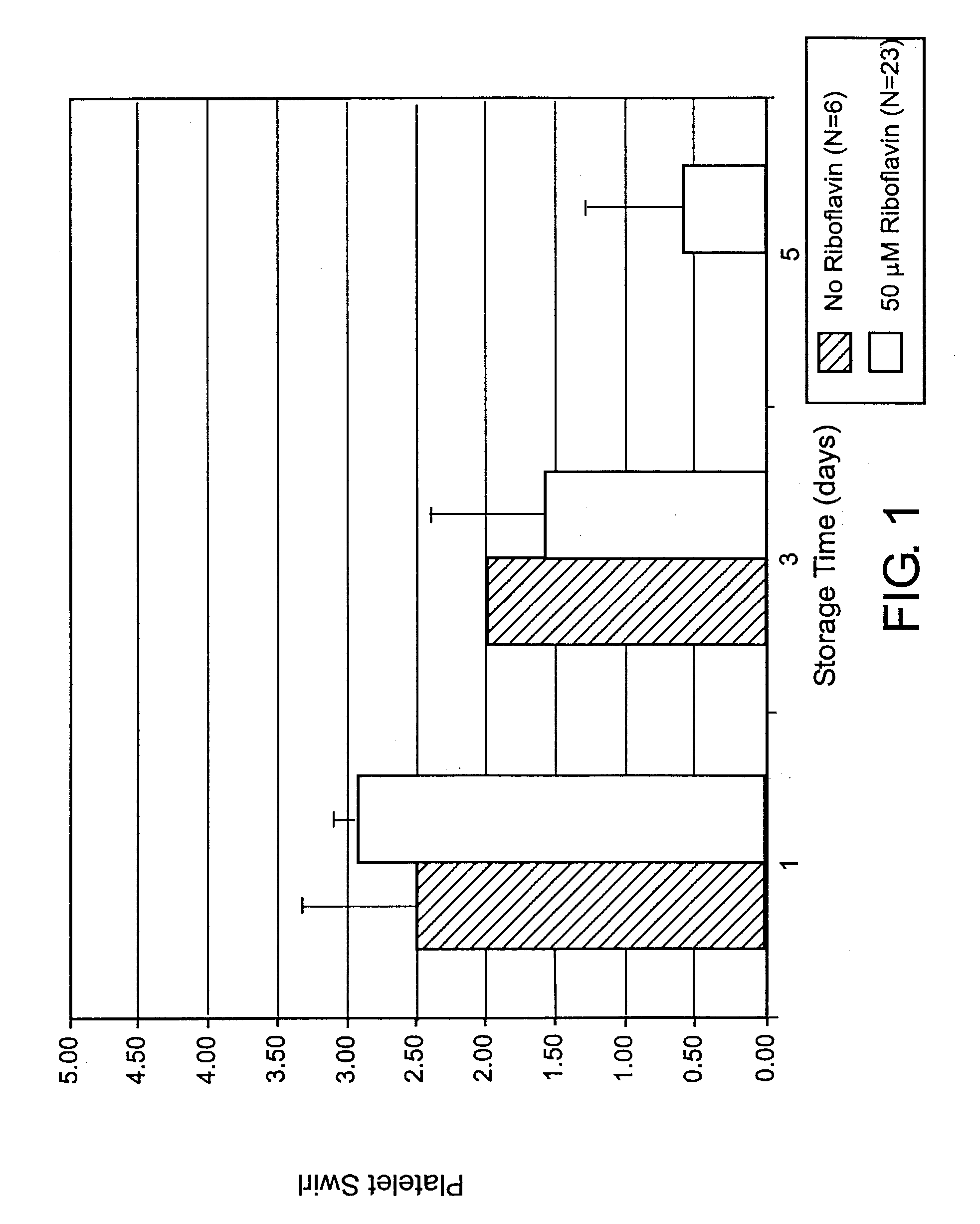
Such changes may be indications of platelet damage due to the storage conditions.
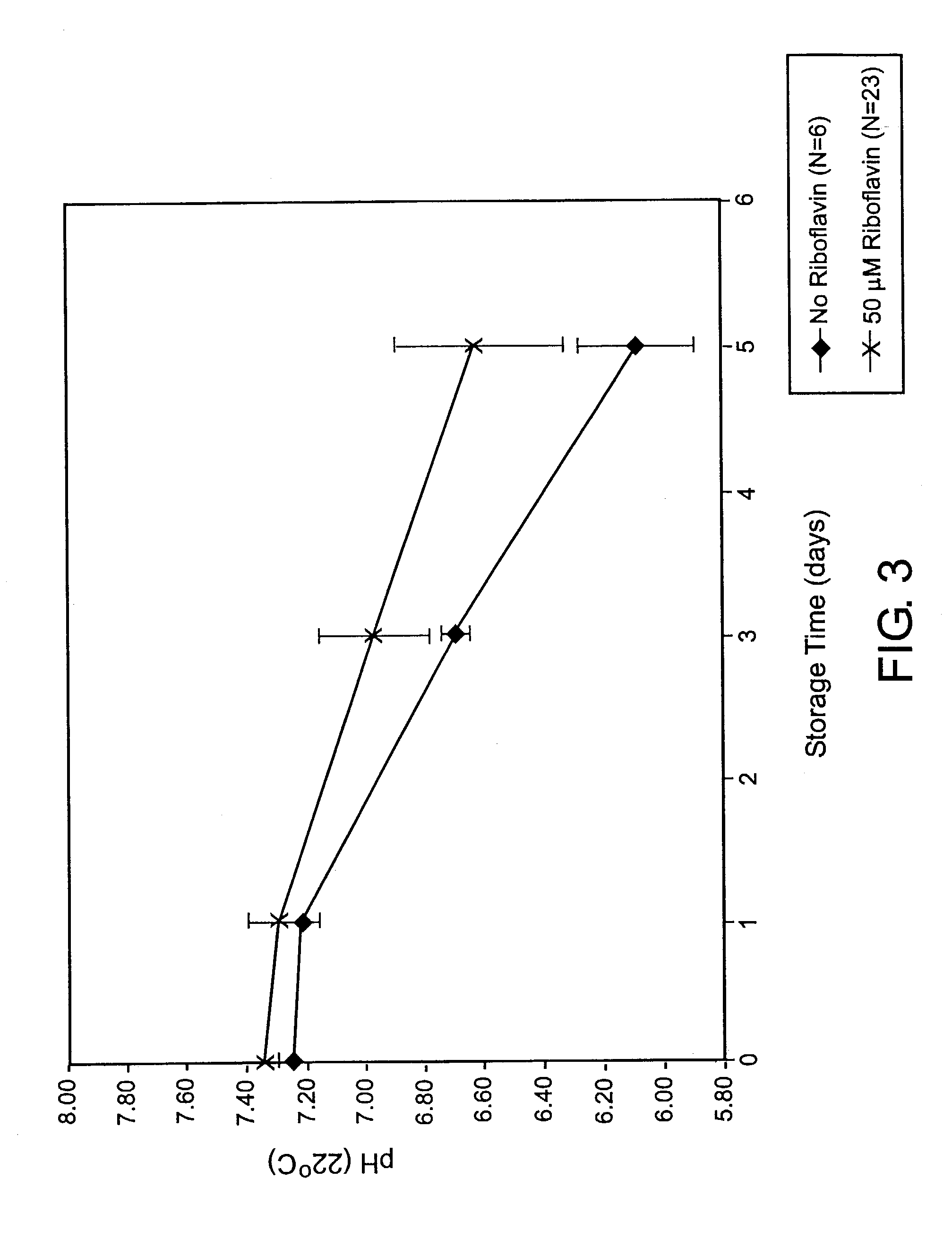
Such a drop in pH indicates as well as contributes to a decrease in cell quality during cell storage.
Currently used methods to sterilize pathogenic contaminants which may be present in blood or blood components can cause damage to the mitochondria of the cells being treated.
If this occurs, the cells can only make ATP through the glycolysis pathway, causing a buildup of lactic acid in the cell and a subsequent drop in pH during storage.
Mitochondria are sensitive to endogenous and exogenous influences and may be easily damaged or destroyed.
Dysfunctional energy metabolism and, more severely, damaged mitochondria, lead to a decline in platelet quality and eventual cell death.
Possible causes of damage to blood components may be storage, pathogen inactivation, and pathogen reduction processes.
It has been observed that a side effect of a pathogen reduction process is that when platelets are subjected to UV light, the mitochondria of the platelets have a greater chance of suffering at least some damage than when they have been subjected to visible light.
A substantial byproduct of ATP generation is the formation of potentially toxic oxygen radicals.
Furthermore, photochemical methods for pathogen inactivation and reduction of blood products which generate singlet oxygen species in the process of photolysis of the photosensitizer cause further damage to mitochondrial membranes.

Method used


Image

  • Method for preventing damage to or rejuvenating a cellular blood component using mitochondrial enhancer
  • Method for preventing damage to or rejuvenating a cellular blood component using mitochondrial enhancer
  • Method for preventing damage to or rejuvenating a cellular blood component using mitochondrial enhancer

Examples


example 1

Treatment of Platelets with Riboflavin

[0118]Platelets were collected using standard collection methods using a COBE® Spectra™ apheresis machine (manufactured by Gambro BCT, Lakewood, Colo., USA). Fresh platelets were less than 24 hours old after collection via apheresis. Other apheresis machines useful for collecting transfusion-quality platelets are useful in the practice of this invention. Collected platelets were diluted in a solution containing 0.9% sodium chloride with either 0 or 10 μM riboflavin. Samples were saturated with air via vigorous mixing. Platelets were exposed to no photoradiation greater than ambient light. Results are shown in Table 1.

[0119]

TABLE 1O2ConsumptionLactateGlucose(μM / hr / 1012RiboflavinPhotoradiationProductionConsumptionplts)pHConcentrationEUVEVISRateRateDayDay(μM)J / cm2J / cm2(μM / hr)(μM / hr)024 hrs00003118921141000291913612400050876.921000681866.91

Cell quality indicators were improved or unaffected by the addition of riboflavin.

example 2

Treatment of Platelets with Riboflavin and Visible Light

[0120]Platelets were collected using standard collection methods using a COBE® Spectra™ apheresis system (manufactured by Gambro BCT, Lakewood, Colo., USA) and TRIMA® apheresis system (available from Gambro BCT, Lakewood, Colo., USA). Fresh platelets were less than 24 hours old after collection via apheresis. Other apheresis machines useful for collecting transfusion-quality platelets are useful in the practice of this invention. Collected platelets were diluted in a solution containing 0.9% sodium chloride with either 0 or 10 μM riboflavin. Samples were saturated with air via vigorous mixing. The samples were irradiated with a Dymax light source using one bulb at 15.5 mW / cm2 for varying times. The UV component of the light was filtered out using a polycarbonate filter. Exposure time was 31 minutes. Results are shown in Table 2.

[0121]

TABLE 2O2ConsumptionLactateGlucose(μM / hr / 1012RiboflavinPhotoradiationProductionConsumptionplts)...

example 3

Treatment of Platelets with Riboflavin and Ultraviolet and Visible Light

[0122]Platelets were collected using standard collection methods using a COBE® Spectra™ apheresis machine (manufactured by Gambro BCT, Lakewood, Colo., USA). Fresh platelets were less than 24 hours old after collection via apheresis. Other apheresis machines useful for collecting transfusion-quality platelets are useful in the practice of this invention. Collected platelets were diluted in a solution containing 0.9% sodium chloride with either 0 or 10 μM riboflavin. Samples were saturated with air via vigorous mixing. The samples were irradiated with a Dymax light source using one bulb at 15.5 mW / cm2 for varying times. Results are shown in Tables 3 and 4.

[0123]

TABLE 3O2ConsumptionLactateGlucose(μM / hr / 1012RiboflavinPhotoradiationProductionConsumptionplts)pHConcentrationEUVEVISRateRateDayDay(μM)J / cm2J / cm2(μM / hr)(μM / hr)024 hrs002940327115200102940603955111

When riboflavin was added, lactate production rate was decre...


PUM

PropertyMeasurementUnit
Fractionaaaaaaaaaa
Fractionaaaaaaaaaa
Timeaaaaaaaaaa

Abstract

This invention provides methods for treating cellular blood components and other cells containing mitochondria to improve vital qualities of the cells by contacting the cells with a mitochondrial enhancer to the cells. Mitochondrial enhancers prevent damage to and rejuvenate mitochondria and cells containing mitochondria. Mitochondrial enhancers include alloxazines and related compounds, such as riboflavin. Cells are optionally treated with photoradiation to reduce pathogens with may be present, before, after, and / or during treatment with mitochondrial enhancer. Treating with mitochondrial enhancer enables utilization of higher photoradiation energies, which achieves better pathogen reduction. When platelets are treated with mitochondrial enhancer, treated platelets may be stored for longer times than untreated platelets before they are administered to patients.

Description

CROSS-REFERENCE TO RELATED APPLICATIONS[0001]This application is a continuation-in-part of U.S. application Ser. No. 09 / 586,147, filed Jun. 2, 2000 now abandoned which is a continuation-in-part of U.S. application Ser. No. 09 / 357,188, filed Jul. 20, 1999, now U.S. Pat. No. 6,277,337, issued Aug. 21, 2001, which is a continuation-in-part of U.S. application Ser. No. 09 / 119,666, filed Jul. 21, 1998, now U.S. Pat. No. 6,258,577, issued Jul. 10, 2001. This application is a continuation-in-part of U.S. application Ser. No. 09 / 777,727, filed Feb. 5, 2001 now U.S. Pat. No. 6,828,323, which is a continuation of U.S. application Ser. No. 09 / 420,652, filed Oct. 19, 1999, now U.S. Pat. No. 6,268,120, issued Jul. 31, 2001. This application claims priority to U.S. provisional application Ser. No. 60 / 378,374 filed May 6, 2002. The documents cited above are incorporated herein by reference to the extent not inconsistent herewith.BACKGROUND OF THE INVENTION[0002]Whole blood collected from volunteer...

Claims


Application Information

Patent Timeline
IPC IPC(8): A01N43/60C07D239/00A61K31/525A61K35/14A61K35/19A61K41/00A61L2/00A61L2/08A61L2/10A61M1/02A61M1/36
CPCA61K31/525A61K35/19A61K41/0019A61L2/0005A61L2/0011A61L2/0082A61L2/0088A61L2/08A61L2/10A61M1/3681A61M1/0213A61M1/3683A61M1/3693A61M2202/0413A61M2205/123A61M2202/0078A61M2202/0427A61M2205/051A61K41/17
Inventor DUMONT, LARRY JOEGOODRICH, JR., RAYMOND P.
Owner TERUMO BCT BIOTECH
Learn More