Ultrasound contrast agents

a contrast agent and ultrasonic technology, applied in ultrasonic/sonic/infrasonic diagnostics, echographic/ultrasound-imaging preparations, applications, etc., can solve the problems of preventing the left side of the heart from being useful for many purposes, and affecting the use of the left side of the heart. , to achieve the effect of low attenuation, large acoustic changes, and high reflectivity

Inactive Publication Date: 2008-05-20
LIEBEL FLARSHEIM CO
View PDF25 Cites 0 Cited by
  • Summary
  • Abstract
  • Description
  • Claims
  • Application Information

AI Technical Summary

Benefits of technology

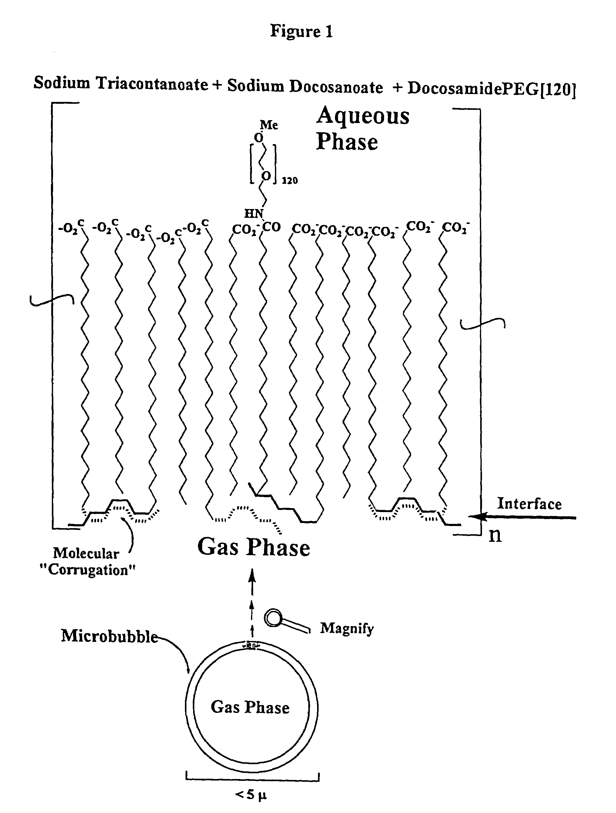
[0029]The microbubbles made using the above compounds are useful as ultrasound contrast agents in diagnostic and therapeutic medical procedures because they have exceptionally high reflectivity, have very low attenuation, and can exhibit large acoustic changes with frequency for only a slight pressure variation. They are stable during the manufacturing and sterilization process and are stable for long storage periods. Because of their inherit low atte...

Problems solved by technology

Although non-encapsulated gas microbubbles may be used for some purposes, they tend to change size very rapidly.
Those that are small enough to pass through the lungs are effectively to small and dilute to provide useful contrast upon reaching the heart and are thus not practical for imaging the left side of the heart.
These known agents are satisfactory for some applications but have limitations that prevent them from being useful for many purposes.
They are often not stable when subjected to sterilization procedures and have relatively short half-lives after formulation or reconstitution.
Also, the physical properties of the microbubbles at effective concentrations limit the depth in the tissue where a diagnostic image can be obtained.
Often useful images can only be acquired for tissue a few centimeters from the ultrasound transmitter/receiver thus limiting the “penetration” of the procedure and the depth in the body from which images can be obtained.
Further, the physical properties of the microbubbles cause relatively high attenuation that limits the usefulness of the agent to obtain certain types of images, e.g. myocardial perfusion and images that must be obtained immediately after injection.
Additionally, kno...

Method used


Image

  • Ultrasound contrast agents
  • Ultrasound contrast agents
  • Ultrasound contrast agents

Examples


experimental examples

Example 1

Synthesis of Methoxypolyoxyethylene(˜5,000)docosamide-{DocosamidePEG(5,000), (C21CONPEG[120])}

[0090]Docosanoic acid (0.203 g, 5.96×10⁻⁴ mol, Aldrich®) was weighed into a round bottomed flask and dissolved into anhydrous chloroform (5 mL, Aldrich®) and hexanes (1 mL, Aldrich®). To the solution was added excess dicyclohexylcarbodiimide (0.15 g, 7.0×10⁻⁴ mol, DCC, Aldrich®). After the solution was stirred a few minutes, an equivalent of N-hydroxysuccinimide (0.081 g, 7.0×10⁻⁴ mol, Aldrich®, 97%) was added. The docosanoyl-N-succinimide ester was filtered through a small plug of glass wool contained in a pipette to remove the byproduct urea after stirring for a two hours. The N-succinimide ester was introduced directly into a concentrate of methoxypolyethylene(˜5,000) amine (3.01 g, 6.0×10⁻⁴ mol, Sigma®) in toluene left over from the distillation to remove any water. The reaction medium was stirred overnight under nitrogen (Air Products, High Purity Grade). The solution was refl...

example 2

Synthesis of N,N-Bis[2-(docosanoyl)oxyethyl]aminoacetic acid

[0091]Bicine (0.95 g, 5.82×10⁻³ mol, Aldrich®, 99+%) was added to chloroform (30 mL, anhydrous, Aldrich®, 99+%). A concentrate of docosanoyl chloride, prepared by the reaction of docosanoic acid (4.12 g, 1.14×10⁻² mol, Aldrich®) with excess oxalyl chloride (Sigma) in chloroform, was added dropwise to the suspension. After stirring for a few minutes, excess pyridine (1.5 mL, anhydrous, Aldrich®, 99+%) was added to the flask. The flask was refluxed overnight. The reaction mixture was acidified with dilute HCl saturated with sodium chloride (Mallinckrodt, AR®). The aqueous phase was separated. Any remaining volatiles were removed from the organic phase under reduced pressure with mild heating. The glassy product was recrystallized from ethanol (Mallinckrodt, AR®). The product was recovered by filtration as an amorphous white solid. The product weighed 2.86 g after vacuum drying for a 61% yield; mp 73-75° C.; Rf=0.23 (silica ge...

example 3

Synthesis of 1-N′,N′-Bis[2-(docosanoyl)oxyethyl]aminoacetamide-3-N-[methoxypolyethylene glycol(˜5,000)], (C22BicinePEG[120])

[0092]N,N-Bis[2-(docosanoyl)oxyethyl]aminoacetic acid (0.221 g, 2.73×10⁻⁴ mol) was weighed into a round bottomed flask and dissolved into chloroform (7 mL, anhydrous, Aldrich®, 99+%). To the solution was added dicyclohexylcarbodiimide (DCC, 0.0625 g, 3.03×10⁻⁴ mol, Aldrich®). After stirring for about ten minutes, N-hydroxysuccinimide (0.0345 g, 3.03×10⁻⁴ mol, Aldrich®, 97%) was added. Stirring was continued for two hours. The N-hydroxysuccinimide ester was filtered through a small plug of glass wool contained in a pipette to remove the byproduct urea and introduced directly into a concentrate of methoxypolyethylene(˜5,000) amine (1.52 g, 3.03×10⁻⁴ mol, Sigma®) in toluene left over from the distillation to remove any water. The solution was stirred overnight under nitrogen. The solution was heated to reflux under a flow of nitrogen for one hour. The product was ...


PUM


Abstract

New ultrasound contrast agents that comprise microbubbles encapsulating a gas within a shell made from a blend of bipolar compounds having mixed carbon chain length. The compounds have hydrophobic groups selected from the group consisting of straight-chained alkyls, alkylethers, alkylthiolethers, alkyldisulfides, polyfluoroalkyls, and polyfluoroalkylethers having a carbon chain length greater than or equal to 16 and less than or equal to 32. The polar groups are connected to the hydrophobic groups by means of a linker. The polar head groups are selected from the group consisting of CO2-M+, SO3M+, SO4M+, PO3M+, PO4M+2, N(R)4+, a pyridinium or substituted pyridinium group, and a zwitterionic group; R is selected from the group consisting of —H, —CH3, alkyl, cycloalkyl, substituted cycloalkyls containing one or more heteroatoms, and benzyl and can be the same or different; and Z′ is a nonionic and M is a cation.

Description

APPLICATION CROSS-REFERENCE[0001]This application is a continuation application of prior U.S. patent application Ser. No. 09 / 806,680 filed on Apr. 3, 2001, now abandoned which is a national phase application under 35 U.S.C. Section 371 of International Application No. PCT / US99 / 23685 filed Oct. 12, 1999, which claims benefit of U.S. Provisional Patent Application No. 60 / 103,860, filed Oct. 12, 1998.BACKGROUND OF THE INVENTION[0002]Ultrasound contrast is a very useful and widely used medical diagnostic technique. The technique takes advantage of the fact that the various fluids and tissues in the body reflect sound waves differently. This results in a contrast between reflected waves that can be detected and used to form an image of the tissue. Ultrasound is used for many different diagnostic purposes, e.g., prenatal imaging or to image bloodflow in the heart and arteries and observe blockages in blood circulation.[0003]It has been discovered that an ultrasound image can be greatly en...

Claims


Application Information

Patent Timeline
IPC IPC(8): A61B8/00A61K49/00A61K9/50A61K49/22
CPCA61K49/223A61K9/5026
Inventor CANTRELL, GARY L.
Owner LIEBEL FLARSHEIM CO
Why Eureka
Try Eureka