Methods for producing and using silk nanofiber nerve conduits

a technology of silk nanofiber and nerve conduit, which is applied in the direction of filament/thread forming, prosthesis, surgery, etc., can solve the problems of inability to apply a specific length of nerve, inability to achieve motor skills, sensory paralysis, etc., and achieve excellent biocompatibility, excellent nerve regeneration effect, and recovery of motor skills and sensory functions

Active Publication Date: 2015-07-07
SEOUL NAT UNIV R&DB FOUND
View PDF13 Cites 2 Cited by
  • Summary
  • Abstract
  • Description
  • Claims
  • Application Information

AI Technical Summary

Benefits of technology

[0015]Silk nanofiber nerve conduit of the present invention has excellent biocompatibility; allows the body fluid to be exchanged in and out of conduit through pores of the conduit; and has a proper elasticity, tensile strength, and tear strength. Due to these properties, the silk nanofiber nerve conduit of the present invention helps the regeneration of the nerve injury to recover a motor skill and a sensory function, and shows an excellent effect of nerve regeneration. Therefore, the silk nanofiber nerve conduit of the present invention may be used in treating a nerve injury as a replacement of an existing synthetic polymeric nerve conduit.BRIEF DESCRIPTIONS OF DRAWINGS
[0016]FIG. 1 presents an image of silk nanofiber nerve conduit according to the present invention;
[0017]FIG. 2 presents electro-microscopic images of silk nanofiber nerve conduit according to the present invention, in which ((a) is a cross-section image, (b) is a ×100 magnified cross-section image, (c) is a ×1000 magnified cross-section image, and (d) is a fiber structure of silk nanofiber nerve conduit surface);
[0018]FIG. 3 presents electro-microscopic images of inside of the nerve conduit after 10 weeks from when silk nanofiber nerve conduit was autografted into mouse, in which ((a) is inside of the nerve conduit, (b) is an magnified image of (a) in which an arrow indicates regenerated nerve bundle, (c) is an image magnified the cross section of regenerated nerve bundle indicated by an arrow in (b), (d) is an image magnified the side of regenerated nerve bundle indicated by an arrow in (b)); and
[0019]FIG. 4 presents a histological analyzed image of inside of the nerve conduit after 10 weeks from when silk nanofiber nerve conduit was autografted into mouse acquired by ((a) hematoxylin-eosin staining as basic staining, (b) Luxol fast blue staining image specifically staining nerve bundle, and (c) is an image magnified

Problems solved by technology

The injury of nerve tissue results in incapacity of a motor skill and sensory paralysis of the muscle which used to be controlled by the injured nerve.
This method cannot be applied to the specific length of nerve which is often treated in the practical operations.
Also, this method has difficulty for fitting the thickness and shape of the autografted nerve tissue into those of the injured nerve tissue.
In this regard, the usefulness of these methods is not trustworthy.
First, the method can block penetration of scar tissue which disturbs nerve generation from the environment.
However, if body tissues are used as the materials of nerve conduit, the supply of the tissues are not enough and if body tissues are not obtained by autograft, the usage of nerve conduit is limited due to an immune reaction.
Also, if natural polymers are used, the polymers are easily absorbed in the body and the properties are hard to control.
Accordingly, natural polymers have many unsolved problems to be used as a material of nerve conduit.
In clinics, non-degradable silicon tube is commonly used, but since this tube is remained in the body even though nerves are regenerated, it causes disadvantages such as chronic inflammation, calcium products of silicon tube and several pains originated from the pressed regenerated nerves; therefore, use thereof has been gradually decreasing.
Among the currently available nerve conduits in the market, although a collagen-based nerve conduit has excellent biodegradability, the nerve conduit has following problems.
Preparation and storage of the collagen is complicate since the collagen of the nerve conduit has to be extracted from animals.
Producing large quantity of the collagen is not suitable and the cost of the nerve conduit is very expensive.
These disadvantages limit utilizing the nerve conduit in clinics.

Method used


Image

  • Methods for producing and using silk nanofiber nerve conduits
  • Methods for producing and using silk nanofiber nerve conduits
  • Methods for producing and using silk nanofiber nerve conduits

Examples


example 1

Producing Silk Nanofiber Nerve Conduit

[0041]Step 1: Preparing Fibrous Spinning Solution

[0042]40 g of dried cultivated cocoon was submerged in 1 L of mixed solution wherein 0.3% of sodium oleate and 0.2% (w / v) of sodium carbonate was dissolved, heated to boil for 1 hour, and washed repeatedly to remove sericin. The sericin-removed cocoon was dried again and dissolved in mixed solution at the ratio of 1 / 20 at 85° C. for 3 min, wherein the mixed solution included calcium chloride / water / ethanol at mole ratio of 1 / 8 / 2. This dissolved solution was placed in the dialyzing diaphragm (MWCO: 12-14 kDa) and underwent dialysis with distilled water for 3 days, and the silk fibroin solution having approximately 2% (w / w) concentration was obtained. After the solution was lyophilized, silk fibroin sponge which was appropriate for the secondary dissolution was prepared and perfectly dissolved in 13% concentration of the formic acid (over 98% purity); therefore, fibrous spinning solution was prepared...

experiment example 1

Measurement of Structural Characteristics

[0047]The following experiment was performed in order to find out the structural characteristics of silk nanofiber nerve conduit of the present invention.

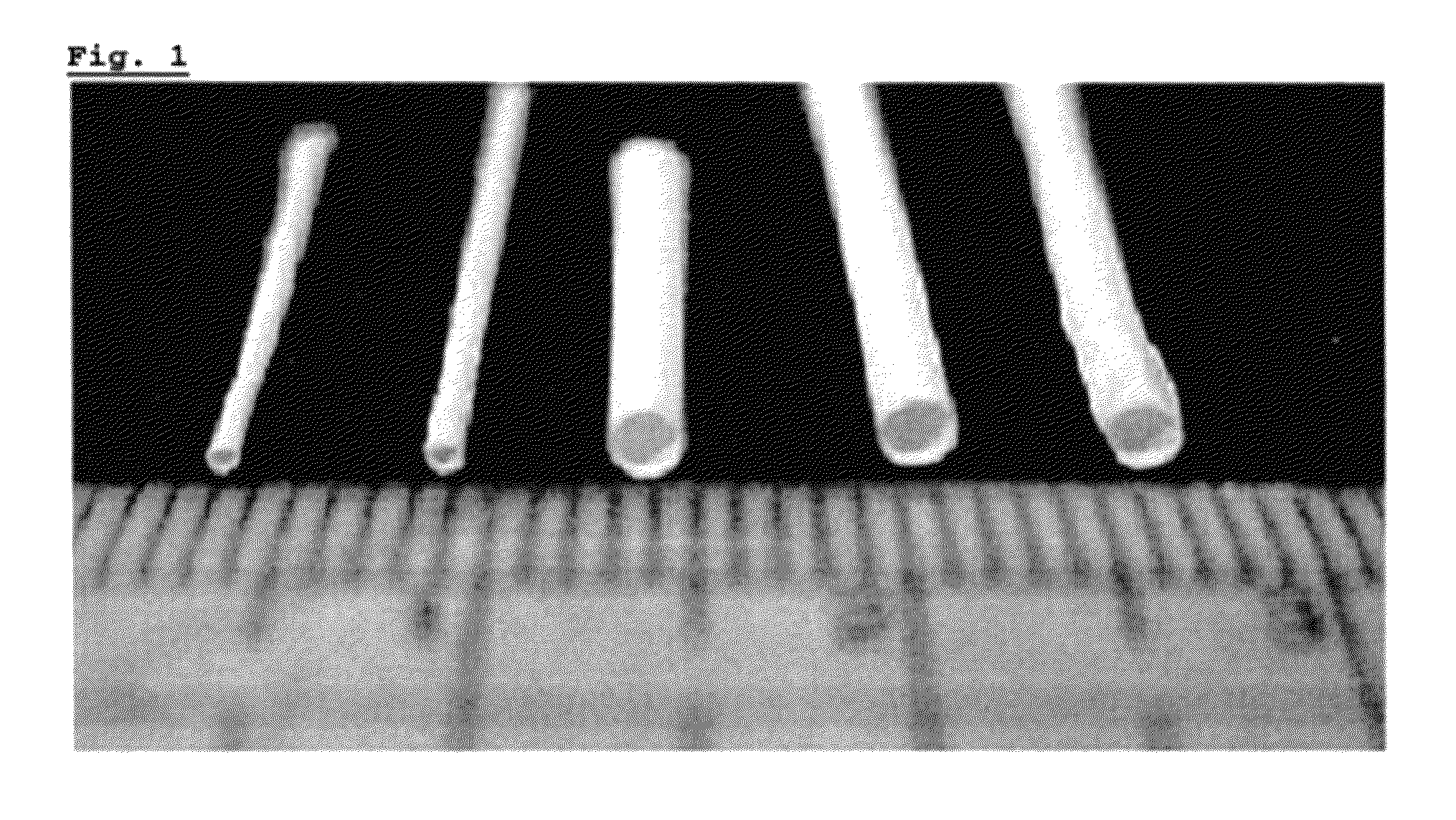
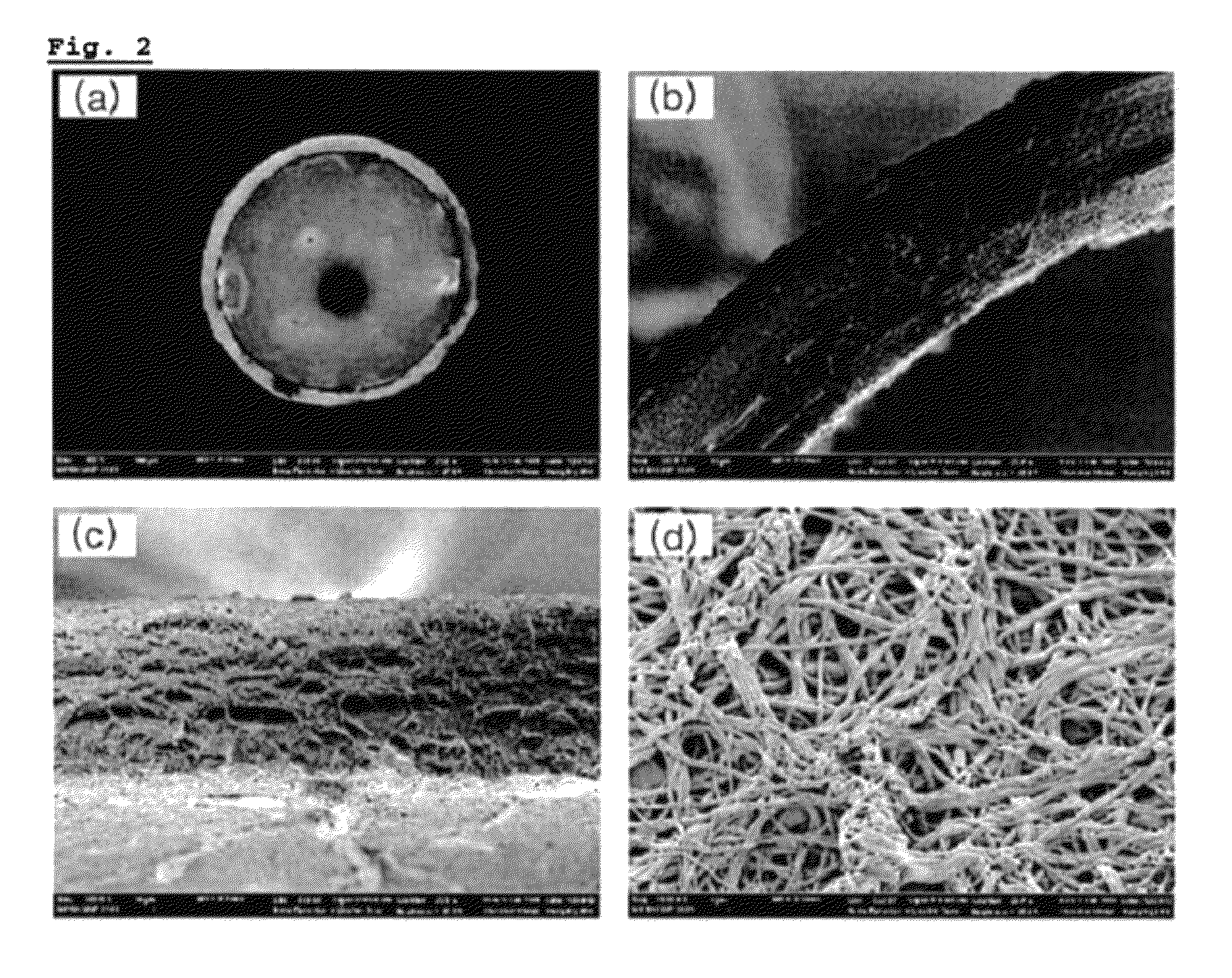
[0048]FIG. 1 presents photographic images of silk nanofiber nerve conduit of the Example 1. FIG. 2 presents electro-microscopic images of silk nanofiber nerve conduit regarding the present invention. For more details, (a) in FIG. 2 presents the cross-section of silk nanofiber nerve conduit, (b) and (c) of FIG. 2 present magnified cross section images, and (d) of FIG. 2 presents the fabric structure of the silk nanofiber nerve conduit surface.

[0049]According to FIG. 1, silk nanofiber nerve conduit of Example 1 of the present invention had circle-shaped cross section. The inside of the silk nanofiber nerve conduit was hollow and the thickness of the conduit was approximately 0.2 mm.

[0050]According to the (a),(b),(c) and (d) of FIG. 2, the cross section of silk nanofiber nerve conduit of Exampl...

experiment example 2

Measurement of Mechanical Property

[0052]The following experiment was performed in order to recognize the mechanical property of silk nanofiber nerve conduit of the present invention.

[0053]In order to test tensile strength of the nerve conduit, the conduit having 4 cm length was prepared. Inner diameter of the nerve conduit was 1.65 mm and the thickness of conduit wall was average 200 μm. Therefore, cross sectional area was 1.16 mm2([{(1.65+0.4) / 2}2−(1.65 / 2)2]*π). Tensile strength test was performed after submerging the conduit in normal saline solution for 1 hour. The ending of the sample which was swollen in the solution was fixed on test device. With 2 cm gauge length and 10 mm / min speed, the strength and elongation of severance was measured until desired position.

[0054]The result is as shown in Table 1 below.

[0055]

TABLE 1Example 1Cutting elongation (%)243 ± 15Cutting strength (MPa) 4.92 ± 0.07Yield point (%)20

[0056]As referred in Table 1, elongation of severance was approximately...


PUM

PropertyMeasurementUnit
outer diameteraaaaaaaaaa
sizeaaaaaaaaaa

Abstract

The present invention relates to a silk nanofiber nerve conduit characterized in that fibroin nanofibers having a diameter of 200 to 400 nm, originated from silk fiber, are stacked layer upon layer to form a porous conduit-shape; and a method for producing thereof, more specifically, to a method for producing a silk nanofiber nerve conduit comprising: (Step 1) preparing a fibrous spinning solution; (Step 2) producing a silk nanofiber of conduit-shape by electrospinning the fibrous spinning solution prepared in step 1 into the cylindrical collecting part coated with polyethyleneoxide; and (Step 3) separating a silk nanofiber of conduit-shape produced in step 2 from the collecting part. The silk nanofiber nerve conduit of the present invention has excellent biocompatibility; allows the body fluid to be exchanged inter in and out of conduit through pores of the conduit, as well; has a proper elasticity, tensile strength, and tear strength. Due to these properties, the silk nanofiber nerve conduit of the present invention helps the regeneration of the nerve injury to recover a motor skill and a sensory function, and thus shows an excellent effect of nerve regeneration. Therefore, the silk nanofiber nerve conduit of the present invention can be used in treating a nerve injury instead of an existing synthetic polymeric nerve conduit.

Description

CROSS-REFERENCE TO RELATED APPLICATIONS[0001]This application is a U.S. National Stage Application under 35 U.S.C. §371 of International Application No. PCT / KR2010 / 005280, filed Aug. 11, 2010, which claims the benefit of Korean Application No. 10-2009-0074161, filed Aug. 12, 2009. Both of these applications are hereby incorporated by reference in their entireties.TECHNICAL FIELD[0002]The present invention relates to a silk nanofiber nerve conduit characterized in that fibroin nanofibers having a diameter of 200-400 nm, originated from silk fiber, are stacked layer upon layer to form a porous conduit-shape and a method for producing thereof.BACKGROUND ART[0003]Injury of nerve tissue is mainly caused by car accidents emerged from the development of the transportation, cancer according to the environmental pollution and side effects of the operations related to nerve system (B. Schlosshauer et al., “Rat Schwann cells inbioresorbable nerve guides to promote and accelerate axonal regener...

Claims


Application Information

Patent Timeline
Patent Type & Authority Patents(United States)
IPC IPC(8): D01D5/06A61L27/36D01F4/02D01D5/00D01D1/06A61F2/04D01D1/02A61L27/56
CPCA61F2/04D01D1/02D01D1/06D01D5/003D01D5/0046D01D5/0076D01F4/02A61L27/3604A61L27/56A61L2400/12A61L2430/32A61F2/02
Inventor PARK, YOUNG HWANKI, CHANG SEOKKIM, HYUN JEUNGPARK, SOOK YOUNG
Owner SEOUL NAT UNIV R&DB FOUND
Why Eureka
Try Eureka