Supplemented and unsupplemented tissue sealants, methods of their production and use

a technology of tissue sealant and supplement, which is applied in the direction of powder delivery, peptide/protein ingredients, and macromolecule non-active ingredients, etc. it can solve the problems of inconsistent use of fgf growth factor to promote wound healing, inability to establish if fgf growth factor is chemotactic for fibroblasts, and inability to possess true wound healing properties. , to achieve the effect of increasing the longevity and stability of fg, increasing stability, and increasing the shelf li

Inactive Publication Date: 2006-07-18
AMERICAN NAT RED CROSS
View PDF154 Cites 33 Cited by
  • Summary
  • Abstract
  • Description
  • Claims
  • Application Information

AI Technical Summary

Benefits of technology

[0095]A sixth advantage is that the AB-supplemented FG of this invention, such as TET-FG, has unexpectedly increased the longevity and stability of the FG compared to that of the unsupplemented FG. This increased stability continues even after appreciable quantities of the AB are no longer remaining in the FG. For example, soaking a newly formed FG clot in a saturated solution of TET produced from free base TET, or in a solution of CIP HCl, produces a FG clot which is stable and preserved even after substantially all the TET or CIP has left the FG clot. While not wishing to be bound by any theory as to how this effect is produced, it is believed that the AB, such as TET or CIP, inhibits plasminogen which is in the TFC and breaks down the FG. Once the plasminogen is inhibited, its continued inhibition does not appear to depend on appreciable quantities of the TET or CIP remaining in the FG. As a result of this stabilizing effect, one can expect an increased storage shelf life of the TS, and possi

Problems solved by technology

However, it has not been established if any FGF growth factor is chemotactic for fibroblasts.
However, their use to promote wound healing has yielded inconsistent results (see, e.g., Carter et al., in Growth Factors and Other Aspects of Wound Healing: Biological and Clinical Implications, Alan R. Liss, Inc., New York, N.Y., pp.
The reasons for such inconsistent results are not known, but might be the result of difficulty in applying growth factors to a wound in a manner in which they can exhibit their normal array of biological activities.
However, this heat inactivation method may produce denatured proteins in the FG which may also be allergenic.
In addition, there is concern that this inactivation method will not inactivate prions which cause bovine spongiform encephalopathy, “mad cow disease,” which may be present in the TS due to the use of bovine proteins therein.
Although FG maintains hemostasis and reduces blood loss, it has not yet been shown to possess true wound healing properties.
Unfortunately, DBM materials have little clinical use unless combined with particulate marrow autografts.
There is a limit to the quantity of DBM that can be surgically placed into a recipient's bone to produce a therapeutic effect.
Soft-tissue collapse into the wound bed may likewise inhibit the proper migration of osteocompetent stem cells into the wound bed.
However, DBM in powder form is difficult to use.
Therefore, these results are inconsistent and confusing.
However, commercially available preparations of FG and other TSs are too dense to allow cell migration into and through them.
This limits their effectiveness in some in vivo uses.
However, the source of bone autografts is usually limited and the use of allogeneic bones involves a high risk of viral contamination.
However, one general problem with these techniques is that nonautologous cells

Method used


Image

  • Supplemented and unsupplemented tissue sealants, methods of their production and use
  • Supplemented and unsupplemented tissue sealants, methods of their production and use
  • Supplemented and unsupplemented tissue sealants, methods of their production and use

Examples


example 1

Preparation of HBGF-=1 for Supplementation of FG

[0249]An 800 ml culture of recombinant E. coli containing a plasmid that included DNA encoding HBGF-1β was prepared. After induction and culturing for 24 hours at 37° C., the cells were centrifuged and the supernatant was discarded. The cell pellet was resuspended in 25 mls of 20 mM phosphate buffer, containing 0.15 M NaCl, pH 7.3. The suspended cells were disrupted with a cell disrupter and the cell debris was separated from the resulting solution by centrifugation at 5000 g for 20 min.

[0250]The pellet was discarded and the supernatant containing the solubilized HBGF-1β and other bacterial proteins was loaded onto a 2.6 cm diameter by 10 cm high column of Heparin-Sepharose™ (Pharmacia Fine Chemicals, Upsala, Sweden). The column was washed with 5 column volumes of 0.15 M NaCl in 20 mM phsophate buffer, pH 7.3, and then was eluted with a 0.15 M NaCl in 20 mM phosphate buffer to 2.0 M NaCl gradient.

[0251]The eluate was monitored by UV ab...

example 2

Stability of HBGF-1

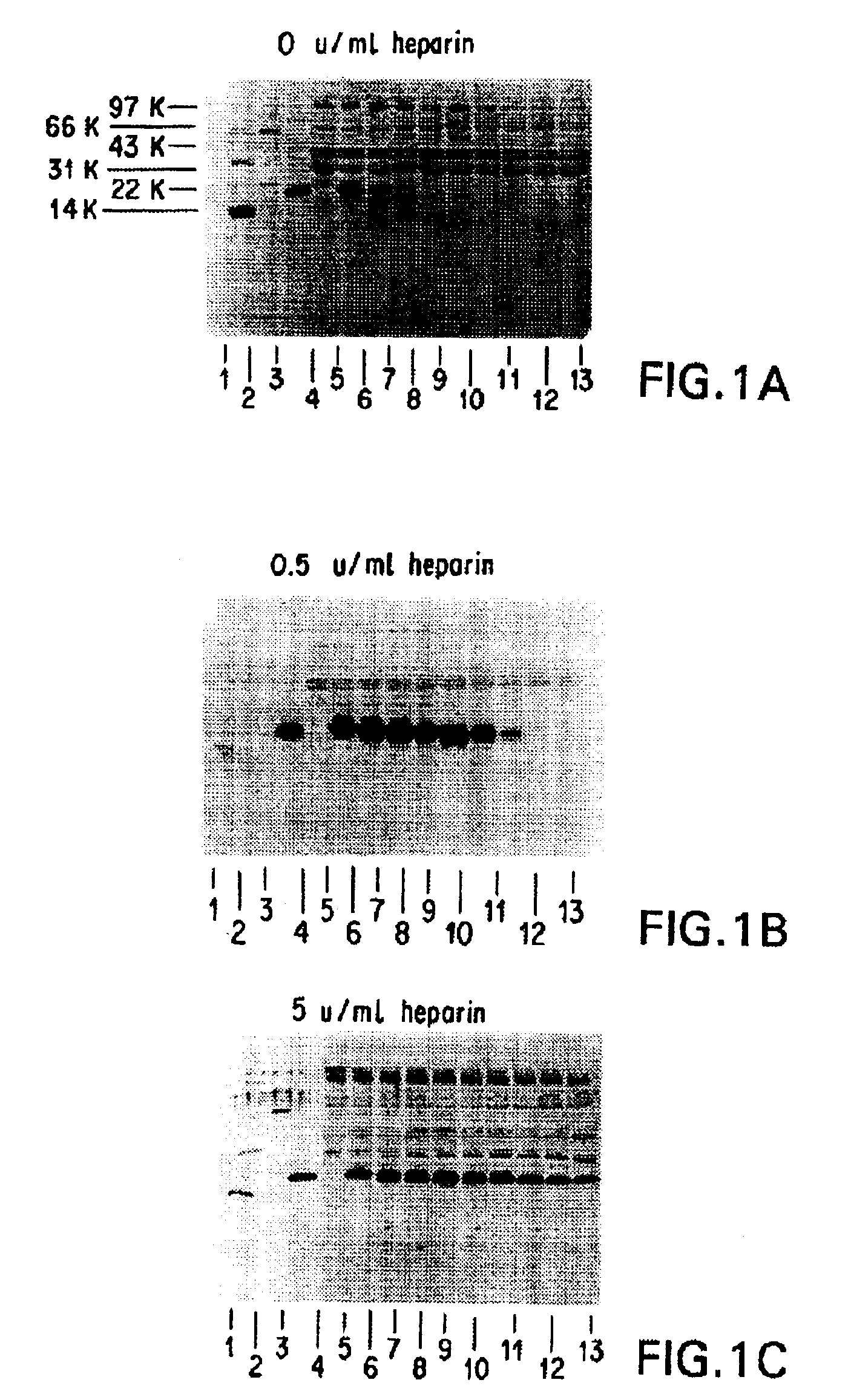
[0254]It was necessary to add an ingredient to the FG that would inhibit or prevent the digestion of HBGF-1β by thrombin (Lobb, Biochem. 27:2572-2578 (1988)), which is a component of FG. Heparin, which absorbs to HBGF-1, was selected and tested to determine whether it could protect HBGF-1 from digestion by thrombin and any other proteolytic components of the FG. The stability of HBGF-1 in the presence of increasing concentrations of heparin was assessed.

[0255]Solutions containing HBGF-1β (10 μg / ml), thrombin (250 U / ml), and increasing concentrations of heparin (0, 0.5, 5, 10, 20, and U / ml) were incubated at 37° C. Aliquots were periodically removed from the incubating solutions and were frozen and stored at −70° C. for further testing.

[0256]After the incubation was complete, the samples were thawed and separated on 15% SDS polyacrylamide gels under reducing conditions according to the method of Laemmli (Nature 227:680 (1970)). The gel was then electroblotted onto ...

example 3

The Biological Activity of HBGF-1β after Incubation in the Presence of Heparin and Thrombin

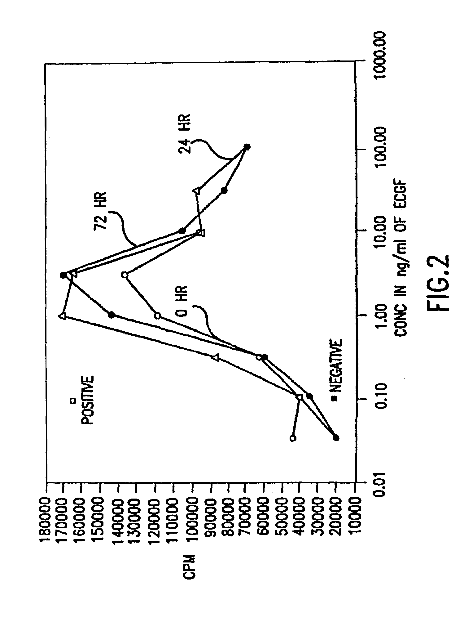
[0257]The biological activity of HBGF-1 in the incubation mixture that contained 5 U / ml of heparin, and was described in Example 2, was measuring using an 3H-thymidine incorporation assay with NIH 3T3 cells.

[0258]NIH 3T3 cells were introduced into 96 well plates and were incubated at 37° C. under starvation conditions in Dulbecco's Modified Medium (DMEM; GIBCO, Grand Island, N.Y.) with 0.5% et al bovine serum (BCS; GIBCO, Grand Island, N.Y.) until the cells reached 30 to 50% confluence. Two days later, varying dilutions of HBGF-1 from the samples prepared in Example 2 were added to each well without changing the medium. Diluent (incubation buffer) was added in place of growth factor for the negative controls and DMEM with 10% BCS, which contains growth factors needed for growth, was added in place of the HBGF-1 sample for the positive controls.

[0259]After incubation at 37° C. for 18 hours, 0.2...


PUM

PropertyMeasurementUnit
Volumeaaaaaaaaaa
Volumeaaaaaaaaaa
Volumeaaaaaaaaaa

Abstract

This invention provides supplemented tissue sealants, methods for their production and use thereof. Disclosed are tissue sealants supplemented with at least one cytotoxin or cell proliferation inhibiting composition. The composition may be further supplemented with, for example, one or more antibodies, analgesics, anticoagulants, anti-inflammatory compounds, antimicrobial compositions, cytokines, drugs, growth factors, interferons, hormones, lipids, deminearlized bone or bone morphogenetic proteins, cartilage inducing factors, oligonucleotides polymers, polysaccharides, polypeptides, protease inhibitors, vasoconstrictors or vasodilators, vitamins, minerals, stabilizers and the like.

Description

CROSS REFERENCE TO RELATED APPLICATIONS[0001]This application is a Continuation-in-Part Application of U.S. application Ser. No. 08 / 351,006, filed Dec. 7, 1994, abandonded, which is a Continuation-in-Part Application of U.S. application Ser. No. 08 / 328,552, filed Oct. 25, 1994, abandoned, which is a Continuation Application of U.S. application Ser. No. 08 / 031,164, filed Mar. 12, 1993, abandoned, which is a Continuation-in-Part Application of U.S. application Ser. Nos. 07 / 618,419 and 07 / 798,919, filed Nov. 27, 1990, and Nov. 27, 1991, respectively, both of which are abandoned, all of which are herein incorporated by reference.RIGHTS OF THE UNITED STATES GOVERNMENT IN THIS INVENTION[0002]Under a Cooperative Research and Development Agreement between The American National Red Cross and The U.S. Army Institute of Dental Research, the U.S. Government may have a non-exclusive, irrevocable, paid-up license in one or more embodiments of this invention.FIELD OF INVENTION[0003]This invention ...

Claims


Application Information

Patent Timeline
IPC IPC(8): A61K38/48A61K9/70A61L15/16A61K38/17A61K38/18A61K38/36A61K47/42A61L15/32A61L24/00A61L24/10A61L26/00A61L27/34A61L27/50C07K14/50C12N5/00C12N5/071
CPCA61K38/18A61K38/1875A61K38/363A61K47/42A61L15/32A61L24/0015A61L24/106A61L26/0042A61L26/0052A61L26/0066A61L26/0085A61L27/34A61L27/507A61L2300/256A61L2300/402A61L2300/404A61L2300/414A61L2300/416A61L2300/418A61L2300/602C07K14/501C07K14/503C12N5/0062C12N5/069C12N2533/56A61K38/00A61K38/1841A61K38/45A61K38/4833C08L89/06C08L89/00C08L5/08A61K2300/00
Inventor MACPHEE, MARTIN JAMESDROHAN, WILLIAM NASHLIAU, GENENUNEZ, HERNANBURGESS, WILSON H.MACIAG, THOMAS
Owner AMERICAN NAT RED CROSS
Why Eureka
Try Eureka