Three-dimensional large aperture tissue engineering scaffold based on nano-fibers and application thereof

A tissue engineering scaffold and nanofiber technology, applied in medical science, prosthesis, etc., can solve the problems of affecting tissue regeneration, easy collapse, poor excretion, etc., to achieve good mechanical properties, reduce degradation products, and reduce the effect of the demand

Inactive Publication Date: 2009-06-03
ZHEJIANG UNIV
View PDF0 Cites 38 Cited by
  • Summary
  • Abstract
  • Description
  • Claims
  • Application Information

AI Technical Summary

Problems solved by technology

At present, collagen membrane materials (such as BioGide) and hyaluronic acid (such as HYAFF 11) have been used clinically, but this scaffold can only be used to repair the surface of defective tissue, and cannot form a three-dimensional structure so that the regenerated tissue can fully grow into the scaffold. The main disadvantages are: 1) the gap is small, and the cells cannot grow in depth; 2) the transmission or exchange of macromolecular nutrients and cytokines is blocked, which affects tissue regeneration; 3) the mechanical properties are poor in the non-axial direction, and it is easy to collapse; 4) More local degradation products, poor excretion, affecting tissue formation

Method used


Image

  • Three-dimensional large aperture tissue engineering scaffold based on nano-fibers and application thereof
  • Three-dimensional large aperture tissue engineering scaffold based on nano-fibers and application thereof
  • Three-dimensional large aperture tissue engineering scaffold based on nano-fibers and application thereof

Examples


Embodiment 1

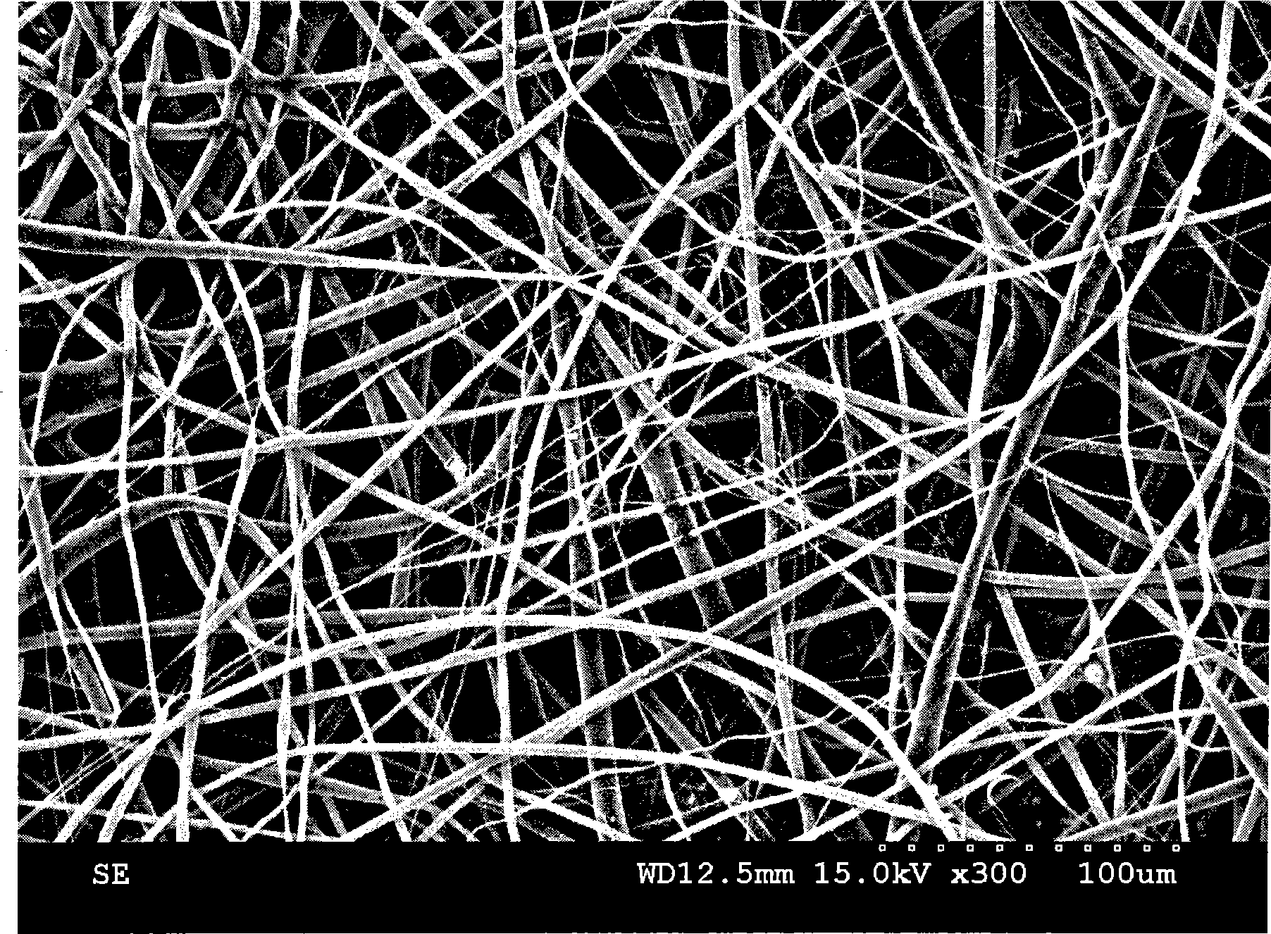
[0028] First prepare a PLLA solution with a concentration (10 mg / ml), and the solvent is chloroform / ethanol (v / v=3:1). The nanofiber membrane is prepared by electrospinning (control flow rate is 0.5ml / h~1.0ml / h, voltage is 14KV~16KV) technology (see the electron microscope photo figure 1 ), cut the prepared nanofiber membrane into thin strips with a width of 50-200 μm and a thickness of 50-200 μm to obtain micro-fibers. The micro-fibers are staggered and stacked to form a three-dimensional configuration. PCL nano-filaments are added and heated and melted at a temperature above 65 degrees for 1 minute. After cooling, a three-dimensional large-pore tissue engineering composite scaffold composed of nanofibers can be obtained (the resulting nanofiber layer has a pore size of 100-500 nm, and the micron fiber layer has a pore size of 50-200 μm). The bone marrow mesenchymal stem cells were planted on the fiber scaffold, and the cells were observed under the electron microscope after 7 d...

Embodiment 2

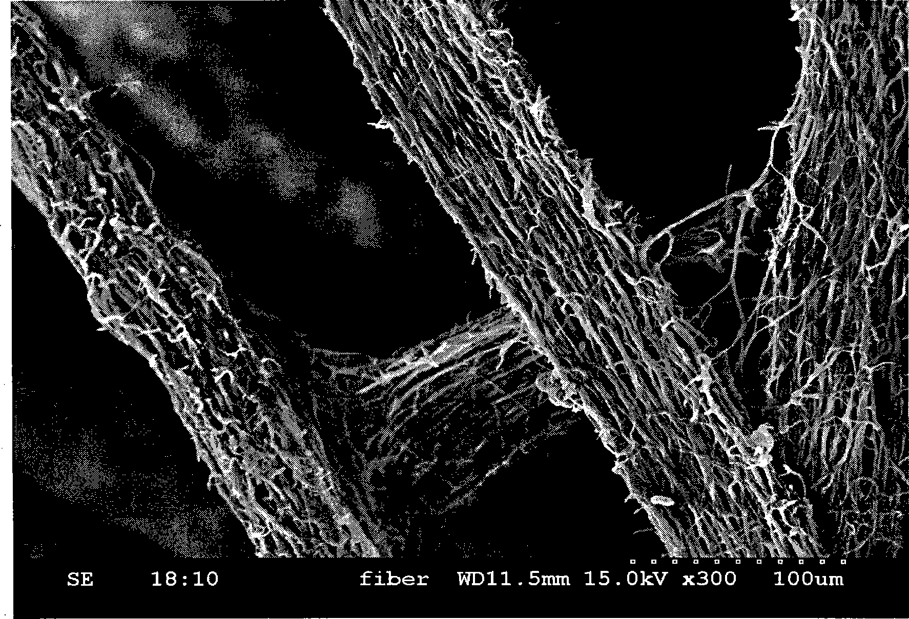
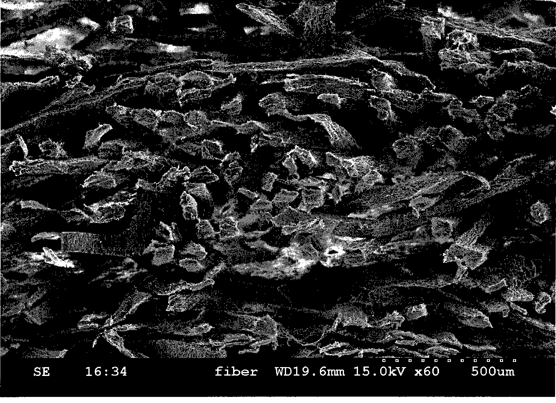
[0030]First prepare a PGA solution with a concentration (15 mg / mL), and the solvent is chloroform / ethanol (v / v=3:1). The nanofiber membrane is prepared by electrospinning (control flow rate is 1.5ml / h~2.0ml / h, voltage is 16KV~18.7KV) technology, and the film thickness is 200~500μm. The obtained nanofiber membrane is cut along a certain axial direction, and the cutting distance can be 200-500 μm to produce micron fibers with a primary structure. Then assemble according to the required block shape, soak in 3mg / ml collagen solution, lift up and filter dry for 1 minute, freeze at -70℃, freeze-dry it, and obtain nanofibers as the base layer, three-dimensional assembled by microfibers Micron pore size tissue engineering composite scaffold. (see figure 2 , image 3 ) (The diameter of the obtained nanofibers is 100-1000 nm, and the pore diameter of the scaffold is 100-500 μm).

Embodiment 3

[0032] First prepare a mixed solution of PLLA (10mg / mL) and TCP (5mg / ml), the solvent is chloroform / ethanol (v / v=3:1). Prepare nanofiber membranes by electrospinning (control flow rate is 0.5ml / h~1.0ml / h, voltage is 14KV~16KV) technology, and cut the prepared nanofiber membrane into thin strips with a width of 50~200μm and a thickness of 50~200μm , To obtain micro-fibers, the micro-fibers are staggered to form a three-dimensional configuration, (add PCL nanowires, heat and melt at 65 degrees or more for 3 minutes, after cooling, a three-dimensional large-pore tissue engineering composite scaffold composed of nanofibers can be obtained (the scaffold has a 50 ~200μm).


PUM

PropertyMeasurementUnit
diameteraaaaaaaaaa
diameteraaaaaaaaaa
diameteraaaaaaaaaa

Abstract

The invention discloses a three-dimensional large aperture tissue engineering scaffold based on nano-fibers which is prepared in the following steps: a nano-fiber membrane which is made from electrostatic spinning is cut into fiber bundles with the diameter of 10mum-1mm. The fiber bundles are overlapped and the pore spacing between the fibers is 10mum-1mm. A three-dimensional structure is assembled and is bonded to be fixed. Thus, the three-dimensional large aperture tissue engineering scaffold which comprises the nano-fibers is obtained. The three-dimensional large aperture tissue engineering scaffold based on the nano-fibers has the beneficial effects that: (1) a nano-fiber secondary structure macroporous layer of the scaffold can promote the growth, the proliferation, the differentiation, and the like of target cells to form certain tissues, and a micron fiber primary structure macroporous layer of the scaffold can provide full tissue ingrowths space to defect internal cell tissues; (2) with good mechanical property, the scaffold can greatly reduce the requirement of polymeric biomaterials and degradation products; (3) the scaffold can be applied to bulk defect repairing and organizing engineering of tissues including bones, tendons, cartilages, skins, etc.

Description

(1) Technical field [0001] The invention relates to a nanofiber-based three-dimensional large-aperture tissue engineering scaffold and its application. (2) Background technology [0002] Injuries to various tissues of the human body are extremely common, especially connective tissue injuries such as bone and cartilage defects, tendons, ligaments, etc. (accounting for 50% of sports injuries), and skin injuries are countless. Statistics show that in 2001, there were 408,000 bone transplant operations in Europe, while there were 605,000 cases in the United States alone. At present, the global population over 65 is growing at a rate of 2 to 3% every year. And due to the improvement of people's material living standards, changes in lifestyles, and improvements in medical standards, these have led to more demands and wider applications for bone transplantation and bone repair surgery. In my country, according to incomplete statistics, there are 3 million patients with bone defects or b...

Claims


Application Information

Patent Timeline
Patent Type & Authority Applications(China)
IPC IPC(8): A61L27/24A61L27/22A61L27/20A61L27/18A61L27/54
Inventor 欧阳宏伟蔡友志张国荣
Owner ZHEJIANG UNIV
Why Eureka
Try Eureka