Magneto-optical dual-mode molecular image probe and preparation method thereof

A molecular imaging and dual-mode technology, which is applied in the fields of nuclear magnetic resonance/magnetic resonance imaging contrast agents, pharmaceutical formulations, preparations for in vivo tests, etc., can solve the problem of imperfect molecular imaging technology, inability to provide clinical diagnosis, low resolution, etc. problems, to achieve the effects of excellent thermodynamic stability, excellent photostability, and high quantum yield

Inactive Publication Date: 2010-09-08
SHANGHAI NORMAL UNIVERSITY
View PDF1 Cites 7 Cited by
  • Summary
  • Abstract
  • Description
  • Claims
  • Application Information

AI Technical Summary

Problems solved by technology

Currently common molecular imaging techniques include X-ray tomography (CT), ultrasound imaging (US), optical imaging (OI), magnetic resonance imaging (MRI), positron emission tomography (PET) and single photon emission computed tomography. (SPECT), etc., but the single-mode molecular imaging technologies that have been developed are not perfect, and all have their defects, which canno

Method used


Image

  • Magneto-optical dual-mode molecular image probe and preparation method thereof
  • Magneto-optical dual-mode molecular image probe and preparation method thereof
  • Magneto-optical dual-mode molecular image probe and preparation method thereof

Examples


Embodiment 1

[0020] Preparation of Ir-Gd complexes: Take the compound phenylpyridine-o-phenanthroline-1,4,7-tri(tert-butyl ester)-1,4,7,10-tetraazacyclododecane iridium complex 74.7mg (0.05mmol) was placed in a three-necked flask, N 2Add 2mL of dichloromethane under the atmosphere, then add 1mL of trifluoroacetic acid dropwise therein, stir overnight, track the plate until the end of the reaction, spin off the solvent and unreacted acid to obtain a brownish-yellow oil, and then take 22.55mg (0.05 mmol)Gd(NO 3 ) 3 .6H 2 O was dissolved in 1.5mL of water, added to the oil, adjusted to pH 7 with 1M NaOH, stirred for 24 hours, filtered with suction to obtain a brownish-yellow solid, and purified by reverse-phase HPLC to obtain a yellow powder. MS (ESI + ) m / z: 1237.3 ([M+H] + ).

Embodiment 2

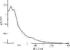
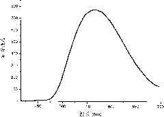
[0022] UV-visible spectrum and phosphorescence spectrum test of Ir-Gd complex: UV-visible and phosphorescence spectrum tests are all carried out in aqueous solution, wherein phosphorescence spectrum is excited by 405nm ultraviolet light, the test results are shown in figure 1 with figure 2 .

Embodiment 3

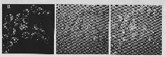
[0024] Cell fluorescence confocal imaging experiments of Ir-Gd complexes: KB cells were provided by the Chinese Institute of Biochemistry and Cell Biology. The cells were cultured in medium (MEM) containing 10% fetal bovine serum (FBs) at 37°C in 5% CO 2 in progress. The cells were plated for 24 hours, and the cell concentration was 5×10 8 / L. The confocal imaging of the cell imaging experiment was completed under an OLYMPUS FV1000 laser scanning microscope and a 60x objective lens. A 405nm semiconductor laser is used to excite the phosphorescence emission of the Ir-Gd complex, and then the emission with a wavelength ranging from 520nm to 620nm is collected. Before the experiment, the cells were washed with PBS buffer solution, and then incubated with PBS buffer solution of Ir-Gd complex (16.7 μM) at 37°C for 30 minutes; after that, the cell imaging experiment was performed after washing with PBS buffer solution. see results image 3 .


PUM


Abstract

The invention relates to the field of multi-mode molecular image probes, in particular to a heteronuclear (Ir-Gd) metal complex containing two functional units, namely an iridium phosphorescent complex and a rare-earth macrocyclic polyamine polyacid complex for magnetic resonance-phosphorescent dual-mode imaging of a living body. The cell fluorescent confocal imaging experiment proves that the Ir-Gd complex in the invention can penetrate a cell membrane to enter the cell, specifically colors the cytoplasm region of the cell, and has high water solubility and lower cell toxicity; and in vitro magnetic resonance imaging data proves that the Ir-Gd complex has higher longitudinal relaxation rate, and further research finds that the complex can be used as a magnetic resonance imaging probe for the living body.

Description

technical field [0001] The invention relates to the field of multi-mode molecular imaging probes, in particular to a heteronuclear (Ir-Gd) metal complex containing two functional units of an iridium phosphorescent complex and a rare-earth macrocyclic polyamine polyacid complex for magnetic imaging in living bodies. Resonance-phosphorescence dual-mode imaging. Background technique [0002] Molecular imaging is a science that uses imaging methods to display specific molecules at the tissue level, cells and subcellular levels, reflects the changes at the molecular level in the living body, and conducts qualitative and quantitative research on its biological behavior in terms of imaging. It is a new frontier discipline produced by the combination of molecular biology technology and modern medical imaging. Molecular medical imaging technology is a medical imaging method that displays human life information that cannot be or is difficult to recognize with the naked eye or other t...

Claims


Application Information

Patent Timeline
IPC IPC(8): A61K49/06A61K49/00C07F19/00
Inventor 杨红钱俊杰杨仕平
Owner SHANGHAI NORMAL UNIVERSITY
Why Eureka
Try Eureka