
3D printing preparation method of bond defect repair stent

A 3D printing and bone defect technology, which is applied in the preparation of biomedical tissue engineering scaffold materials, and the 3D printing preparation of new multi-level microstructure calcium-phosphorus-silicon-based active bone repair material scaffolds, can solve the toxic and side effects of pore-forming agents, lack of Problems such as pore structure space direction, connectivity control, lack of manufacturing complex shapes, etc., to achieve optimized mechanical properties, good biological activity and osteogenesis effect, and facilitate the effect of adherent proliferation

Inactive Publication Date: 2016-03-09
QINGDAO UNIQUE PRODS DEV
View PDF3 Cites 12 Cited by
  • Summary
  • Abstract
  • Description
  • Claims
  • Application Information

AI Technical Summary

Problems solved by technology

Although the traditional methods of preparing porous scaffold materials have different advantages, they are all manual operations with poor repeatability; pore-forming agents have potential side effects; lack of control over pore structure (such as pore size, spatial orientation, connectivity, etc.) control, and the lack of ability to manufacture complex shapes

Method used

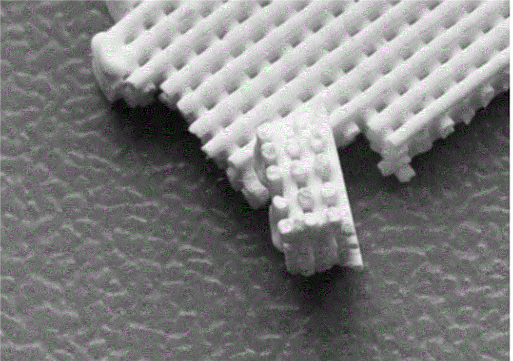
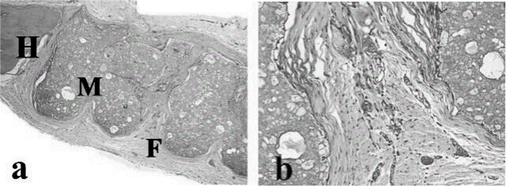
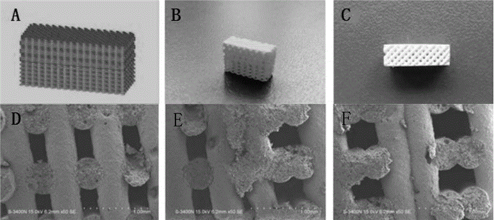

Image

  • 3D printing preparation method of bond defect repair stent
  • 3D printing preparation method of bond defect repair stent
  • 3D printing preparation method of bond defect repair stent

Examples


preparation example Construction

[0024] In the preparation method of the calcium-containing mesoporous silicon, the calcium source is added twice, and the calcium source is added for the first time to play the role of imprint, forming a reserved space in the material, and in step 1), the deionized water can be washed to remove the calcium source. The calcium source is cleaned, and after adding the calcium source in step 2), it is sintered to form a stable structure.

[0025] In the above preparation method of calcium-containing mesoporous silicon, the hydrothermal reaction temperature, time, sintering temperature, and heating rate play a key role in the performance of mesoporous silicon materials: as the temperature of the hydrothermal reaction increases, the average pore size of the sample increases , the specific surface area first increases and then decreases; as the hydrothermal reaction time increases, the average pore diameter of the sample increases first and then decreases, and the specific surface are...

Embodiment 1

[0029] A 3D printing preparation method of a bone defect repair bracket of the present invention comprises the following steps:

[0030] 1) Preparation of mesoporous silicon material: 4.0 g of polyoxyethylene-polyoxypropylene-polyoxyethylene triblock copolymer P123 was dissolved in 150 mL of hydrochloric acid solution with a concentration of 1.6 mol / L and stirred until the system was clear. 8.5g of TEOS was added dropwise to the solution. After TEOS was completely added and pre-hydrolyzed for 15 minutes, 2.6g of calcium nitrate tetrahydrate was gradually added to the solution. Stir at room temperature for 5 hours to disperse the system evenly. Set for aging for 24 hours, and then transfer to a hydrothermal reactor at 100°C for 48 hours of constant temperature reaction to obtain a precipitate. The precipitate was filtered, washed thoroughly with deionized water, and dried under vacuum at 60 °C to obtain a white powder. Disperse the dried powder in 150mL of deionized water, sti...


PUM

PropertyMeasurementUnit
Particle sizeaaaaaaaaaa

Abstract

The invention provides a 3D printing preparation method of a bone defect repair stent. The method comprises the following steps: taking calcium phosphate bone cement and calcium-containing mesoporous silicon as raw materials, mixing, and grinding to obtain calcium phosphorus silicon-based active bone repair material powder; and then uniformly mixing the calcium phosphorus silicon-based active bone repair material powder with curing liquid, and constructing the obtained mixture to obtain the bone defect repair stent by virtue of a 3D printing rapid prototyping method. The bone repair material stent is prepared by virtue of the 3D printing method, so that the shape, the structure and the porosity of the stent can be accurately controlled, and the cell attachment proliferation can be facilitated; and meanwhile, the selected calcium phosphorus silicon-based active bone repair material has good biological activity and bone forming effect and is suitable for being used as a novel bone defect repair stent.

Description

technical field [0001] The invention belongs to the technical field of tissue engineering, and is particularly suitable for the preparation of scaffold materials for biomedical tissue engineering, specifically a 3D printing preparation method for a novel multi-level microstructure calcium-phosphorus-silicon-based active bone repair material scaffold. Background technique [0002] Bone tissue defects caused by trauma, infection, tumor, congenital deformity and other factors have brought great inconvenience to the lives of patients. Bone grafting is a common treatment for bone defects. Autologous bone transplantation has the advantages of good curative effect and no immunogenicity. However, due to the limited amount of autologous bone available for transplantation, the damage to the bone donor site brings more pain to patients. Bone allografts are limited by the body's immune response and carry the risk of disease transmission. As an important research content of bone tissue...

Claims


Application Information

Patent Timeline
IPC IPC(8): A61L27/42A61L27/58A61L27/56
CPCA61L27/425A61L27/56A61L27/58A61L2400/08A61L2430/02
Inventor 王红张楠张莉
Owner QINGDAO UNIQUE PRODS DEV
Learn More