Preparation method of new biological patches

A biological patch, a new type of technology, applied in tissue regeneration, medical science, prosthesis, etc., can solve problems such as excessive cell residue, poor decellularization effect, and immune rejection

Active Publication Date: 2016-09-28
拜欧迪赛尔成都生物科技有限公司
View PDF7 Cites 6 Cited by
  • Summary
  • Abstract
  • Description
  • Claims
  • Application Information

AI Technical Summary

Problems solved by technology

[0004] There are a variety of commercialized small intestine decellularized products abroad, such as Surgisis, Restore and other products; in my country, there are also related patents such as "decellularized small intestinal submucosa biological material" (application number 200810150792.5) and "a kind of decellularized biological patch and Its preparation method and device” (Application No. 201310117569.1), etc. The problem with these products is that the decellularization method is not perfect. For example, the treatment method only uses low-concentration peracetic acid treatment, and there are many residual cells, and the decellularization effect is not good. Initiate the immune rejection of the body; or add ethanol to decellularize; or apply long-term immersion; or use ultrasound, repeated freezing and thawing, and nuclease for decellularization. Growth factors contained in decellularized tendon and intestinal tissue material are ineffective

Method used


Image

  • Preparation method of new biological patches
  • Preparation method of new biological patches
  • Preparation method of new biological patches

Examples


Embodiment 1

[0025] Preparation of acellular protection solution for tendon and small intestinal serosa patches:

[0026] 1. Take 8g of DMEM powder and add 500ml of deionized water, dissolve and sterilize under high pressure;

[0027] 2. Take 30g of chondroitin sulfate, add 275ml of deionized water, heat, dissolve and autoclave to make a chondroitin sulfate solution; add 35g of low-molecular-weight dextran to 100ml of water for injection to dissolve and autoclave to make a low-molecular-weight dextran solution; L -Histidine hydrochloride 3.83g, add 100ml of water for injection to dissolve and autoclave to make L-histidine hydrochloride solution;

[0028] 3. Take a sterile container and add 500ml of DMEM culture solution, 275ml of autoclaved chondroitin sulfate, 100ml of low-molecular dextran solution, 100ml of L-histidine hydrochloride solution, and 25ml of Hepes buffer; add 0.5g of allopurinol, hydroxy Propyl methylcellulose 6g, reduced glutathione 1.5g, dexamethasone injection 1.8mg, le...

Embodiment 2

[0031] Preparation of decellularized porcine tendon patches.

[0032] 1. Take fresh (obtained within 3 hours after slaughter) pig thigh tendon tissue, and ensure that the outer tendon membrane is intact. Cut the aponeurosis to make a 1×3cm piece of tendon tissue, and seal it in a plastic bag filled with protective solution;

[0033] 2. Under the condition of 400MP high static pressure, treat 4 times, each time is 2 minutes;

[0034] 3. After taking out the tendon, place it in a protective solution containing 2% TritonX-100+600U / ml DNase at a temperature of 25°C, set the speed of the shaker at 100 rpm, and treat for 2 hours;

[0035] 4. After the detergent and enzyme treatment, take out the decellularized tendon and rinse it in the protective solution for 2 hours;

[0036] 5. The prepared decellularized tendon was attached to the nitrocellulose mold, put into anhydrous calcium chloride molecular sieve for dehydration, bagged, sealed and marked, and then irradiated with cobalt...

Embodiment 3

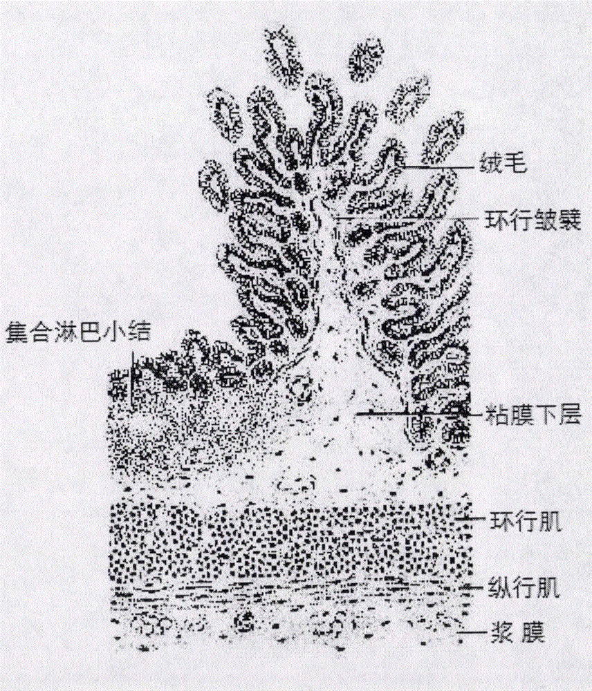
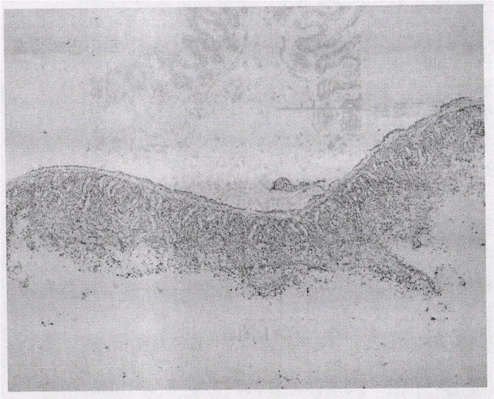
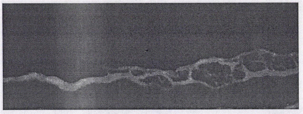
[0039] Preparation of serosal patch of decellularized porcine small intestine.

[0040] 1. Take fresh (obtained within 3 hours after slaughter) porcine small intestine (including jejunum and ileum) tissue, rinse with water, cut into 5cm sections; bluntly scrape the small intestinal mucosa, submucosa and muscular layer, leaving only the fibrous serosa layer , rinsed with PBS; the tissue of the fibrous serosa layer of the small intestine was made into a tissue piece of 1 × 3 cm, and sealed in a plastic bag filled with the above-mentioned protective solution;

[0041] 2. Under the condition of 300MP high static pressure, treat 4 times, each time is 1 minute;

[0042] 3. After taking out the small intestine slices, place them in a protective solution containing 1.5% TritonX-100+500U / ml DNase at a temperature of 25°C, set the shaker speed to 100 rpm, and treat for 1 hour;

[0043] 4. After the detergent and enzyme treatment, take out the decellularized intestinal fibrous serous me...


PUM


Abstract

The invention relates to a preparation method of two types of new biological patches, namely a muscle tendon patch and a small intestine serosa patch, and provides a natural biological patch material with favorable biomechanical characteristic and biocompatibility. The method comprises the following steps: acquiring muscle tendons and small intestine serosa of a pig as raw materials; utilizing a protective solution with a supporting structure capable of protecting the muscle tendons and the small intestine serosa during a whole decellularization process; performing predissociation on cells in tissues of the muscle tendons and the small intestine serosa by use of a high hydrostatic pressure technology; digesting residual cell nucleus by use of a composite nuclease method; removing loose and broken cell sheets by use of a detergent; rinsing with the protective solution. On the basis of thorough cell nucleus removal, the method is capable of being used for reducing the immunogenicity in the tissues of the muscle tendons and the small intestine serosa to the greatest extent and meanwhile reserving the three-dimensional structure and arrangement polarity of normal collagenous fibers of the tissues, and is applicable to tissue defect repair, tissue material filling and the like. Because of the characteristics of a biological fiber material, the new biological patches can also be widely applied to other biomedical domains.

Description

technical field [0001] The invention belongs to the technical field of tissue engineering, in particular to a preparation method of a novel tendon and small intestine serosa tissue patch. Background technique [0002] The biggest problem in the repair of tissue defects such as nerves, bladder, blood vessels, tendons, abdominal wall and eye conjunctiva is the repair material. At present, most of the materials for surgical patches at home and abroad are synthetic materials, such as polypropylene patch (PP) and polyester patch (PE). Its biggest defect is low anti-infection rate, poor histocompatibility, sequelae caused by aseptic inflammation caused by physical stimulation and chronic rejection, and even causes infection of repaired organs and adhesion with surrounding tissues. Semi-absorbent composite materials developed in recent years, such as polyglycolic acid (PGA) and polylactic acid (PLA), often lose their curative effect due to rapid degradation. However, the use of s...

Claims


Application Information

Patent Timeline
Patent Type & Authority Applications(China)
IPC IPC(8): A61L27/36A61L27/50
CPCA61L27/3604A61L27/3629A61L27/3687A61L27/3691A61L27/50A61L2430/40
Inventor 史真史伟云
Owner 拜欧迪赛尔成都生物科技有限公司
Why Eureka
Try Eureka