Lactate dehydrogenase isoenzyme electrophoretic separation method

A lactate dehydrogenase and electrophoresis separation technology, which is applied in the field of tissue and cell activity enzyme electrophoresis detection, can solve the problems that barbiturate buffer is not easy to store, the experimental results are not very good, and it is not suitable for human tumor cells. Experimental results, stable and experimental results, the effect of stable results

Inactive Publication Date: 2016-11-09
SOUTHWEST MEDICAL UNIVERISTY
View PDF2 Cites 4 Cited by
  • Summary
  • Abstract
  • Description
  • Claims
  • Application Information

AI Technical Summary

Problems solved by technology

There are four deficiencies in this method at least: one, barbiturate and barbital sodium have gradually become national control drugs in recent years, and it is more and more difficult to buy, and the price is more expensive; It is not easy to preserve; three, when adopting barbiturate buffer solution to carry out electrophoresis separation, the fever is serious, which easily leads to poor banding of experimental results; four, the prior art is extremely small in terms of separation and detection of lactate dehydrogenase isoenzyme: On the one hand, due to the particularity of the isoelectric points of different species, the agarose gel method that can be quantitatively analyzed in the prior art is only suitable for species with narrow and moderate isoelectric points such as mice, and is not suitable for such species as human tumor cells. Species with a wide range of isoelectric points
On the other hand, although the cellulose acetate film method is not limited by the isoelectric point, its sensitivity is low and cannot be quantified, so it is only suitable for general experimental teaching

Method used


Image

  • Lactate dehydrogenase isoenzyme electrophoretic separation method
  • Lactate dehydrogenase isoenzyme electrophoretic separation method
  • Lactate dehydrogenase isoenzyme electrophoretic separation method

Examples


Embodiment 1

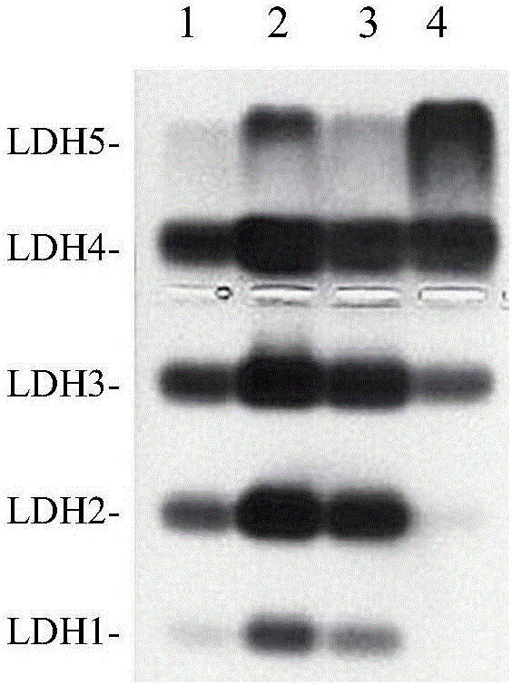
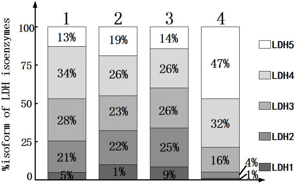
[0030] Agarose gel electrophoresis separation of lactate dehydrogenase isozymes in different human tumor cells.

[0031] 1. Materials

[0032] 1.1 Equipment

[0033] Centrifuge, constant temperature cell incubator, inverted microscope, ultra-clean bench, cell culture bottle, cell brush, pipette, agarose gel horizontal electrophoresis tank, electrophoresis apparatus, glue mold, constant temperature water bath, oven, electronic balance , stirrer, microwave oven, gel imaging system, EP tube, pipette gun, pH meter

[0034] 1.2 Reagent preparation

[0035] Phosphate buffered saline (PBS): Na 2 HPO 4.7 h 2 O 22.55g, NaH 2 PO 4 2.16g, add distilled water to dissolve, dilute to 1000ml, pH 7.4;

[0036] Trypsin digestion solution: 0.5g Trypsin, 0.04g Na 2 EDTA·2H 2 O, dissolved in PBS, add PBS to 200ml, pH 7.2;

[0037] 10 times concentrated TBE (Tris-Boric acid-EDTA) buffer: Tris 108g, Na 2 EDTA·2H 2 O 7.44g, boric acid 55g, add distilled water to dissolve, and dilute to 1...

Embodiment 2

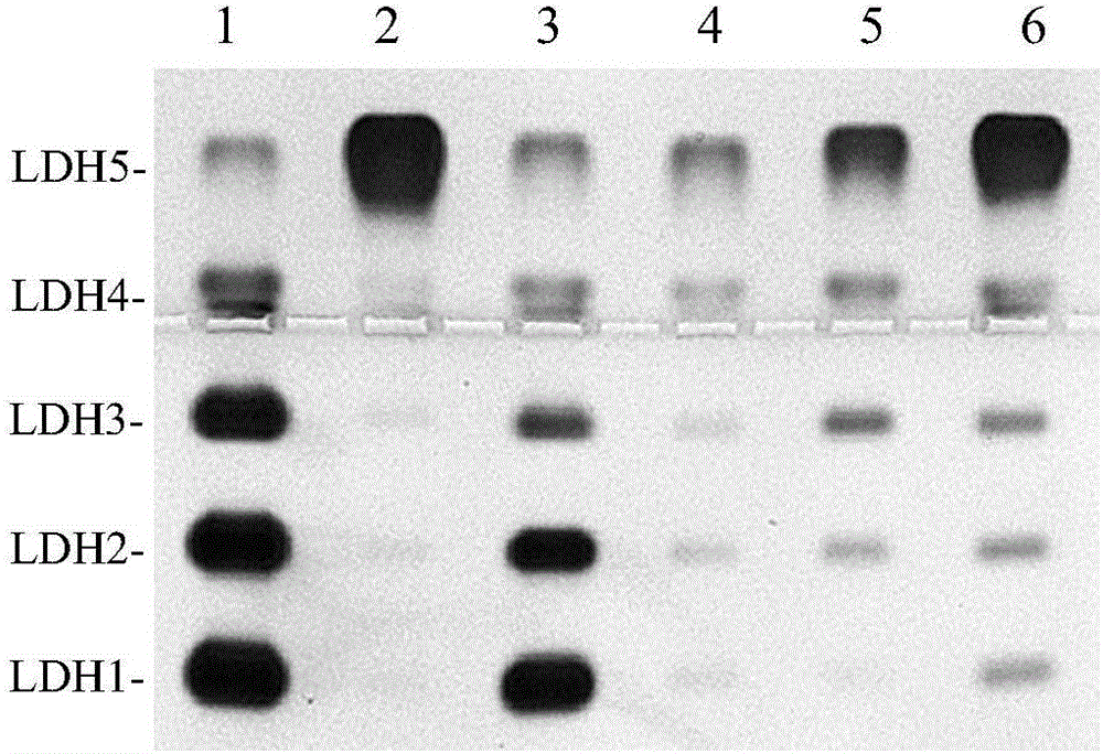
[0072] Agarose gel electrophoresis separation of lactate dehydrogenase isozymes in different tissues of rats.

[0073] It does not repeat the same part as Embodiment 1, and its difference is:

[0074] 1. Materials

[0075] 1.1 Equipment

[0076] A homogenizer is also required; no constant temperature cell incubator, inverted microscope, ultra-clean bench, cell culture bottle, cell brush, pipette, constant temperature water bath, oven

[0077] 1.2 Reagent preparation

[0078] 10 times concentrated TBE (Tris-Boric acid-EDTA) buffer solution: the preparation operation is the same as in Example 1, and the pH is adjusted to 8.0;

[0079] Trypsin digestion solution, 1‰ Triton X-100, DMEM medium, and 1640 medium are not required (the rest are the same as in Example 1).

[0080] 1.3 Sample and preparation

[0081] 1.3.1 Experimental animals

[0082] the rat

[0083] 1.3.2 Tissue acquisition (no cell culture required)

[0084] Prepare 3% anesthetics with normal saline and amoba...

Embodiment 3

[0096] Agarose gel electrophoresis separation of lactate dehydrogenase isozymes in different tissues of guinea pigs.

[0097] Its similarities with Embodiment Two are not repeated, and its difference is:

[0098] 1. Materials

[0099] 1.1 equipment (same as embodiment two)

[0100] 1.2 Reagent preparation

[0101] 10 times concentrated TBE (Tris-Boric acid-EDTA) buffer solution: the preparation operation is the same as in Example 1, and the pH is adjusted to 9.2.

[0102] 1.3 Sample and preparation

[0103] 1.3.1 Experimental animals

[0104] Guinea Pig 1.3.2 Tissue Acquisition (Same as Example 2, no need for cell culture)

[0105] 1.3.3 Tissue sample preparation (same as Example 2, no cell sample preparation required)

[0106] 2. Agarose gel electrophoresis separation operation

[0107] 2.1 Agarose gel (5g / L) preparation (same as embodiment two)

[0108] 2.2 Electrophoresis

[0109] Clean the electrophoresis tank, pour TBE buffer solution with pH 9.2, gently and slow...


PUM


Abstract

The invention discloses a lactate dehydrogenase isoenzyme electrophoretic separation method. Aiming at the defects existing in the separation methods taking barbital as an electrophoretic buffer solution in the prior art, the invention provides the lactate dehydrogenase isoenzyme electrophoretic separation method taking TBE buffer solution as the electrophoretic buffer solution, wherein the TBE buffer solution is obtained by dissolving 10.8g of Tris, 0.744g of Na2EDTA.2H2O and 5.5g of boric acid in distilled water, and making the volume constant to be 1000ml. The method can be optimized from two aspects, namely, controlling the electrophoresis temperature and/or controlling the pH of the electrophoretic buffer solution. The invention further provides a human cancer cell lactate dehydrogenase isoenzyme electrophoretic separation method realized by utilizing the lactate dehydrogenase isoenzyme electrophoretic separation method, and an application method of the TBE buffer solution in the lactate dehydrogenase isoenzyme electrophoretic separation. With the adoption of the method, the technical defects generated due to the use of the barbital buffer solution in the prior art are effectively avoided, and the application values in scientific research, clinical test and experiment teaching of the lactate dehydrogenase isoenzyme electrophoretic separation method are expanded.

Description

technical field [0001] The invention relates to an enzyme electrophoresis separation method, in particular to a lactate dehydrogenase isozyme electrophoresis separation method. Belongs to the field of tissue and cell activity enzyme electrophoresis detection Background technique [0002] Lactate dehydrogenase isoenzyme (lactate dehydrogenase, LDH) is an enzyme that can reversibly catalyze the dehydrogenation of lactate to generate pyruvate, which has an important relationship with sugar metabolism and energy supply. [0003] Lactate dehydrogenase is widely present in the cytoplasm of various tissue cells in the body, and is most abundant in heart, skeletal muscle and kidney, followed by liver, spleen, pancreas, brain and lung. It contains 5 types of isozymes composed of A subunit (skeletal muscle type) and B subunit (cardiac muscle type), namely LDH1 (B4), LDH2 (B3A), LDH3 (B2A2), LDH4 (BA3), LDH5 (A4). LDH isozymes are distributed differently in different tissues, and th...

Claims


Application Information

Patent Timeline
Patent Type & Authority Applications(China)
IPC IPC(8): C12N9/04
CPCC12N9/0006
Inventor 曾凡才李培娟张黎
Owner SOUTHWEST MEDICAL UNIVERISTY
Why Eureka
Try Eureka