Magnetic nanometer material used for imaging and photothermal therapy and preparation method and application of magnetic nanometer material

A photothermal therapy, magnetic nanotechnology, applied in the field of medicine, can solve problems such as shedding

Active Publication Date: 2017-02-15
THE THIRD AFFILIATED HOSPITAL OF THIRD MILITARY MEDICAL UNIV OF PLA
View PDF3 Cites 7 Cited by
  • Summary
  • Abstract
  • Description
  • Claims
  • Application Information

AI Technical Summary

Problems solved by technology

However, since metal ions are adsorbed on the surface of nanoparticles by chelation, there is a risk of shedding

Method used


Image

  • Magnetic nanometer material used for imaging and photothermal therapy and preparation method and application of magnetic nanometer material
  • Magnetic nanometer material used for imaging and photothermal therapy and preparation method and application of magnetic nanometer material
  • Magnetic nanometer material used for imaging and photothermal therapy and preparation method and application of magnetic nanometer material

Examples


Embodiment 1

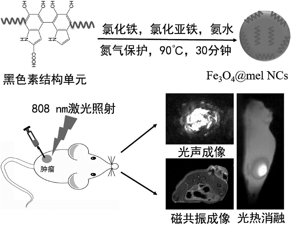
[0042] Embodiment 1 co-precipitation method synthesizes Fe 3 o 4 @mel NCs

[0043] 100umol, 19.881mg of FeCl 2 4H 2 O with 200umol, 54.058mg of FeCl 3 ·6H 2 O was dissolved in 5mL of 1M HCl solution, heated to 90°C under nitrogen gas, and 0.5mL of 28vol.%NH was quickly added under vigorous stirring. 4 OH solution, react for 20 seconds. Subsequently, 5 mL of melanin solution (1 mg / mL, pH=9.0) was added within 1 minute, stirred at 90° C. for 30 minutes, cooled to room temperature, and magnetically separated until the pH of the solution reached neutral. Collect the precipitate and obtain the Fe 3 o 4 @mel NCs are dispersed in water, see figure 1 .

Embodiment 2

[0044] Example 2 Fe 3 o 4 Synthesis and characterization of @mel NCs

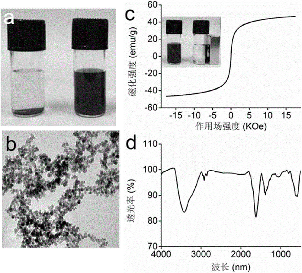
[0045] Morphology observed by transmission electron microscope: pipette 50uL Fe 3 o 4 The @mel NCs solution was added dropwise on the carbon-coated copper grid, and the excess solution was absorbed with filter paper, and air-dried at room temperature. The morphology, particle size and dispersion of nanoparticles were observed under the condition of 200kV voltage by transmission electron microscope.

[0046] Vibrating the sample magnetometer to test the magnetic properties: Weigh about 10 mg of the dried powder sample, put it in a special micro test tube for testing, and compact it with absorbent cotton plug. Use a vibrating sample magnetometer to test the normal temperature performance: the power supply operating frequency is 86Hz, the input power is 5W, and the X-axis records the magnetic field change on the computer, and the Y-axis records the magnetization intensity change.

[0047] Fourier transfor...

Embodiment 3

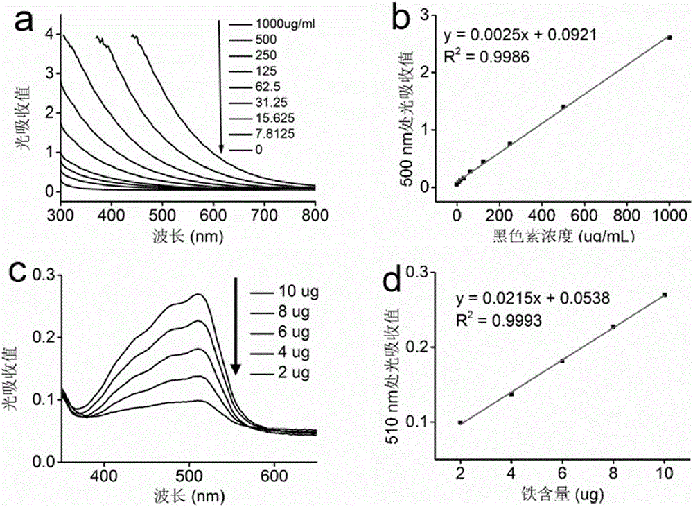
[0049] Embodiment 3 detects Fe 3 o 4 Melanin and iron content in @mel NCs

[0050] Different concentrations of melanin solutions were prepared, and their optical absorption was measured with a full-wavelength microplate reader. Take the light absorption value at 500nm, make the absorbance value-melanin solution concentration curve, and measure the synthesized Fe 3 o 4 Melanin content in @mel NCs. with (NH 4 ) 2 Fe(SO 4 ) 2 ·6H 2 O prepares the iron standard bath solution, adopts the standard o-phenanthroline method to make the iron concentration standard absorption curve at 510nm, and measures the synthesized Fe 3 o 4 Iron content in @mel NCs. Calculate Fe 3 o 4 The ratio of melanin and iron content in @mel NCs.

[0051] Absorption standard curves were made at 500nm and 510nm by full-wavelength microplate reader and o-phenanthroline method, respectively, to detect the content of melanin and iron in the product. It is measured that every 1mg Fe corresponds to 0.2...


PUM


Abstract

The invention relates to a magnetic nanometer material used for imaging and photothermal therapy and a preparation method and application of the magnetic nanometer material. The magnetic nanometer material used for imaging and photothermal therapy refers to nanometer iron oxide particles coated with melanin. The magnetic nanometer material is non-toxic, high in biocompatibility and capable of imaging. According to the preparation method, a one-step coprecipitation method is adopted to composite water-soluble Fe3O4@mel NCs. The magnetic nanometer material has the advantages of mildness and simplicity.

Description

technical field [0001] The invention relates to the technical field of medicine, in particular to a magnetic nanometer material for imaging and photothermal therapy, a preparation method and application thereof. Background technique [0002] With the rapid development of biotechnology and nanoscience, near-infrared laser-mediated photothermal therapy has gradually been used as a promising alternative therapy for tumor treatment. Photothermal therapy is a less invasive topical treatment modality. When the tumor area is exposed to the range of near-infrared laser irradiation, the photothermal conversion reagent enriched in the tumor area can convert light energy into heat energy and effectively kill tumor cells. Compared with traditional therapies (such as surgical resection, chemotherapy, radiotherapy, etc.), photothermal therapy has the advantages of non-invasiveness, high selectivity, high treatment efficiency, and less damage to surrounding healthy tissues. At present, t...

Claims


Application Information

Patent Timeline
Patent Type & Authority Applications(China)
IPC IPC(8): A61K41/00A61K49/18A61K49/10A61K49/22
CPCA61K41/0052A61K49/1833A61K49/225
Inventor 张伟国刘恒陈晓薛巍张俊峰
Owner THE THIRD AFFILIATED HOSPITAL OF THIRD MILITARY MEDICAL UNIV OF PLA
Why Eureka
Try Eureka