Tumor-targeting nanoparticles co-loading chemotherapy drug and nucleic acid and preparation method thereof

A chemotherapeutic drug and nanoparticle technology, applied in the direction of antineoplastic drugs, drug combinations, pharmaceutical formulations, etc., to achieve the effect of non-immunogenicity, easy operation, and inhibition of cell proliferation

Active Publication Date: 2017-05-10
TIANJIN TUMOR HOSPITAL
View PDF3 Cites 4 Cited by
  • Summary
  • Abstract
  • Description
  • Claims
  • Application Information

AI Technical Summary

Problems solved by technology

[0007] At present, there is no report on the use of hyaluronic acid to co-load chemotherapy drugs and nucleic acids to make tumor-targeting nanoparticles

Method used


Image

  • Tumor-targeting nanoparticles co-loading chemotherapy drug and nucleic acid and preparation method thereof
  • Tumor-targeting nanoparticles co-loading chemotherapy drug and nucleic acid and preparation method thereof
  • Tumor-targeting nanoparticles co-loading chemotherapy drug and nucleic acid and preparation method thereof

Examples


Embodiment 1

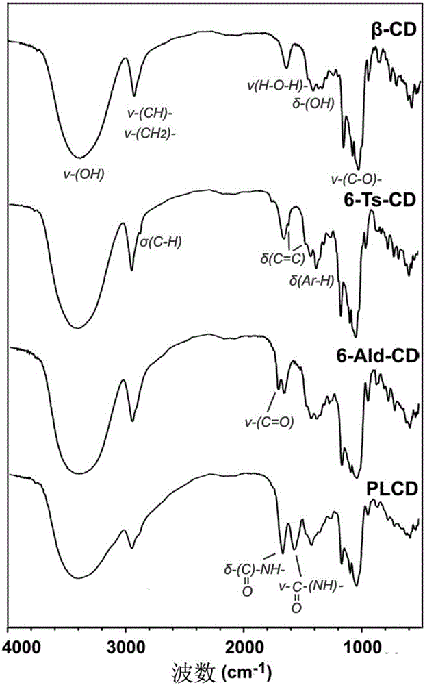
[0050] (1) Synthesis of polylysine grafted β-cyclodextrin derivative (PLCD)

[0051] Dissolve 282mg (0.25mmol) 6-Ald-CD, 57mg (where the lysine structural unit is 0.25mmol) poly-L-lysine hydrobromide in 5mL acetate buffer solution (0.2M) pH 4.4 Stir the reaction at room temperature for 1h, add 62.8mg (1mmol) sodium cyanoborohydride, continue stirring at room temperature for 72h, add 2M NaOH aqueous solution to adjust the system to neutral, and then transfer the reaction system to a dialysis bag (the molecular weight cut-off is 7000Da ), dialysis in ultrapure water for 2 days, the dialysate is freeze-dried, and the obtained white flocculent product is polylysine grafted β-cyclodextrin derivative (PLCD), in which the degree of substitution of β-CD is 12.9%.

[0052] The viscosity average molecular weight of the poly-L-lysine hydrobromide is 15,000 to 30,000 Da;

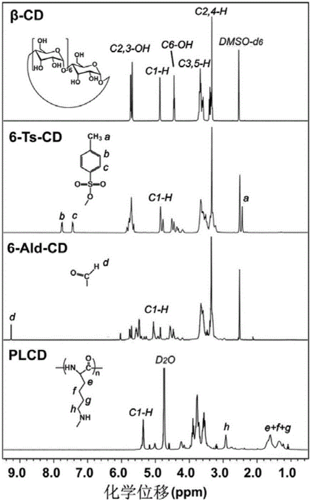
[0053] The chemical structure of PLCD was characterized by infrared spectrometer and nuclear magnetic resonance spectromete...

Embodiment 2

[0067] In vitro drug release test

[0068] Take three PDR and HPDR prepared in Example 1, each 1mL, transfer to a dialysis bag (the molecular weight cut-off is 8000~14000Da), respectively place them in 10mL pH 5.0, 6.5 and 7.4 PBS solution at 37℃ Light oscillation (100rpm), at different time points (see Figure 8 ) Take out 1.5 mL of the medium for testing, and add an equal volume of fresh dialysis medium; use an ultraviolet spectrophotometer to detect the release of DOX (detection wavelength is 480 nm). The cumulative drug release rate is calculated according to the following formula: DOX cumulative release rate=(DOX release amount / total DOX input)×100%. See DOX release curve Figure 8 , Showing slow release characteristics, and has significant pH sensitivity.

Embodiment 3

[0070] Cell Uptake Research

[0071] Replace Control RNA in Example 1 with FAM-RNA. Others are the same as Example 1. The prepared PDR and HPDR are named PDR respectively FAM And HPDR FAM .

[0072] Use the liver cancer cell line MHCC-97H to examine PDR FAM And HPDR FAM The ability and location to enter cells; the above cells are at 37℃, 5% CO 2 Cultivate in an incubator, the medium is high-sugar DMEM medium containing 10% FBS; when the cells grow in the logarithmic growth phase, digest the cells according to 5×10 4 The cell / well density is seeded in a 12-well plate pre-laid with a special glass sheet for laser confocal. After 24 hours of culture, free DOX, FAM-RNA, PDR diluted in serum-free medium are added FAM And HPDR FAM , Make the final concentration of DOX in the system 3.3μg / mL and the final concentration of FAM-RNA 1.0μg / mL; after incubating for 4h, the cells are fixed with 4% paraformaldehyde, the nucleus is stained with DAPI, the glass slide is taken out, and the lase...


PUM

PropertyMeasurementUnit
viscosity average molecular weightaaaaaaaaaa
molecular weightaaaaaaaaaa
particle diameteraaaaaaaaaa

Abstract

The invention discloses tumor-targeting nanoparticles co-loading chemotherapy drug and nucleic acid and a preparation method thereof; the preparation method comprises: (1) preparing polylysine-grafted Beta-cyclodextrin derivative; (2) preparing nanoparticles loading chemotherapy drug; (3) preparing nanoparticles co-loading chemotherapy drug and nucleic acid; (4) preparing tumor-targeting nanoparticles co-loading chemotherapy drug and nucleic acid. The preparation process of the invention is simple and easily operable and saves time and power, and a load material used herein is high in bio-safety and is good in bio-compatibility and biodegradability, zero in toxicity and free of immunogenicity; the tumor-targeting nanoparticles co-loading chemotherapy drug and nucleic acid have typical core-shell structure, are 150-200 nm in particle size, are effective in loading both chemotherapy drug and nucleic acid to cells that highly express CD44 molecules, can inhibit cell proliferation, and have significant in-vivo and in-vitro tumor targeting property.

Description

Technical field [0001] The invention belongs to the field of nanomedicine, and specifically relates to a tumor-targeting nanoparticle co-carrying a chemotherapeutic drug and nucleic acid and a preparation method thereof. Background technique [0002] The incidence and mortality of malignant tumors are increasing year by year, which is a serious threat to human health. In recent years, with the continuous improvement of liver surgery technology, anesthesia technology and perioperative management level, surgical treatment has become the first choice for the treatment of malignant tumors. However, the vast majority of clinical patients have already lost the opportunity for surgery at the time of diagnosis. Chemotherapy is also one of the common strategies for the treatment of malignant tumors. However, due to the heterogeneity of tumors, a single chemotherapy often fails to achieve the desired effect, and patients tend to tolerate chemotherapy. [0003] In the late 1960s, American s...

Claims


Application Information

Patent Timeline
Patent Type & Authority Applications(China)
IPC IPC(8): A61K9/51A61K47/40A61K31/704A61K31/4745A61K31/337A61K31/7105A61K31/711A61K48/00A61P35/00
CPCA61K9/5161A61K31/337A61K31/4745A61K31/704A61K31/7105A61K31/711A61K2300/00
Inventor 宋天强熊青青
Owner TIANJIN TUMOR HOSPITAL
Why Eureka
Try Eureka