Preparation method of artificial skin with controllable drug release

An artificial skin and drug technology, applied in the field of artificial skin and drug controlled release, can solve the problems of complex process, difficult cell adhesion, affecting wound healing efficacy, etc., and achieve the effect of improving in vivo biological effects and promoting blood vessel regeneration.

Active Publication Date: 2020-10-30
THE FIRST AFFILIATED HOSPITAL OF SUN YAT SEN UNIV
View PDF5 Cites 0 Cited by
  • Summary
  • Abstract
  • Description
  • Claims
  • Application Information

AI Technical Summary

Problems solved by technology

However, these microspheres are difficult to be well immobilized
[0006] Second, most of the materials of artificial skin products are hydrophobic, which makes it difficult for cells to adhere, which in turn affects the efficacy of wound healing
Unfortunately, however, the aforementioned material modification techniques are often expensive, complex, and not widely applicable to all biological materials.

Method used


Image

  • Preparation method of artificial skin with controllable drug release
  • Preparation method of artificial skin with controllable drug release
  • Preparation method of artificial skin with controllable drug release

Examples


Embodiment 1

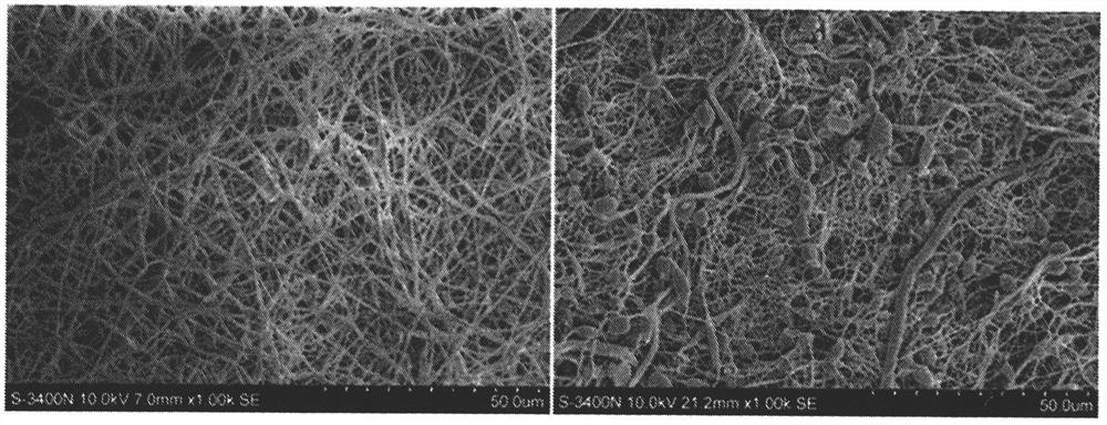
[0046] Embodiment 1: Preparation of artificial skin, observation by scanning electron microscope, confirmation of microsphere morphology (preparation method effect test).

[0047] Use dichloromethane / dimethylformamide solution, the volume ratio of dichloromethane / dimethylformamide is 5:1, configure 20% polycaprolactone solution, prepare three-dimensional porous material by freeze-drying method, cross-link and fix, Dry and set aside. First, immerse in a fibronectin solution, incubate in a 37°C incubator for 18 hours, rinse with pure water for 3 times, the fibronectin is 5 mg / ml, and the pH is 7.5. Then, immerse in platelet endothelial growth factor (VEGF) solution, 37 ° C, 12h, rinse once with pure water, platelet endothelial growth factor (VEGF) is 1 mg / ml, pH7.5. Finally, immerse in acetone for 5 minutes. After drying, its structure was observed by scanning electron microscope ( figure 1 ).

Embodiment 2

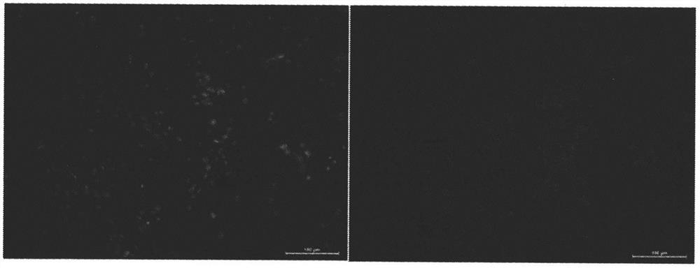
[0048] Example 2: Inoculate cells into artificial skin, and observe the morphology and distribution of cells in artificial skin with a fluorescence microscope (biological safety and compatibility testing).

[0049] The primary mouse dermal fibroblasts were isolated and cultured. When they were cultured to the fifth generation, they were digested with trypsin, diluted, and suspended by pipetting to prepare a fibroblast suspension with a concentration of about 10×10 5 / ml. The cell suspension was inoculated into the sterilized artificial skin, and cultured in the cell box until the 8th day. First, fix with 10% paraformaldehyde solution, then use rhodamine-labeled phalloidin (R415, Molecular Probes) to stain the fibrous actin in the cytoplasm respectively, and use 4′,6-diamidine Cell nuclei were stained blue with 2-phenylindole (DAPI). Finally, the slides were sealed with anti-fluorescence quenching mounting reagent and stored in the dark. Observe and take pictures with a fluor...

Embodiment 3

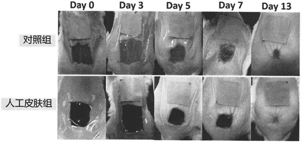
[0050] Embodiment 3: Using animal experiments, detect the effect of the artificial skin on the wound healing speed of mice (detect the curative effect of the artificial skin at the level of the animal body).

[0051] In the first step, the experimental mice are anesthetized. The experimental mice were injected intraperitoneally with 0.8% pentobarbital for anesthesia. Anesthetics were used at a dose of 0.02 mg / g body weight. In the second step, hair removal cream was used to remove the hair on the back of the mouse, and disinfected with alcohol; a full-thickness skin defect wound was made on the back of the mouse to ensure that the size and depth of each mouse wound were basically the same. The third step is to cover the wound with the prepared artificial skin graft, take pictures of the wound, and calculate the wound healing rate. On the 0th, 3rd, 5th, 7th, and 13th day after artificial skin transplantation, a Canon digital camera was used perpendicular to the wound surface....


PUM


Abstract

The invention discloses artificial skin capable of controllably releasing a drug and a preparation method. The preparation method comprises the steps: (1) preparing a biological material support of a three-dimensional porous structure; (2) coating bipolar viscosity molecules on the surface of the biological material support; (3) utilizing the bipolar viscosity molecules to adsorb drug; (4) soaking the materials into a solvent which can dissolve the support material, wherein a combined body of the support material, the viscosity molecules and the drug which are originally combined together is eluted, and microspheres with appropriate diameters can be generated on the surface of the support in situ. The microspheres are prepared from the support material, the viscosity molecules and the drug. By regulating a degradation rate of the microspheres, a release rate of the drug can be regulated.

Description

technical field [0001] The invention belongs to the field of artificial skin and drug controlled release, in particular to a method for preparing artificial skin with controllable drug release. Background technique [0002] The controlled release of drugs is an important research direction in the field of pharmacology and pharmacokinetics. Controlled drug release not only enables the drug to achieve and maintain an effective drug concentration and exert good pharmacological effects, but also avoids the toxic and side effects caused by intermittent large-dose administration. Therefore, realizing the controlled release of drugs is an important topic in the field of pharmacy and clinical medicine. [0003] For the field of artificial skin, artificial skin that releases fungicides, growth factors, vitamins and other drugs in a controlled manner is expected to improve the effect of wound treatment, improve the quality of healing, and then approach the physiological requirements ...

Claims


Application Information

Patent Timeline
Patent Type & Authority Patents(China)
IPC IPC(8): A61L27/34A61L27/18A61L27/54A61L27/56A61L27/60
CPCA61L27/18A61L27/34A61L27/54A61L27/56A61L27/60A61L2300/252A61L2300/414A61L2300/602A61L2300/622C08L67/04C08L89/00
Inventor 王玉振吴军祁少海舒斌石富胜石德光张晨阳
Owner THE FIRST AFFILIATED HOSPITAL OF SUN YAT SEN UNIV
Why Eureka
Try Eureka