Fluorescence immunochromatographic assay test paper for kanamycin residues and preparation method of fluorescence immunochromatographic assay test paper

A technique of fluorescence immunochromatography and kanamycin is applied in the field of fluorescence immunochromatography test paper for detecting kanamycin and its preparation, which can solve the problems of insufficient quantitative detection and sensitivity, low cost and the like, and achieve obvious economic benefits and advantages. Social benefits, improved sensitivity, and the effect of ensuring food safety and protecting consumers' health

Active Publication Date: 2017-12-08
CHINA INSPECTION LAB TECH CO LTD
View PDF9 Cites 6 Cited by
  • Summary
  • Abstract
  • Description
  • Claims
  • Application Information

AI Technical Summary

Problems solved by technology

At present, the colloidal gold immunoassay strip method is widely used in the on-site quick detection method. This method is simple, fast and low in cost, but it has shortcomings in quantitative detection and sensitivity.

Method used


Image

  • Fluorescence immunochromatographic assay test paper for kanamycin residues and preparation method of fluorescence immunochromatographic assay test paper
  • Fluorescence immunochromatographic assay test paper for kanamycin residues and preparation method of fluorescence immunochromatographic assay test paper
  • Fluorescence immunochromatographic assay test paper for kanamycin residues and preparation method of fluorescence immunochromatographic assay test paper

Examples


Embodiment 1

[0046] Embodiment 1: prepare the kanamycin antibody conjugate (Eu 3+ -Kana-ab)

[0047] (1) 200 mg of dextran was dissolved in 5 ml of 0.03 M sodium periodate solution, protected from light, and reacted overnight at room temperature. Then, it was dialyzed overnight with ultrapure water to remove unreacted small molecules, and stored at 4°C for future use.

[0048] (2) The kanamycin antibody was taken and dialyzed overnight with 25mM pH 8.0 carbonic acid buffer solution (CBS for short).

[0049] (3) Take the prepared Eu 3+ -BHHCT@SiO 2 4 mg of fluorescent nanoparticles were suspended in 400 μL of 25 mM carbonate buffer solution with pH 9.6, and then 400 μL of aldylated dextran was added, and reacted in the dark for 7 hours. Wash 3 times with 25mM carbonate buffer of pH 9.6, and suspend in 200 μL of 25mM carbonate buffer of pH 9.6.

[0050] (4) Mix 200 μL of the dialyzed Kanna antibody with the above-mentioned aldehyddextran-modified fluorescent nanoparticles, and react ove...

Embodiment 2

[0052] Example 2: Preparation of Kanamycin Residual Fluorescent Immunoassay Test Strips

[0053] (1) Immobilization of fluorescently labeled antibodies: Dilute the prepared fluorescently labeled antibodies with 200mM pH 7.8 Tris-HCl buffer at a certain ratio; , and then freeze-dried for later use.

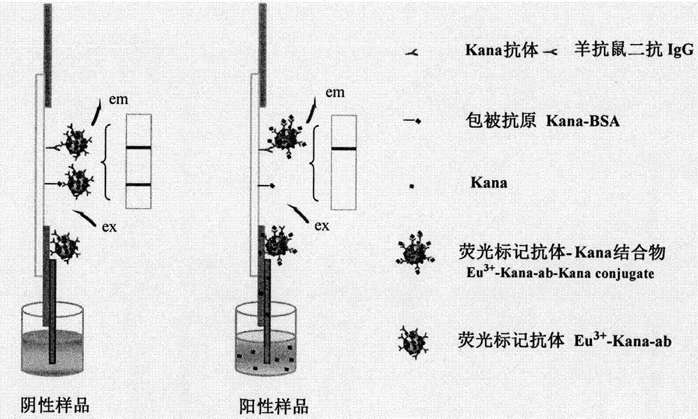
[0054] (2) Coating of quality control line (C line) and detection line (T line): on the NC membrane, use a dot film device to spot the coated antigen Kana-BSA into a strip as the detection line (T line); On the 0.5cm membrane of the detection line, draw a control line with goat anti-mouse secondary antibody IgG as the quality control line (C line), and dry it for later use.

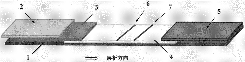
[0055] (3) Assembly of test strips: paste NC film on the middle of a rectangular bottom plate, mark the line with a film spotter, paste glass fiber on one end of the film, and lay an antibody-binding pad on the transfer side of the film. Glue absorbent paper to the other end. Then cut into 4mm strip test st...

Embodiment 3

[0058] Example 3: Preparation of Kanamycin Residual Fluorescent Immunoassay Strips, Different Fluorescently Labeled Antibodies Eu 3+ -Research experiment on the effect of Kana-ab dilution factor on the fluorescence performance of test strips

[0059] Antibodies labeled with fluorescent rare earth silica nanoparticles (Eu 3+ -Kana-ab) solution was diluted 1:1000, 1:1500, 1:2000, 1:4000, added 150 μL of blank sample, reacted in the dark, and carried out gel electrophoresis imaging detection and fluorescence card reader measurement.

[0060] Gel electrophoresis ( Figure 4 a) shows that, with Eu 3+ - As the dilution ratio of Kana-ab increases, the brightness of C-line and T-line gradually becomes lighter. When the dilution ratio is 1000 times, 1500 times and 2000 times, there is little difference in brightness visually. In the process of making fluorescent test strips, the ratio of the fluorescence intensity of the T line to the C line is close to 1, which is the best, such as...


PUM

PropertyMeasurementUnit
diameteraaaaaaaaaa

Abstract

The invention discloses a fluorescence immunochromatographic assay detection test paper for kanamycin residues and a preparation method of the fluorescence immunochromatographic assay detection test paper. The fluorescence immunochromatographic assay detection test paper comprises a sample cushion, an antibody binding cushion, an NC cellulose membrane and a bibulous paper, wherein the sample cushion, the antibody binding cushion, the NC cellulose membrane and the bibulous paper are sequentially and mutually overlapped through a bottom plate with adhesives from bottom to top; a coating detection line, namely a T line and a quality control line, namely a C line are sequentially arranged at one end close to the antibody fixation cushion, of the NC cellulose membrane; the antibody fixation cushion is used for bearing conjugates of fluorescence labeling antibodies; the T line which is sprayed and drawn by a coupling coating antigen Kana-BSA and a C line which is sprayed and drawn by a goat anti-rat secondary antibody IgG are separately arranged on the NC cellulose membrane from top to bottom; a fluorescence labelling probe is made from rare earth fluorescence silicon dioxide nanometer particles, and the fluorescence labeling probe is bound to the antibodies by using aldehyde group glucan as a cross-linking reagent; the preparation conditions of the fluorescence immunochromatographic assay test paper strip are inspected by a gel imaging system and a fluorescence chart reader; and the fluorescence intensity of the T line is detected by the fluorescence chart reader, and the quantitative detection is completed through a standard curve of the kanamycin antigen concentration.

Description

technical field [0001] The invention relates to an immunochromatographic test paper, in particular to a fluorescent immunochromatographic test paper for detecting kanamycin and a preparation method thereof. Background technique [0002] Kanamycin (Kanamycin, referred to as Kana), belongs to a kind of aminoglycoside antibiotics, has a strong inhibitory effect on common pathogenic bacteria, such as Escherichia coli, Mycobacterium tuberculosis and other Gram-negative bacteria. Bacterial 30S ribosomal RNA interacts to inhibit its protein synthesis and play a bactericidal effect. Once the dosage is not standardized, excessive intake of human body through food or drugs will cause serious side effects, including nephrotoxicity, hearing loss, allergies, etc. However, due to its low price and wide antibacterial properties, kanamycin is widely used, and it is easy to cause its residue in animal-derived foods, endangering human health. The EU stipulates that the residue of kanamycin i...

Claims


Application Information

Patent Timeline
Patent Type & Authority Applications(China)
IPC IPC(8): G01N33/533
CPCG01N33/533Y02A50/30
Inventor 邹明强齐小花赵兵洁
Owner CHINA INSPECTION LAB TECH CO LTD
Why Eureka
Try Eureka