Method for building vascularized tissue engineering periosteum

A tissue-engineered bone and vascularization technology, applied in the field of materials, can solve problems such as the inability of vascularized fibrous membrane to meet clinical needs, and achieve the effects of shortening the healing process, simple method, and shortening integration time.

Active Publication Date: 2018-02-02
WUHAN UNIV
View PDF12 Cites 10 Cited by
  • Summary
  • Abstract
  • Description
  • Claims
  • Application Information

AI Technical Summary

Problems solved by technology

Therefore, the vascularized fibrous membrane prepared b

Method used


Image

  • Method for building vascularized tissue engineering periosteum
  • Method for building vascularized tissue engineering periosteum
  • Method for building vascularized tissue engineering periosteum

Examples


Embodiment 1

[0032] Step 1. Preparation of chitosan / cellulose acetate cellulose membrane:

[0033] Chitosan and cellulose acetate (mass ratio 2:1) were dissolved in a mixed solvent of dimethylacetamide and acetone to obtain 8% composite spinning solution. Then the chitosan / cellulose acetate composite fiber membrane was prepared by electrospinning technology, and then the resulting chitosan / cellulose acetate composite fiber membrane was vacuum-dried in a vacuum oven for 24 hours to fully volatilize the solvent; the electrospinning voltage 16kV, propel the spinning solution at a speed of 1.2mL / h, set the distance to the receiver at 12cm, the spinning temperature at 25°C, and the relative humidity at 40%;

[0034] Soak in 100% alcohol for 30 minutes, soak in 95% alcohol for 5 minutes, soak in 90% alcohol for 5 minutes, soak in 85% alcohol for 5 minutes, soak in 80% alcohol for 5 minutes, soak in 70% alcohol for 5 minutes, soak in 60% alcohol for 5 minutes, 50 Soak in % alcohol for 5 minutes,...

Embodiment 2

[0041] Step 1. Preparation of acetate / cellulose micro / nanofibrous membrane:

[0042] Cellulose acetate was dissolved in a mixed solution of acetone and dimethylacetamide (mass ratio 2:1) to obtain a 16 wt% acetic acid / cellulose mixed solution. Then, the acetate / cellulose micro / nanofibrous membrane was prepared by electrospinning. Parameter setting: temperature is 25°C, relative humidity is 50%, voltage is 16kV, receiving distance is 15cm, solution flow rate is 1mL / h;

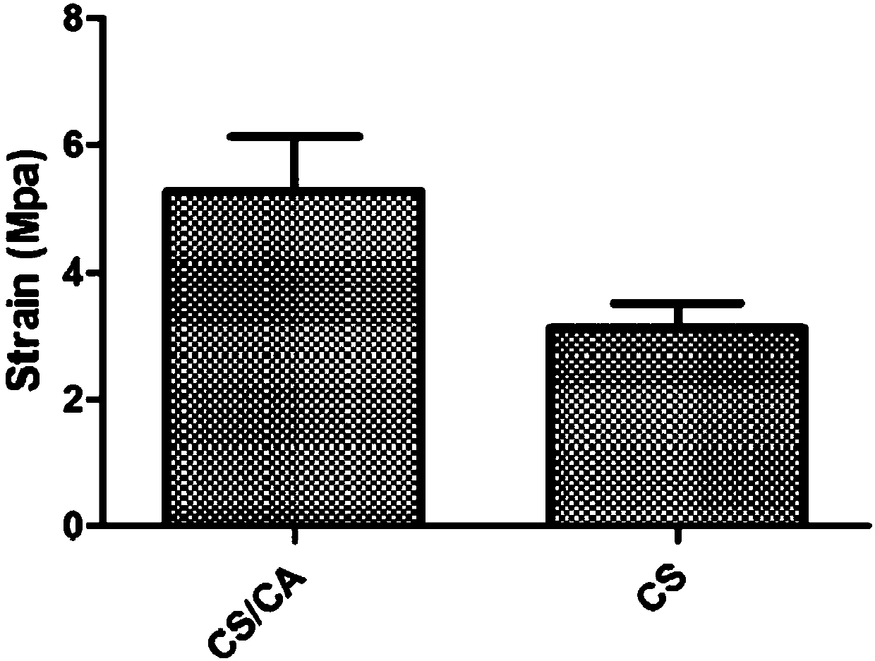
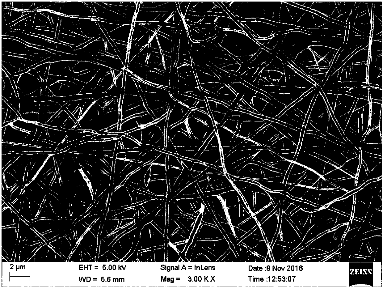
[0043] Vacuum-dry the prepared cellulose acetate micro / nanofiber composite membrane at 60°C to fully evaporate the residual solvent; place the dried micro / nanofiber membrane in 0.05mol / L sodium hydroxide solution for 7 days and dry it at room temperature Ultraviolet radiation disinfection for 24 hours; figure 2 As shown, the tensile test shows that the tensile force of the acetate / cellulose micro / nanofiber membrane is greater than that of the pure chitosan nanofiber membrane; it proves that the mechanical pro...

Embodiment 3

[0049] Step 1. Preparation of silk fibroin / polycaprolactone composite nanofiber membrane:

[0050] Take a certain amount of silk fibroin and polycaprolactone and dissolve them in hexafluoroisopropanol to prepare a mixed solution with a mass fraction of 3% of silk fibroin and polycaprolactone at 4:1; then use electrospinning technology to Preparation of silk fibroin / polycaprolactone composite nanofiber membrane, parameter settings: temperature 25°C, relative humidity 40%, voltage 15kV, receiving distance 12cm, solution flow rate 1mL / h;

[0051] Soak in 100% alcohol for 30 minutes, soak in 95% alcohol for 5 minutes, soak in 90% alcohol for 5 minutes, soak in 85% alcohol for 5 minutes, soak in 80% alcohol for 5 minutes, soak in 70% alcohol for 5 minutes, soak in 60% alcohol for 5 minutes, 50 Soak in % alcohol for 5 minutes, soak in double distilled water for 10 minutes;

[0052] The obtained silk fibroin / polycaprolactone composite nanofiber membrane was vacuum-dried at 60°C and ...


PUM


Abstract

The invention provides a method for building a vascularized tissue engineering periosteum. The method is characterized by comprising the following steps: 1, preparing a macromolecular micro/nano-fibermembrane by an electrostatic spinning method; 2, inoculating seed cells to the nano-fiber membrane, performing cultivation in vitro and performing directional induction for a period of time; 3, implanting a seed cell/nano-fiber membrane composite body subjected for in-vitro directional induction for a period of time in vivo, performing in-vivo vascularization for a period of time and then takingout to obtain the tissue engineering periosteum. By the method, the rejection reaction of an autologous tissue can be effectively reduced and the vascularization degree of the tissue engineering periosteum can be greatly increased; furthermore, the tissue engineering periosteum is vascularize in vivo in advance, so that the integration time of a blood vessel at the defect position and a blood vessel of the implanted tissue engineering periosteum can be shortened, the healing process of the bone defect is shortened, and the method has a very wide application prospect in the fields of wound healing and tissue engineering.

Description

technical field [0001] The invention belongs to the technical field of materials, and in particular relates to a method for constructing vascularized tissue engineering periosteum. technical background [0002] Craniomaxillofacial bone and long bones of extremities are susceptible to large-scale bone defects caused by trauma, infection, tumor and other iatrogenic factors. Traditional treatment methods mainly include autologous bone graft, allogeneic bone graft, and allogeneic bone graft repair, etc., but the source of autologous bone is limited and additional trauma is added. Allogeneic bone and allogeneic bone also have limited sources, poor biological activity, and defects such as immune rejection and infectious diseases, which limit their wide clinical application. In recent years, the rapid development of bone tissue engineering has brought hope for the clinical treatment of large-scale bone defects. [0003] However, the existing bone tissue engineering scaffolds focu...

Claims


Application Information

Patent Timeline
IPC IPC(8): A61L27/38A61L27/22A61L27/20A61L27/18A61L27/50D04H1/728D01D5/00
CPCA61L27/18A61L27/20A61L27/227A61L27/3821A61L27/3834A61L27/50A61L2430/02A61L2430/40D01D5/003D04H1/728C08L89/00C08L67/04C08L5/08C08L1/12
Inventor 邓红兵程谷施晓文杜予民
Owner WUHAN UNIV
Why Eureka
Try Eureka