Small-bore artificial blood vessel with micropatterned inner wall

An artificial blood vessel and micro-patterning technology, applied in coatings, medical science, prostheses, etc., can solve the problems of inability to guide the oriented growth of endothelial cells, poor mechanical properties of small-caliber artificial blood vessels, etc., and solve the problem of poor biocompatibility Poor, solve the effect of insufficient mechanical properties

Inactive Publication Date: 2018-03-13
BEIHANG UNIV
View PDF14 Cites 6 Cited by
  • Summary
  • Abstract
  • Description
  • Claims
  • Application Information

AI Technical Summary

Problems solved by technology

[0007] In order to solve the problem that the small-diameter artificial blood vessel prepared by the prior art has poor mechanical properties and cannot guide the oriented growth of endothelial cells, thereby inhibiting the function of endothelial cells, the present invention provides an artificial blood vessel that can guide the oriented growth of endothelial cells and has good mechanical properties and biological phase. Capacitive inner wall micropatterned small-bore artificial blood vessel

Method used


Image

  • Small-bore artificial blood vessel with micropatterned inner wall
  • Small-bore artificial blood vessel with micropatterned inner wall
  • Small-bore artificial blood vessel with micropatterned inner wall

Examples


example 1

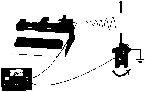
[0027] 1. Cover the AR-P 3250 positive photoresist evenly on the glass sheet with a coater, and after curing at 95°C, use a UV lithography machine to prepare parallel stripe micropatterns with a stripe width and spacing of 20 μm and a depth of 2 μm. Engraving;

[0028] 2. Use a coating machine to evenly cover the surface of the above micropattern photoresist with polydimethylsiloxane, place it on a heating plate at 95°C for 2 hours, and use tweezers to carefully peel off the film on the surface after curing to obtain the micropattern Polydimethylsiloxane film;

[0029] 3. Boil 20 g silkworm silk in 2 L of 0.02 mol / L sodium carbonate deionized aqueous solution for 30 minutes for degumming treatment, wash the boiled silk 6 times with deionized water, loosen it and place it in an oven at 37°C to dry , to obtain degummed silk;

[0030] 4. Dissolve degummed silk in 9.3mol / L lithium bromide aqueous solution at the ratio of 27g:100mL, stir and dissolve in a 60°C oven for 4 hours to...

Embodiment 2

[0039] 1. Soak the micro-patterned small-diameter artificial blood vessel on the inner wall prepared in Example 1 in phosphate buffered saline, and fully wet it to obtain a wet small-caliber artificial blood vessel;

[0040] 2. The wet small-diameter artificial blood vessel is cut into a test sample with a length of 3 cm, and the axial tensile mechanical test is carried out using a Japanese Shimadzu universal material testing machine. min;

[0041] 3. Cut the wet small-diameter artificial blood vessel into a test sample with a length of 6 mm, use the above-mentioned material testing machine to carry out a radial tensile mechanical test, select a load cell with a measuring range of 100 N, and set the tensile rate to 3 mm / min;

[0042] 4. The wet small-caliber artificial blood vessel is cut into a test sample with a length of 2 cm, and the suture strength mechanical test is carried out using the above-mentioned material testing machine and 6-0 polyglycolic acid surgical suture w...

example 3

[0046] 1. Cut the micropatterned small-diameter artificial blood vessel on the inner wall prepared in Example 1 into a test sample with a length of 1 cm, soak it in phosphate buffer solution, and sterilize it in an autoclave to obtain a sterile test sample;

[0047] 2. Place the sterility test sample and the utensils used for cell inoculation in an ultra-clean bench, use ultraviolet sterilization for more than 0.5h, move the sterility test sample into a 24-well plate, and place the sterility test sample on the test sample after drying. Inoculation of inner wall 1x10 5 Endothelial cells were cultured in a carbon dioxide incubator for 2 hours after inoculation to make the cells adhere, then added 0.8mL endothelial cell medium, and placed in a carbon dioxide incubator for 3 days;

[0048] 3. Immunofluorescent staining of the cultured small-caliber artificial blood vessels, using fluorescein isothiocyanate-labeled phalloidin to stain the cytoskeleton fibrous actin, and then using ...


PUM

PropertyMeasurementUnit
thicknessaaaaaaaaaa
thicknessaaaaaaaaaa
thicknessaaaaaaaaaa

Abstract

The invention relates to the technical field of biological materials, and provides a small-bore artificial blood vessel with a micropatterned inner wall. The small-bore artificial blood vessel has a two-layer structure. An outer layer is an electrospun microfiber layer, the mechanical properties of the small-bore artificial blood vessel are improved, and the stability of a tubular structure is maintained. An inner layer is a coating layer having a micro-patterned structure, so adhesion, spreading and growth of endothelial cells are facilitated, the extension of an endothelial cytoskeleton canbe promoted and the oriented growth of the endothelial cells can be guided, and the normal exertion of cell functions is ensured. However, the mechanical properties of conventional products and researched small-bore artificial blood vessels are relatively poor, the clinical requirements cannot be completely met, and the oriented growth of the endothelial cells and the normal exertion of the cell functions cannot be promoted. The small-bore artificial blood vessel with the micropatterned inner wall has good mechanical and biological properties, and is suitable for repairing and replacing small-bore blood vessel tissues with injury or lesion.

Description

technical field [0001] The invention relates to the technical field of biomaterials, in particular to a micropatterned small-diameter artificial blood vessel. Background technique [0002] Cardiovascular disease has become the number one killer threatening human health. The occurrence of many cardiovascular diseases is closely related to the narrowing or occlusion of blood vessels. Narrowing or occlusion of blood vessels prevents the normal flow of blood, causing tissue damage due to insufficient nutrient supply. Common diseases include coronary heart disease, cerebrovascular disease, peripheral atherosclerosis and deep vein thrombosis, etc. Vascular reconstruction is an important method for the treatment of such diseases and occupies a very important position in clinical surgery. Up to now, more than 500,000 patients need vascular surgery each year in the United States alone, and most of them need suitable vascular grafts. [0003] At present, the clinically applied sma...

Claims


Application Information

Patent Timeline
Patent Type & Authority Applications(China)
IPC IPC(8): A61L27/40A61L27/18A61L27/22A61L27/34A61L27/50
CPCA61L27/18A61L27/22A61L27/34A61L27/50A61L27/507C08L83/04C08L67/04C08L89/00
Inventor 刘海峰邹同强丁希丽樊瑜波
Owner BEIHANG UNIV
Why Eureka
Try Eureka