Medical biological yarn, medical biological repair mesh and preparation method thereof
A kind of biological thread and biological technology, applied in medical science, tissue regeneration, prosthesis, etc., can solve the problems of tissue regeneration mismatch, cytotoxicity, and insufficient mechanical strength, so as to reduce the incidence of inflammation and infection and achieve good mechanical properties , the effect of large mechanical strength
- Summary
- Abstract
- Description
- Claims
- Application Information
AI Technical Summary
Problems solved by technology
Method used
Image
Examples
Embodiment 1
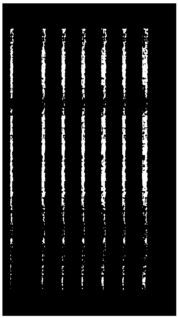
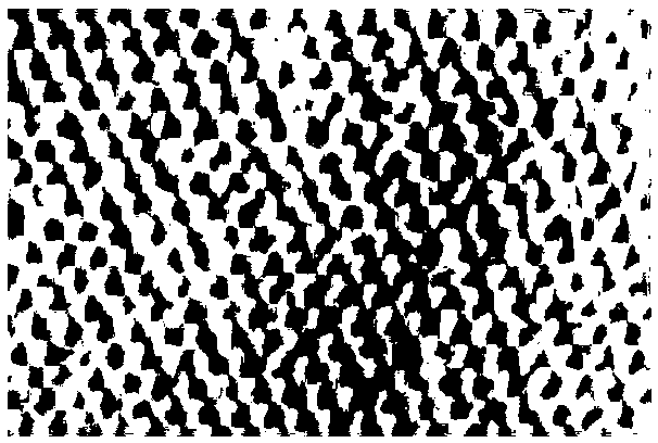
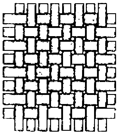
[0056] see Image 6 , Preparation of medical biological thread and medical biological repair mesh of porcine small intestinal submucosa soft tissue
[0057] (1) Pretreatment
[0058] Freshly slaughtered porcine small intestine tissues were cleaned, soaked in 0.5% acetic acid solution for 30 minutes, the ratio of porcine small intestine to acetic acid solution was 1:5, and the mucosal layer, muscular layer and serosa layer of porcine small intestine and jejunum were removed by physical scraping , lymph nodes, the submucosa was separated, cut into thin strips evenly in the longitudinal direction, and washed 3 times with purified water to obtain biological materials, namely small intestinal submucosa, hereinafter referred to as SIS materials.
[0059] (2) Virus inactivation
[0060] Use a mixed aqueous solution containing 1.0% peracetic acid and 15% ethanol, the ratio of the SIS material to the mixed aqueous solution is 1:10, and soak at room temperature for 100 minutes under u...
Embodiment 2
[0071] Preparation of bio-repair mesh for porcine small intestinal submucosa
[0072] (1) Pretreatment
[0073] Freshly slaughtered pig small intestine tissues were cleaned and soaked in 0.01% acetic acid solution for 120 minutes. The ratio of pig small intestine to acetic acid solution was 1:10. The mucosal layer, muscular layer and serosa layer of pig small intestine jejunum were removed by physical scraping , lymph nodes, the submucosa was separated, cut into fragments, and washed 3 times with purified water to obtain a biological material, that is, the submucosa of the small intestine, hereinafter referred to as SIS material.
[0074] (2) Virus inactivation
[0075] Use a mixed aqueous solution containing 0.5% peracetic acid and 25% ethanol, the ratio of the SIS material to the mixed aqueous solution is 1:15, and soak at room temperature for 120 minutes under ultrasonic conditions to inactivate the virus. Afterwards, it was ultrasonically washed 3 times with purified wat...
Embodiment 3
[0086] For the safety of the samples, the samples prepared in Example 1-2 were tested for immunogenic substances.
[0087] (1) Detection method of residual cells: fixed with 10% neutral formalin, embedded in paraffin, cut into thin slices of 0.4 micron, dewaxed with xylene, dehydrated with serial alcohol, stained with hematoxylin-eosin, and microscope Next, observe the cell residue and matrix fiber structure.
[0088] (2) Detection method of DNA content: according to YY / T 0606.25-2014 "Method for determination of DNA residues in biological materials of animal origin: fluorescent staining method".
[0089] (3) Detection method of α-Gal antigen content: After the sample was fixed with paraformaldehyde, it was routinely embedded in paraffin and sectioned, with a thickness of 3 μm. Immunohistochemical reaction was carried out by using the specific affinity between biotin-labeled BSI-B4 and α-Gal antigen. Judgment of staining results: dark brown-yellow particles are strongly posi...
PUM
 Abstract
Description
Claims
Application Information
 - R&D
- Intellectual Property
- Life Sciences
- Materials
- Tech Scout
- Unparalleled Data Quality
- Higher Quality Content
- 60% Fewer Hallucinations
Browse by: Latest US Patents, China's latest patents, Technical Efficacy Thesaurus, Application Domain, Technology Topic, Popular Technical Reports.
© 2025 PatSnap. All rights reserved.Legal|Privacy policy|Modern Slavery Act Transparency Statement|Sitemap|About US| Contact US: help@patsnap.com


