
Amphiphilic polymer nano micelle containing ferric ions chelated with polydopa amino acid and application thereof

An amphiphilic polymer and polydopa amino acid technology, which is applied in the field of amphiphilic polymer nanomicelles, can solve the problems of indistinguishable signal substances, restricted reticuloendothelial system, time-consuming and high cost, and achieves relaxation performance. High, obvious imaging effect, the effect of broadening the scope of medical applications

Active Publication Date: 2019-11-26
ZHEJIANG UNIV
View PDF2 Cites 2 Cited by
  • Summary
  • Abstract
  • Description
  • Claims
  • Application Information

AI Technical Summary

Problems solved by technology

Its disadvantages on the one hand T 2 Imaging contrast agents are difficult to distinguish from low-signal substances such as gas, cortical bone, and iron deposition in the body; on the other hand, the scope of application is limited, limited by the reticuloendothelial system
[0008] However, the surface modification of iron oxide nanoparticles is generally cumbersome, time-consuming and costly, and T 2 Imaging MRI contrast agents have largely been phased out, so there is a need to develop new alternatives to gadolinium for MRI T 1 imaging contrast agent

Method used


Image

  • Amphiphilic polymer nano micelle containing ferric ions chelated with polydopa amino acid and application thereof
  • Amphiphilic polymer nano micelle containing ferric ions chelated with polydopa amino acid and application thereof
  • Amphiphilic polymer nano micelle containing ferric ions chelated with polydopa amino acid and application thereof

Examples


Embodiment 1

[0051] (1) The structural formula of polydopa amino acid-polysarcosine block copolymer (PDOPA-b-PSar) is as follows:

[0052]

[0053] Among them, R 1 Be benzyl; m=5~200, n=5~50;

[0054] Concrete synthetic steps include:

[0055] Add sarcosine NCA to the Schlenk bottle, dissolve it with DMF, then add the DMF solution of benzylamine, the molar ratio of sarcosine NCA to benzylamine is 5-200:1, react at room temperature for 1 day, then add benzyloxycarbonyl ( cbz) protected DMF solution of dopa NCA, the molar ratio of dopa NCA to benzylamine is 5-50: 1, react at room temperature for 1 day, pour the polymer solution into ether to precipitate, filter, and vacuum-dry the obtained polymer In 1 day, a cbz-protected polydopa amino acid-polysarcosine block copolymer was obtained.

[0056] Dissolve 300 mg of block copolymer in 3 mL of trifluoroacetic acid, add 4 times the equivalent of hydrogen bromide in acetic acid solution (33%), react for 3 hours, precipitate with ether, filte...

Embodiment 2

[0063] (1) Other preparation conditions are the same as in Example 1, except that polyethylene glycol amine is used as a macroinitiator, and the prepared polydopa amino acid-polyethylene glycol block copolymer (PDOPA-b-PEG ) has the following structural formula:

[0064]

[0065] Among them, R 2 It is methyl; m=5~200, n=5~50.

[0066] (2) Dissolve the weighed 9.7mg PDOPA-b-PEG in DMF to form a solution, then slowly add 3.27mg Fe(NO 3 ) 3 9H 2 O in DMF and dialyzed against deionized water for 48 hours. The obtained micellar solution was filtered with a filter membrane with a pore size of 0.45 μm and used at constant volume.

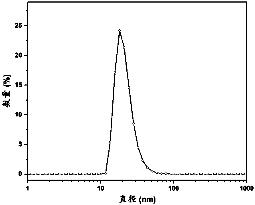
[0067] Other performance test conditions are the same as in Example 1, the average particle size of the micelles is 30nm, and it has MRI enhancing effect in vitro.

Embodiment 3

[0069] (1) The structural formula of polydopa amino acid-polyethylene glycol methacrylate graft polymer (POEGMA-g-PDOPA) is as follows:

[0070]

[0071] Among them, R 1 It is n-butyl; m=5~200, n 1 =5~50;

[0072] Concrete synthetic steps include:

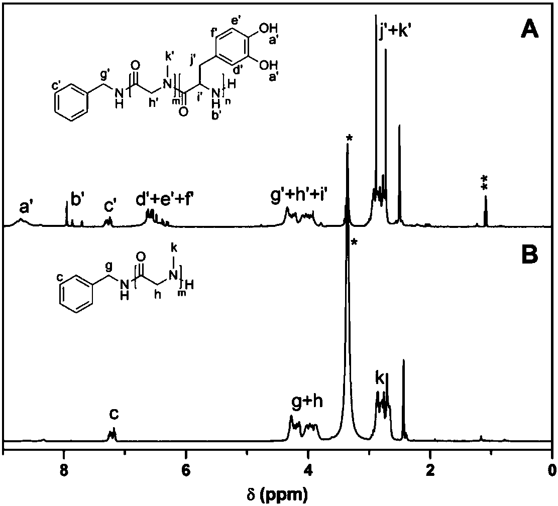
[0073] PDOPA was prepared by ring-opening polymerization and deprotection of cbz-protected dopa NCA initiated by n-butylamine, and the conditions were the same as in Example 1; poly(polyethylene glycol methacrylate) (POEGMA) was prepared by RAFT polymerization. Dissolve 247.4mg POEGMA and 134.0mg PDOPA in 1mL DMF, place in 35oC oil bath to react for 4 days, pour the polymer solution into ether to precipitate, filter, and vacuum dry for 1 day to obtain polydopa amino acid-polymethacrylic acid Oligoethylene glycol ester graft polymer. The H NMR spectrum of the polymer is shown in Figure 7 shown.

[0074] (2) Dissolve the weighed 22.7mg POEGMA-g-PDOPA in DMF to form a solution, then slowly add 5.83mg Fe(NO 3 ) 3 9H 2 O in D...


PUM

PropertyMeasurementUnit
particle sizeaaaaaaaaaa

Abstract

The invention discloses an amphiphilic polymer nano micelle containing ferric ions chelated with polydopa amino acid. The iron ions are chelated with the catechol structure of the biodegradable polydopa amino acid block side chain. The invention also provides a preparation method of the micelle. The method comprises the following steps: complexing an amphiphilic polymer containing the polydopa amino acid with a ferric ion compound, and acquiring the amphiphilic polymer nano micelle containing the ferric ions chelated with the polydopa amino acid by a solvent replacement method. The micelle prepared by the method is used as the Fe3+magnetic resonance T1 imaging contrast agent, and can avoid the toxic and side effects caused by a traditional gadolinium reagent. The longitudinal relaxation rate (r1) of the micelle is 5.6 mM<-1> s <-1>, the circulation time of the micelle in a mouse body can reach 150 minutes, the imaging effect is obvious, the comprehensive performance of the micelle is much higher than that of a commercial gadolinium contrast agent, and the micelle has a good application prospect.

Description

technical field [0001] The invention belongs to the field of magnetic resonance imaging, and in particular relates to an amphiphilic polymer nanomicelle containing polydopa amino acid chelated ferric ions and its application. Background technique [0002] Magnetic Resonance Imaging (MRI) uses the principle of nuclear magnetic resonance plus a high-frequency gradient magnetic field to detect electromagnetic waves emitted from the body, and based on this, it can draw an image of the internal structure of the object. Due to its advantages of non-invasiveness, real-time monitoring, and ability to provide high-resolution soft tissue information and three-dimensional structural imaging, it is one of the most advanced clinical medical examination technologies (Nat. Rev. Neurosci. 2007, 8, 700-711). [0003] In clinical applications, more than 30% of diagnoses require the use of MRI contrast agents in order to improve the contrast between normal and diseased tissues. The magnetic r...

Claims


Application Information

Patent Timeline
Patent Type & Authority Applications(China)
IPC IPC(8): A61K49/18A61K49/12
CPCA61K49/1809A61K49/124C08G69/00A61K49/128B82Y30/00A61B5/055A61K49/101A61K49/1857
Inventor 孙继红凌君岑佳宇苗岳栋
Owner ZHEJIANG UNIV
Learn More