Epitope identification method of thallus antibody

A technology of antigenic epitopes and bacterial antibodies, applied in chemical instruments and methods, specific peptides, anti-bacterial immunoglobulins, etc., to achieve the effects of improving research efficiency, reducing the number of synthesis, and reducing workload

Pending Publication Date: 2020-09-04
JIANGSU OCEAN UNIV
View PDF2 Cites 2 Cited by
  • Summary
  • Abstract
  • Description
  • Claims
  • Application Information

AI Technical Summary

Problems solved by technology

At present, it is known at home and abroad that most of the antibodies recognize the O157 lipopolysaccharide and H7 flagellar antigen of E. coli

Method used


Image

  • Epitope identification method of thallus antibody
  • Epitope identification method of thallus antibody
  • Epitope identification method of thallus antibody

Examples


Embodiment 1

[0033] The recognition epitope identification of implementation example 1 monoclonal antibody 2G12

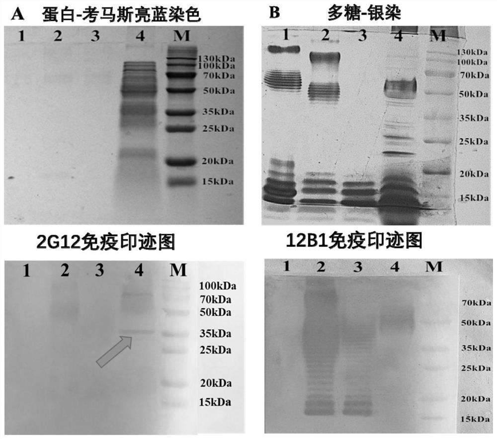
[0034] (1) Identification of the type of antigen recognized by monoclonal antibody 2G12

[0035] Bacterial whole bacterial protein was prepared by ultrasonic crushing method. The ultrasonic conditions were fully mixed in an ice bath, ultrasonic crushed in an ice bath at 4°C, and the intensity was 50%. Centrifuge the sonicated bacterial solution at 12,000 g at 4°C for 10 min, and collect the supernatant. The whole bacterial protein samples were separated by SDS-PAGE protein gel electrophoresis, and the protein was transferred to PVDF membrane by electroporation and semi-dry method (current was 40mA / cm, electroporation was 30min), and immunoblotting was performed (primary antibody and secondary antibody Concentrations were 0.5 μg / mL and 0.2 μg / mL).

[0036] Bacterial lipopolysaccharide was extracted by the hot phenol method, and E.coli O157:H7 was cultured at 37°C for 24 hours,...

Embodiment 2

[0059] Cross-reactivity test of embodiment 2 monoclonal antibody 2G12

[0060] The cross-reaction of monoclonal antibody 2G12 to 68 strains of bacteria was detected by indirect ELISA coated with bacteria. ① Packing plate: Take the boiled and inactivated E.coli O157:H7 bacteria and dilute it with CBS carbonate buffer to a concentration of 108 CFU / ml, add 100uL / well to the microtiter plate. Afterwards, the plate was sealed and placed in a constant temperature incubator, and incubated at 37°C for 2 hours. ②Washing: shake off the liquid and pat dry, add washing solution at 230uL / well, oscillate on a shaker at a constant speed for 3min, spin dry, repeat the steps of washing 3 times, and pat dry. ③Sealing: Add 200uL / well of blocking solution, seal the plate and put it into a constant temperature incubator to seal for 2h. ④ Add serum: After washing according to step (2), dilute the monoclonal antibody (2G12, 12B1 in this article) with antibody diluent (take it out of the refrigerat...

Embodiment 3

[0061] Example 3 Affinity Determination of Monoclonal Antibody 2G12

[0062] The bacteria liquid to be tested was from 10 8 / mL began to coat with six different concentrations of 3-fold dilution of the coating solution, and each well was made in triplicate. Starting from 4 μg / mL, monoclonal antibody was diluted 8 times by 3 times, and the titer of monoclonal antibody (mAb) was determined by indirect enzyme-linked immunosorbent method. Then, with the logarithmic value of mAb concentration (μg / mL) as the abscissa and the absorbance value Abs (450nm) as the ordinate, four S-shaped curves (R 2 ≥0.99), calculate the corresponding antibody concentration when Abs (450nm) is 50% of the highest absorbance value according to the curve equation, convert the unit mol / L of the antibody concentration, and then calculate the affinity constant (Ka ):

[0063] Ka=(n-1) / 2{n[Ab']t-[Ab]t}

[0064] Among them, n is the ratio of the two coating concentrations (greater than 1); [Ab']t is the cor...


PUM


Abstract

The invention provides an epitope identification method of a thallus antibody, and belongs to the technical field of food safety immunological detection. A plurality of polypeptide fragments are obtained by affinity chromatography in identifying protein epitopes; intramembrane sequences and part of high-homology sequences in the target protein are preliminarily eliminated through topological structure, protein sequence homology and bacterial cross reaction analysis of the target protein; epitope mapping is mainly carried out on an extracellular loop structure sequence synthesis short peptide library with 3-6 amino acid differences, and tests prove that the method significantly reduces the workload and cost of epitope mapping, and improves the efficiency of epitope identification; the bacterial antigen epitope identification method has a wide application prospect in bacterial antibody epitope identification and exploration.

Description

technical field [0001] The invention relates to a method for identifying antigenic epitopes of bacteria antibodies, belonging to the technical field of food safety immunology detection. Background technique [0002] The immunoassay method is a common rapid detection method for pathogenic bacteria. It has the characteristics of simple operation, low cost and easy promotion, and is suitable for the primary screening detection of a large number of samples. The national standard detection method for Escherichia coli O157:H7 is accurate and reliable, but it still relies on plate culture and biochemical reactions, and the operation is cumbersome, time-consuming and laborious. Immunoassay methods and polymerase chain reaction (PCR) and other molecular biology detection methods are currently the main rapid detection methods. PCR detection methods have relatively high technical requirements for equipment and operators. Immunoassay methods have the advantages of simple operation and l...

Claims


Application Information

Patent Timeline
IPC IPC(8): G01N33/68G01N33/569G01N33/577G01N27/447G01N30/02G01N30/72C07K14/245C07K16/12
CPCG01N33/68G01N33/56916G01N33/577G01N27/44747G01N30/02G01N30/72C07K14/245C07K16/1232
Inventor 王文彬桑雨浓盘赛昆高云山梁夏夏刘建欣刘蕾袁倩云唐秀
Owner JIANGSU OCEAN UNIV
Why Eureka
Try Eureka