Method for detecting activity of alpha-glucosidase

A glucosidase and detection method technology, applied in the field of biological enzyme activity detection, can solve problems such as fluorescence quenching, and achieve the effects of low detection limit, simple operation and good selectivity

Active Publication Date: 2020-11-03
ANHUI NORMAL UNIV
View PDF20 Cites 8 Cited by
  • Summary
  • Abstract
  • Description
  • Claims
  • Application Information

AI Technical Summary

Problems solved by technology

The invention uses the fluorescence enhancement phenomenon to detect, and solves the problem that oth...

Method used


Image

  • Method for detecting activity of alpha-glucosidase
  • Method for detecting activity of alpha-glucosidase
  • Method for detecting activity of alpha-glucosidase

Examples


Embodiment 1

[0048] A method for preparing carbon quantum dots, comprising the following steps:

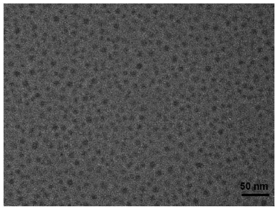
[0049] Dissolve 0.02 g of p-phenylenediamine in 10 mL of ultrapure water, and adjust the pH to 5–6 by adding 10 mL of 1 mM citric acid solution. The mixed solution was then transferred into a polytetrafluoroethylene stainless steel autoclave and 15 mL of ultrapure water was added, then the mixture was placed in an oven and heated at 180 °C for 12 hours. After 12 hours, the mixture was cooled to room temperature, centrifuged to obtain the clear liquid, and then dialyzed with a 500Da dialysis bag for 4 hours to remove unreacted substances, and the carbon dot solution in the dialysis bag was retained. The dialysate during dialysis was ultrapure water. The product was transferred to dry in a vacuum oven, redissolved into 0.35g L -1 The carbon quantum dots (CDs) solution was stored at 4°C for future use.

[0050] figure 1 It is a transmission electron microscope picture of CDs. It can be seen fr...

Embodiment 2

[0055] A quantitative detection of Ce 4+ method, including the following steps:

[0056] (1) Take 100μL 0.35g L -1 Carbon dots (CDs) solution, 100 μL of 1 mM TMB water and a specific concentration of Ce 4+ The aqueous solution was mixed in 100 μL of 0.2M pH 4.0 citrate buffer solution, then water was added to 1.0 mL, and mixed well, Ce 4+ The final concentrations in the reaction system were 0, 10, 20, 30, 40, 50, 60, 70, 80, 90, 100 μM; the Ce 4+ The source of cerium sulfate (Ce(SO 4 ) 2 );

[0057] The mixture was incubated at room temperature for 5 minutes, and then the fluorescence spectrum of each reaction system was tested at an excitation wavelength of 490nm. The fluorescence spectrum of each reaction system was as follows: Figure 5 As shown, it can be seen from the figure that with Ce 4+ As the concentration increases, the fluorescence intensity of the reaction system decreases gradually.

[0058] (2) with Ce 4+ The final concentration is the abscissa, and the...

Embodiment 3

[0061] A method for detecting α-glucosidase activity, comprising the following steps:

[0062] (1) Mix 200μL 0.5mM ascorbyl glucoside (AA-2G) solution with 50μL α-glucosidase solution of different concentrations, then add 10μL 0.2M PBS buffer solution with pH7.0, mix well and incubate at 37℃ 30 minutes;

[0063] (2) Then add 100 μL CDs solution, 100 μL 1mM TMB aqueous solution and 100 μL 1mMCe to the above system in sequence 4+ aqueous solution, then add 100 μL of 0.1M pH 4.0 citric acid buffer solution, and dilute to 1 mL with ultrapure water. After incubating at room temperature for 5 minutes, test the fluorescence spectrum of each reaction system at an excitation wavelength of 490nm, the fluorescence spectrum of each reaction system is as follows: Figure 9 Shown; The final activity of α-glucosidase in the reaction system is 0, 0.5, 1.0, 1.5, 2.0, 2.5, 3.0, 3.5, 4.0, 4.5, 5.0, 5.5, 6 U mL -1 ; The Ce 4+ The source of cerium sulfate (Ce(SO 4 ) 2 );

[0064] (3) With t...


PUM


Abstract

The invention discloses a method for detecting alpha-glucosidase activity, which comprises the following steps: taking p-phenylenediamine as a nitrogen source and citric acid as a carbon source to prepare CDs by a hydrothermal method; detecting the activity of alpha glucosidase based on a CDs/TMB/Ce < 4 + > probe system; ce < 4 + > can oxidize colorless TMB to generate a blue TMB oxide, and due tothe internal filtering effect, the ultraviolet absorption of oxTMB is overlapped with the fluorescence emission of CDs, so that the fluorescence emission intensity of CDs is reduced; the alpha-glucosidase can hydrolyze ascorbic acid glucoside to release ascorbic acid with reducibility, the ascorbic acid can reduce oxTMB into colorless TMB, the internal filtering effect disappears, fluorescence ofCDs is recovered, the fluorescence recovery degree is in direct proportion to the activity of the alpha-glucosidase, and then the activity of the alpha-glucosidase is detected. According to the invention, the fluorescence enhancement phenomenon is used for detection, the problem that the method using the fluorescence quenching phenomenon is easily influenced by false positive signals is solved, and the method has the advantages of low detection limit, high sensitivity and good selectivity.

Description

technical field [0001] The invention belongs to the technical field of biological enzyme activity detection, and relates to a method for detecting α-glucosidase activity, in particular to a method based on CDs / TMB / Ce 4+ A method for detecting α-glucosidase activity with a probe system. Background technique [0002] Biological enzymes control various physiological processes in organisms together with metabolism. Sensitive detection of the activity of biological enzymes is crucial for exploring their biological functions and their impact on organisms, and helps to diagnose related diseases. [0003] α-glucosidase is a membrane-bound enzyme in the epithelium of the small intestine that specifically hydrolyzes α-glucopyranosidic bonds to release individual α-glucose molecules. Excess α-glucose in the blood can cause chronic damage and dysfunction of various tissues. α-glucosidase plays a vital role in controlling postprandial blood sugar levels and keeping glucose levels withi...

Claims


Application Information

Patent Timeline
IPC IPC(8): G01N21/64
CPCG01N21/64G01N21/6428G01N2021/6421G01N2021/6432
Inventor 吴芳菲刘金水
Owner ANHUI NORMAL UNIV
Why Eureka
Try Eureka