Image processing and analysis of individual nucleic acid molecules

a nucleic acid and image processing technology, applied in the field of individual nucleic acid molecules, can solve the problems of inability to obtain sufficient quantities of dna from these sources which are sufficient for detailed analyses, time-consuming and often impractical, and achieve high throughput , the effect of rapid generation of information

Inactive Publication Date: 2003-02-20
WISCONSIN ALUMNI RES FOUND
View PDF0 Cites 64 Cited by
  • Summary
  • Abstract
  • Description
  • Claims
  • Application Information

AI Technical Summary

Benefits of technology

0022] Still further, high throughput methods for utilizing such single nucleic acid molecules in genome analysis are presented. In one embodiment of such high throughput methods, rapid optical mapping approaches are described for the creation of high-resolution restriction maps- In such an embodiment, single nucleic acid molecules are elongated, fixed and gridded to high density onto a solid surface. These molecules can then be digested with appropriate restriction enzymes for the map construction. In an alternative embodiment, the single nucleic acid molecules can be elongated, fixed and gridded at high density onto a solid sur

Problems solved by technology

The analysis of nucleic acid molecules at the genome level is an extremely complex endeavor which requires accurate, rapid characterization of large numbers of often very large nucleic acid molecules via high throughput DNA mapping and sequencing.
The construction of physical maps, and ultimately of nucleotide sequences, for eukaryotic chromosomes currently remains laborious and difficult.
Such electrophoretic methods offer an acceptable level of size resolution, but, especially for purposes of high throughput mapping, suffer from a number of setbacks.
Obtaining quantities of DNA from these sources which is sufficient for detailed analyses, such as restriction mapping, is time consuming and often impractical.
Second, because populations of molecules of like size migrate through the medium at the same rate, it is impossible to separate individual molecules from within a sample of particles by utilizing such a technique.
Further, standard gel electrophoresis techniques involve the sep

Method used


Image

  • Image processing and analysis of individual nucleic acid molecules
  • Image processing and analysis of individual nucleic acid molecules
  • Image processing and analysis of individual nucleic acid molecules

Examples


second embodiment

[0282] In the present invention, the spotted glass surface is rehydrated, after which a teflon block stamp is pressed onto the DNA spots, causing them to spread and fix on the sticky, derivatized surface. Experimental results indicate that this approach is effective for elongating surface mounted DNA without significant breakage. FIG. 29 shows an enlarged view of a DNA spot and the use of a teflon block in accordance with this embodiment of the present invention to spread the molecules onto the derivatized surface.

[0283] In a third, preferred method of the present invention, the deposited droplets of DNA solution are simply let dry on the derivatized surface. Experiments show that as the droplets dry, most of the fixed DNA remains fully elongated, aligned, and primarily deposited within the spot peripheries in a characteristic "sunburst" pattern, clearly observable in FIGS. 26A, B and C. Addition of glycerol to the spotting solution results in well elongated DNA molecules which are ...

example 1

Preparing DNA for Microscopy

[0347] G bacteria was grown as described by Fangman, W. L., Nucl. Acids Res., 5, 653-665 (1978), and DNA was prepared by lysing the intact virus in 1 / 2TBE buffer (1.times.: 85 mM Trizma Baste (Sigma Chemical Co., St. Louis Mo.), 89 mM boric acid and 2.5 mM disodium EDTA) followed by ethanol precipitation; this step did not shear the DNA as judged by pulsed electrophoresis and microscopic analysis.

[0348] DNA solutions (0.1 microgram / microliter in 1 / 2.times.TBE) were diluted (approximately 0.1-0.2 nanogram / al agarose) with 1.0% low gelling temperature agarose (Sea Plague, FMC Corp., Rockport Me.) in 1 / 2.times.TBE, 0.3 micrograms / ml DAPI (Sigma Chemical Co.), 1.0% 2-mercaptoethanol and held at 65.degree. C. All materials except the DNA were passed through a 0.2 micron filter to reduce fluorescent debris. Any possible DNA melting due to experimental conditions was checked using pulsed electrophoresis analysis and found not to be a problem.

example 2

Imaging DNA in a Gel

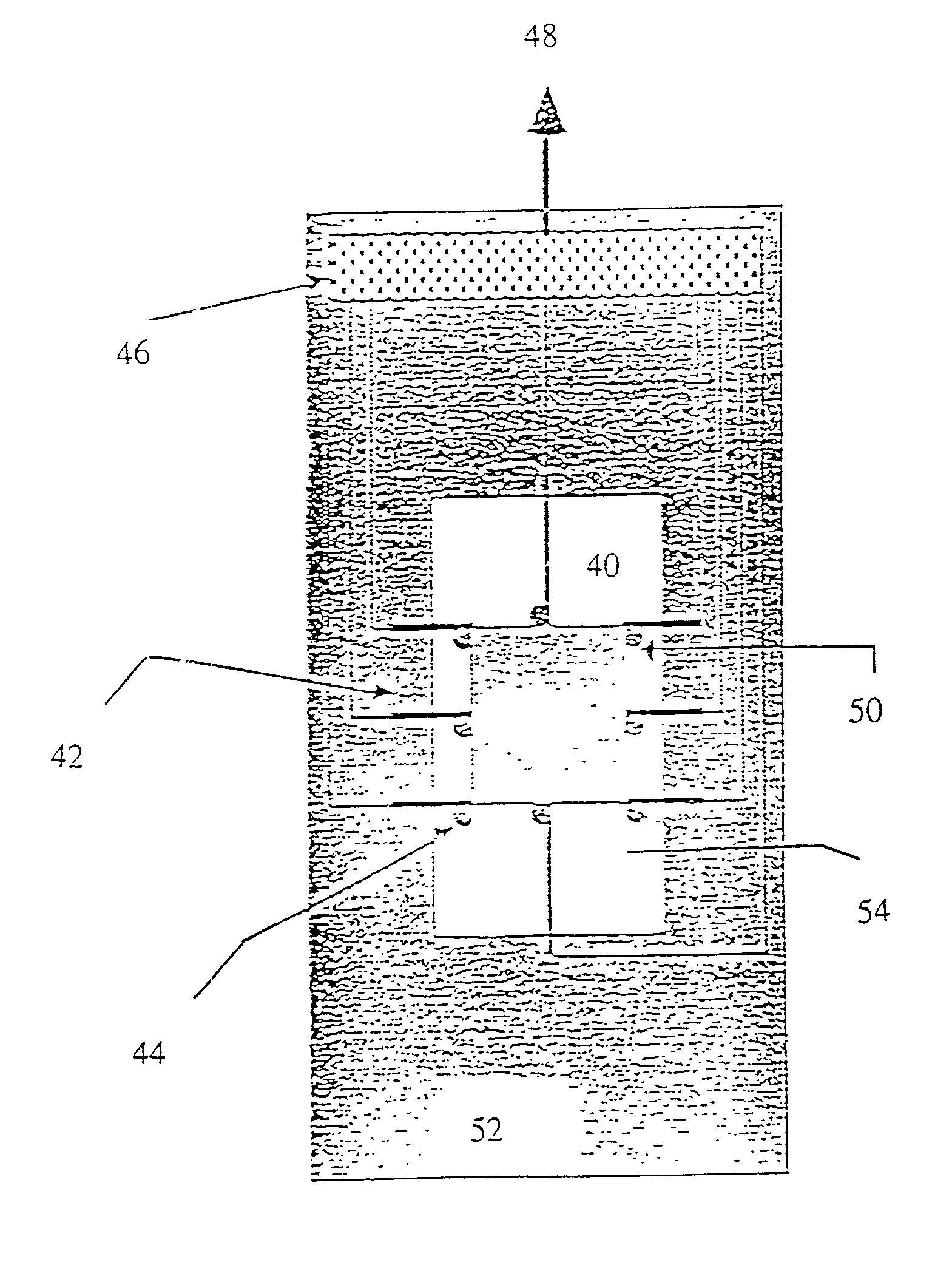
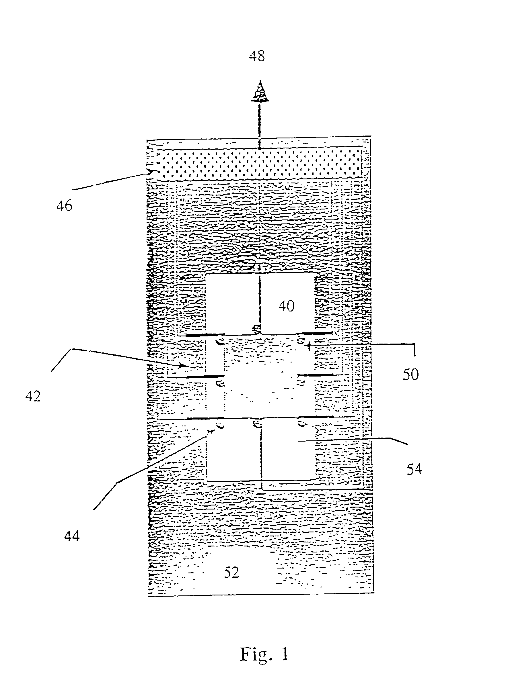
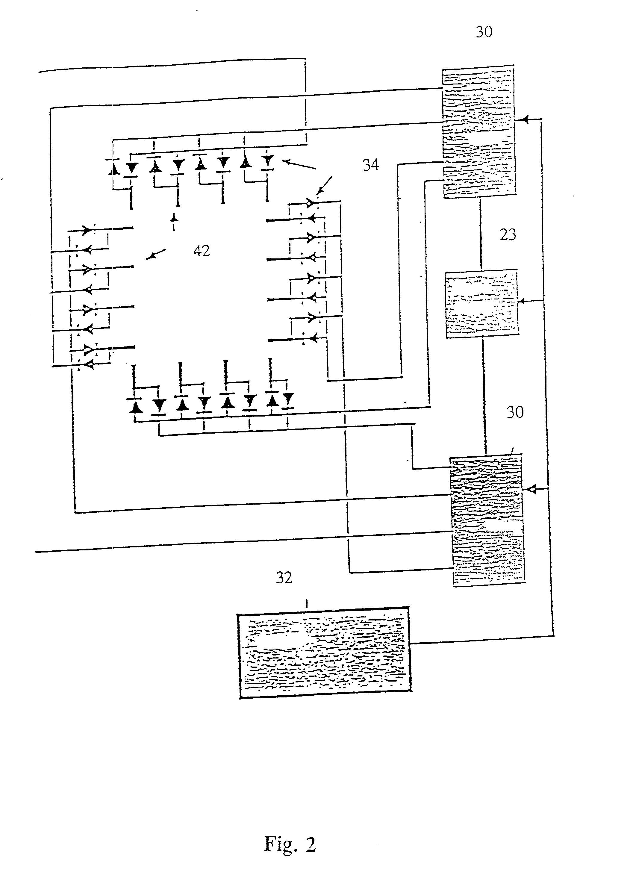
[0349] The sample of Example 1 was placed on a microscope slide. To mount the sample, approximately 3 microliters of the DNA-agarose mixture were carefully transferred to a preheated slide and cover slip using a pipetteman and pipette tips with the ends cut off to reduce Shear. Prepared slides were placed in a miniature pulsed electrophoresis apparatus as shown in FIGS. 1 and 2. All remaining steps were performed at room temperature. Samples were pre-electrophoresed for a few minutes and allowed to relax before any data was collected. Pulsed fields were created with either a chrontrol time (Chrontrol Corp., San diego, Calif.) or an Adtron data generating board (Adtron Corp., Gilbert, Ariz.) housed in an IBM AT computer and powered by a Hewlett Packard 6115A precision power supply. Field Strength was measured with auxiliary electrodes connected to a Fluke digital multimeter (J. Fluke Co., Everett, Wash.). A Zeiss Axioplan microscope (Carl Zeiss, West Germany) equi...


PUM

PropertyMeasurementUnit
Fractionaaaaaaaaaa
Fractionaaaaaaaaaa
Fractionaaaaaaaaaa

Abstract

A method for observing and determining the size of individual molecules and for determining the weight distribution of a sample containing molecules of varying size, which involves placing a deformable or nondeformable molecule in a medium, subjecting the molecule to an external force, thereby causing conformational and/or positional changes, and then measuring these changes. Preferred ways to measure conformational and positional changes include: (1) determining the rate at which a deformable molecule returns to a relaxed state after termination of the external force, (2) determining the rate at which a molecule becomes oriented in a new direction when the direction of the perturbing force is changed, (3) determining the rate at which a molecule rotates, (4) measuring the length of a molecule, particularly when it is at least partially stretched, or (5) measuring at least one diameter of a spherical or ellipsoidal molecule. Measurements of relaxation, reorientation, and rotation rates, as well as length and diameter can be made using a light microscope connected to an image processor. Molecule relaxation, reorientation and rotation also can be determined using a microscope combined with a spectroscopic device. The invention is particularly useful for measuring polymer molecules, such as nucleic acids, and can be used to determine the size and map location of restriction digests. Breakage of large polymer molecules mounted on a microscope slide is prevented by condensing the molecules before mounting and unfolding the molecules after they have been placed in a matrix.

Description

[0002] This application is a continuation-in-part of patent application Ser. No. 08 / 162,379 filed Dec. 7, 1993, which in turn is a continuation of patent application Ser. No. 07 / 333,531 filed Apr. 5, 1989 (abandoned). This application is also a continuation-in-part of patent application Ser. No. 08 / 128,996 filed Sep. 30, 1993, as a continuation-in-part of: (a) patent application Ser. No. 07 / 879,051 filed May 4, 1992 (allowed), which in turn, is a continuation of patent application Ser. No. 07 / 244,897 filed Sep. 15, 1988 (abandoned); and (b) patent application Ser. No. 07 / 333,531 filed Apr. 5, 1989 (abandoned), and patent application Ser. No. 07 / 244,897, filed Sep. 15, 1988 (abandoned). The entire contents of each of the foregoing applications is incorporated by reference herein in its entirety.[0003] 1. FIELD OF THE INVENTION[0004] This invention relates to methods and compositions for manipulating and characterizing individual polymer molecules, especially nucleic acid molecules, a...

Claims


Application Information

Patent Timeline
IPC IPC(8): C12Q1/68G01N15/02G01N15/10G01N15/14G01N27/447
CPCC12Q1/68C12Q1/683C12Q1/6841G01N27/44721G01N27/44773G01N2015/0288G01N2015/1062G01N2015/1081G01N2015/1486G01N2015/149G01N2015/1497
Inventor SCHWARTZ, DAVID C.
Owner WISCONSIN ALUMNI RES FOUND
Why Eureka
Try Eureka